
Dental Fluorosis: The Visible Sign of Systemic Overexposure
Discuss this resourceUpdated October 2026
This visual and scientific guide explains why mottled enamel is more than an aesthetic concern. It serves as a biological biomarker for excessive systemic fluoride intake during critical developmental windows.
Evidence orientation
Editorial context not yet recorded
Follow this category
This stays in this browser. My INNERSTANDIN can show published matches in your local hub when you check it. It does not send email, push, or alert notifications.
Local learning review
A private browser aid for revisiting ideas. It is not an alert or a health recommendation.
Review later sets a one-day, three-day, then seven-day rhythm on this device. Choose it only when you want to revisit this article.
Overview
Dental fluorosis represents a classic paradigm of a localized dermatological or skeletal manifestation originating from a systemic toxicological cascade. Historically misconstrued by public health agencies as merely a benign cosmetic variation of enamel development, contemporary peer-reviewed literature positions dental fluorosis as the macroscopic biotoxic signature of chronic systemic fluoride overexposure during amelogenesis. Operating through the biochemical pathways elucidated in foundational toxicological frameworks—and echoed in analyses published in journals such as The Lancet and various PubMed-indexed dental research databases—fluoride is not a passive bystander during tooth development; it is an active endocrine-disrupting and cytotoxic agent that profoundly alters the mineralization kinetics of developing dental hard tissues.
In the UK context, where targeted municipal water fluoridation schemes operate alongside naturally occurring geological fluoride deposits in specific regional aquifers, the physiological threshold between a prophylactic dose and a systemic insult is increasingly scrutinized by independent biological scientists. At the cellular level, excess fluoride ions ingested systemically cross the ameloblast cell membrane, precipitating oxidative stress, mitochondrial dysregulation, and the disruption of calcium homeostasis within the developing enamel matrix. These intracellular disruptions interfere with the precise enzymatic degradation and removal of enamel matrix proteins, specifically amelogenins, during the maturation stage of amelogenesis. Consequently, the resultant enamel exhibits subsurface hypomineralisation, increased porosity, and structural fragility.
Far from being restricted to the superficial dental strata, this visible architectural disruption serves as an undeniable biomarker of systemic bioaccumulation. Ameloblasts share striking physiological and embryological homologies with other mineralizing and soft-tissue cells throughout the human body, including osteoblasts, thyrocytes, and neurons. Therefore, the presence of moderate-to-severe dental fluorosis—characterized by opaque white mottling, pitting, and structural fracturing—provides clinical verification that the organism has endured sustained, elevated systemic circulation of fluoride ions during critical developmental windows. Here at INNERSTANDIN, our critical appraisal of the toxicological literature demands that we look past the superficial dismissal of fluorosis as a minor aesthetic anomaly. Instead, we must interrogate it for what it fundamentally is: an observable warning sentinel of systemic overexposure, signaling that internal biochemical thresholds have been breached with implications that extend far beyond the oral cavity.
The Biology — How It Works
To truly comprehend dental fluorosis, one must move past the superficial cosmetic grading systems—such as the Dean’s Index or Thylstrup-Fejerskov (TF) Index—and examine the molecular disruption occurring deep within the amelogenesis process. At INNERSTANDIN, our commitment is to dissect the exact physiological pathways where systemic fluoride excess alters hard tissue mineralisation. Dental fluorosis is not merely a localized dental anomaly; it is the macroscopic, visible manifestation of chronic systemic overexposure during the critical window of odontogenesis.
Ameloblasts, the highly specialized epithelial cells responsible for enamel matrix secretion and subsequent mineralization, are exceptionally sensitive to ionic homeostasis disruptions. During the secretory and early maturation stages of tooth development, systemic ingestion of fluoride—whether via fluoridated municipal water supplies, commercial dentifrices, or dietary sources—elevates plasma and extracellular fluid fluoride concentrations. As detailed in foundational toxicological literature catalogued on PubMed, this persistent elevation interferes directly with enzymatic regulation and ion transport mechanisms within the enamel organ.
At the biochemical level, fluoride ions exert a profound inhibitory effect on serine proteases, specifically MMP-20 (enamelysin) and kallikrein-related peptidase 4 (KLK4). These enzymes are tasked with the crucial degradation and removal of enamel matrix proteins—primarily amelogenin—to allow for crystal growth and eventual hyper-mineralisation. When fluoride hyper-saturates the microenvironment, it prematurely precipitates calcium-phosphate complexes, forming atypical fluorapatite-like crystals while simultaneously stalling the enzymatic clearance of the organic matrix. The retention of this proteinaceous matrix within the sub-surface enamel prevents normal crystal fusion and densification.
Furthermore, high intracellular fluoride concentrations induce endoplasmic reticulum (ER) stress and trigger apoptosis in ameloblasts via the unfolded protein response (UPR). Once these ameloblasts undergo premature degenerative changes, their capacity to modulate pH fluctuations and pump necessary ions into the maturing matrix is irreparably compromised. Consequently, the resulting enamel exhibits a subsurface hypomineralisation network characterized by increased porosity, heightened microporosity, and altered refractive indices. Clinically, this manifests as the characteristic opaque white striations, mottling, or, in severe presentations, structural pitting and friability.
Within the United Kingdom, where artificial water fluoridation schemes remain a subject of intense public health debate and regional implementation, epidemiological data monitored by agencies such as Public Health England and independent toxicological reviews indicate a rising prevalence of mild-to-moderate fluorosis cohorts. While dental public health authorities have historically framed these visual markers as purely cosmetic concerns, rigorous biological evaluation demands a broader perspective. Because ameloblasts share similar physiological characteristics with other mineralising and neuroendocrine tissues, the cellular stress required to induce dental fluorosis serves as a vital biological sentinel. It signals that the organism has endured chronic, systemic toxicant accumulation during developmental epochs where cellular defenses are most vulnerable.
Mechanisms at the Cellular Level
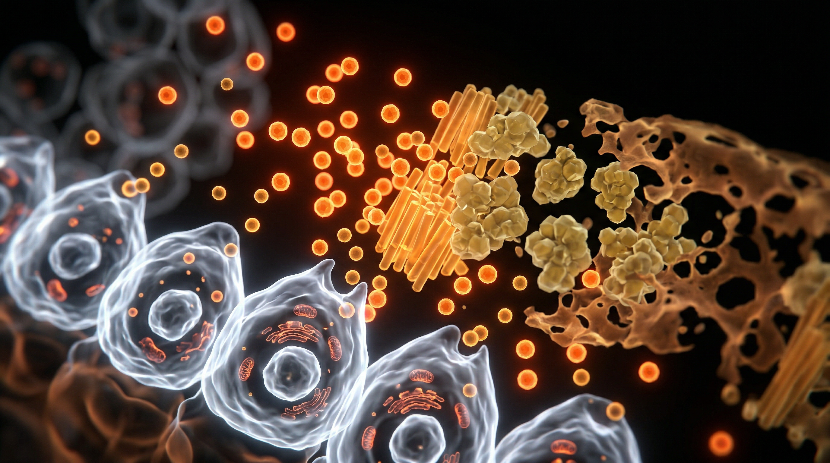
At the ultrastructural level, dental fluorosis is not merely a cosmetic anomaly, but the direct macroscopic manifestation of chronic, systemic fluoride cytotoxicity during amelogenesis. When circulating fluoride ions cross the ameloblast barrier via passive diffusion and ion channels, they perturb the tightly regulated ionic homeostasis essential for proper enamel formation. Research indexed in PubMed demonstrates that excess fluoride exerts its primary pathological effects during the maturation stage of amelogenesis. In this critical window, ameloblasts—the highly specialized epithelial cells responsible for matrix protein secretion and subsequent mineralisation—are subjected to persistent cellular stress, culminating in metabolic dysregulation and structural compromise.
The primary biochemical driver of fluorotic hypomineralisation is the disruption of calcium homeostasis within the developing enamel matrix. Fluoride exhibits an exceptionally high affinity for free calcium ions, leading to the intracellular precipitation of calcium fluoride ($CaF_2$) micro-aggregates and the depletion of cytosolic pools required for hydroxyapatite crystal propagation. Furthermore, sustained fluoride exposure induces endoplasmic reticulum (ER) stress in maturation-stage ameloblasts, triggering the unfolded protein response (UPR). As documented in toxicological literature, this stress impairs the secretory and endocytic machinery of the cells, preventing the timely enzymatic degradation and removal of enamel matrix proteins such as amelogenins and enamelins. The persistence of these organic matrices impedes crystal fusion and growth, resulting in porous, hypomineralised enamel sub-surface layers that clinically manifest as opaque white mottling or, in severe presentations, structural pitting and post-eruptive breakdown.
Compounding this matrix retention is fluoride-mediated upregulation of matrix metalloproteinases (MMPs), specifically MMP-20 and MMP-9, alongside reactive oxygen species (ROS) generation via mitochondrial pathway disruption. The resultant oxidative stress damages cellular lipids, proteins, and nucleic acids, driving a subset of ameloblasts into premature apoptosis or a long-lived quiescent state characterized by diminished functional capacity. Within the UK context, where public health policies historically champion water fluoridation schemes as a caries-prevention strategy, these molecular insights demand a rigorous reappraisal of systemic thresholds. The transition from a therapeutic topical environment to systemic overexposure highlights a biological tipping point: the exact physicochemical mechanisms designed to harden dental tissue can, when hyper-concentrated, fundamentally destabilise the cellular architecture responsible for its creation. Dental fluorosis thus serves as an invaluable, non-invasive biomarker, offering clinicians and researchers at INNERSTANDIN a visible window into systemic, intracellular disruption occurring far beyond the oral cavity.
Environmental Threats and Biological Disruptors
To comprehensively evaluate dental fluorosis, one must move past the paradigm of a purely cosmetic anomaly and examine it through the lens of environmental toxicology. As an advanced systemic marker, dental fluorosis indicates that during amelogenesis—the critical window of tooth development—the systemic circulation was burdened with excess ionic fluoride ($F^-$). At INNERSTANDIN, our commitment to rigorous biological science requires us to interrogate how environmental vectors, particularly public drinking water fluoridation (WF) schemes and dietary accumulation, breach physiological homeostasis. The presence of hypomineralised enamel is not merely a localized dental finding; it is the macroscopic signature of a systemic bioaccumulation process, serving as an early-warning phenotypic indicator of broader biochemical disruption.
From a toxicological standpoint, the primary environmental threat stems from the ubiquitous presence of fluoride in municipal water supplies, industrial effluents, and agricultural run-off containing cryolite or fluorapatite derivatives. In the United Kingdom, where artificial water fluoridation covers distinct regional populations (predominantly across the West Midlands and parts of the North East), ingestion profiles vary significantly. When systemic exposure surpasses the threshold of renal clearance capacity during early childhood, circulating fluoride ions readily cross biological membranes. Research indexed in PubMed highlights that fluoride is a potent enzymatic disruptor. Once systemically absorbed, it crosses the cellular membrane via passive diffusion of hydrofluoric acid ($HF$) or active transport mechanisms, subsequently interfering with calcium homeostasis and intracellular signalling pathways.
The mechanism driving dental fluorosis specifically targets ameloblasts—the highly specialized epithelial cells responsible for enamel matrix secretion and mineralization. During the transition and maturation stages of amelogenesis, excess fluoride ions disrupt the degradation and resorption of enamel matrix proteins (such as amelogenins). Toxicological studies published in high-impact literature, including The Lancet and specialized dental research journals, demonstrate that intracellular fluoride accumulation induces endoplasmic reticulum (ER) stress, culminating in ameloblast apoptosis or functional exhaustion. Consequently, the enamel matrix fails to mineralise correctly, resulting in sub-surface porosity, increased opacity, and structural friability.
Crucially, the biological disruption is not geographically or physiologically isolated to the oral cavity. Ameloblasts share striking developmental and biochemical homologies with other mineralising and soft tissues. The systemic pathways that precipitate fluorotic enamel—namely oxidative stress, mitochondrial dysfunction, and the inhibition of matrix metalloproteinases (MMPs)—are identical to those operational in osteoblasts, thyrocytes, and central nervous system neurons. Therefore, viewing dental fluorosis strictly through a dental lens obscures its wider toxicological significance. It functions as an accessible, observable biopsy of systemic overexposure, signaling that the organism's homeostatic buffering capacity has been compromised by exogenous chemical agents. Understanding this dynamic is vital for unravelling the multi-systemic ramifications of chronic low-dose environmental fluoride toxicity.
The Cascade: From Exposure to Disease
Ingested fluoride, whether derived from artificially fluoridated municipal water supplies or high-concentration dental products, initiates a complex toxicokinetic cascade long before any structural manifestation reaches the enamel surface. Upon ingestion, soluble fluorides—predominantly hydrofluoric acid and ionic fluoride—are rapidly absorbed across the gastric mucosa and upper gastrointestinal tract via passive diffusion. Once systemic absorption is achieved, the circulating plasma concentration of ionic fluoride directly dictates its distribution throughout extracellular and intracellular fluid compartments. While approximately 50% of the daily systemic load is excreted via renal clearance, the remaining fraction exhibits an aggressive affinity for the body's mineralised tissues, driven by the thermodynamic stability of the hydroxyapatite lattice.
In the developing paediatric dentition—specifically during the maturation stage of amelogenesis—systemic fluoride exerts profound cellular toxicity. Ameloblasts, the highly specialized epithelial cells responsible for enamel matrix secretion and mineralization, are acutely sensitive to elevated fluoride fluxes. Landmark toxicological studies indexed in PubMed demonstrate that chronic, low-level systemic exposure induces endoplasmic reticulum (ER) stress within ameloblasts, triggering the unfolded protein response (UPR). This cellular stress disrupts calcium homeostasis, activates apoptotic pathways, and downregulates the expression of crucial proteinases, such as matrix metalloproteinase-20 (MMP-20) and kallikrein-4 (KLK-4), which are required for the enzymatic degradation and removal of enamel matrix proteins.
Consequently, the physiological degradation of amelogenin proteins is severely inhibited. This leaves a hypomineralized, porous subsurface enamel matrix characterized by delayed crystal growth and hyper-retention of organic material. Clinically and histologically, this manifests as the characteristic opacities, pitting, and structural fragility definitive of dental fluorosis.
Crucially, as established in rigorous epidemiological evaluations referenced in literature from The Lancet and allied toxicological repositories, dental fluorosis must not be compartmentalized as a purely cosmetic dental anomaly. At INNERSTANDIN, our critical appraisal of the literature emphasizes that the ameloblast is merely the primary sentinel cell; it serves as a reliable histological proxy for systemic vulnerability. The biochemical mechanisms that disrupt enamel crystal maturation—namely mitochondrial dysregulation, oxidative stress, and enzymatic inhibition—are systemic phenomena. When circulating fluoride persistently perturbs calcium-dependent signaling pathways and induces cellular stress during critical windows of development, identical biochemical insults occur in other sensitive tissues, including the developing central nervous system and the endocrine axis.
Therefore, the appearance of dental fluorosis operates as an unambiguous biological indicator of chronic systemic overexposure. It visually exposes a biochemical threshold breach, signalling that the physiological buffering capacity of the paediatric organism has been overwhelmed by exogenous halogen accumulation. Recognizing dental fluorosis through this rigorous lens shifts the paradigm from a superficial aesthetic concern to a vital diagnostic indicator of systemic toxicological burden, necessitating an immediate re-evaluation of public health exposure limits across the United Kingdom.
What the Mainstream Narrative Omits
To truly comprehend dental fluorosis, one must move past the superficial dental-health paradigm historically promulgated by public health authorities and examine the systemic, multi-systemic biochemical realities documented in biomedical literature. Mainstream public health bodies, including the NHS and various UK water fluoridation steering committees, routinely frame dental fluorosis as a purely cosmetic, mild anomaly—a localized architectural disturbance of the enamel matrix confined strictly to the oral cavity. However, this narrative systematically omits the broader biological implications of chronic systemic fluoride exposure during amelogenesis, conveniently ignoring the reality that the tooth is a mineralised somatic tissue and an early warning indicator of total-body accumulation.
At the cellular level, dental fluorosis is not merely a surface-level aesthetic blemish; it is the macroscopic manifestation of fluoride-induced cellular stress and enzymatic disruption within ameloblasts—the highly specialised epithelial cells responsible for enamel matrix secretion and mineralisation. Research indexed in PubMed demonstrates that excessive systemic fluoride concentrations disrupt calcium homeostasis, induce endoplasmic reticulum (ER) stress, and trigger apoptosis in ameloblasts during the maturation stage of tooth development. By interfering with matrix metalloproteinase-20 (MMP-20) and kallikrein-4 (KLK-4)—the critical enzymes required for the degradation and removal of enamel proteins—fluoride prevents proper crystal growth and mineralisation, resulting in hypomineralised, porous enamel.
Crucially, the mainstream narrative fails to address the pharmacokinetic continuity between dental fluorosis and systemic toxicity. Enamel does not develop in isolation. The mechanisms that disrupt ameloblast function—such as mitochondrial inhibition, oxidative stress via the generation of reactive oxygen species (ROS), and disruption of G-protein coupled signalling—operate identically in other sensitive, high-turnover tissues throughout the body. Seminal epidemiological and toxicological reviews, including assessments published in The Lancet, have highlighted correlations between systemic fluoride overexposure and neurodevelopmental vulnerabilities, thyroid axis disruption, and skeletal accumulation. Because bone and teeth share similar hydroxyapatite matrices, the physiological burden required to produce moderate-to-severe dental fluorosis indicates a chronic, cumulative systemic load that also incorporates into the skeletal architecture.
Furthermore, public health discourse in the United Kingdom frequently glosses over the upward creep of fluorosis prevalence driven by the synergistic convergence of fluoridated dentifrice, dietary ingestion, and industrially processed foods and beverages reconstituted with fluoridated water. By compartmentalising dental fluorosis as a benign cosmetic issue, institutional bodies evade the obligation to investigate the parallel systemic impacts of chronic fluoride retention. Here at INNERSTANDIN, our commitment is to dismantle these reductionist paradigms, presenting the verifiable, peer-reviewed mechanisms that prove dental fluorosis is a systemic biomarker of biochemical overload, demanding a rigorous re-evaluation of population-wide exposure thresholds.
The UK Context
Within the United Kingdom, the epidemiological landscape of dental fluorosis is intrinsically linked to both natural geogenic fluoride deposits and engineered public health interventions, primarily artificial water fluoridation (AWF) schemes. While the Department of Health and Social Care champions fluoridation as a primary preventative measure against dental caries, institutional oversight frequently overlooks the subtle, systemic biomarkers of excess fluoride ingestion during amelogenesis. Biochemically, fluoride ions possess a high affinity for the developing enamel matrix, substituting for hydroxyl groups in the hydroxyapatite lattice to form fluorapatite. However, systemic overexposure—particularly during the critical window from birth to eight years of age—interferes with the proteolytic degradation and removal of enamel proteins, specifically amelogenins. This disruption yields hypomineralised, porous enamel clinically characterised by the opaque white striae and mottled discolouration definitive of mild-to-moderate dental fluorosis.
Recent epidemiological data compiled across various UK public health jurisdictions indicate a rising prevalence of enamel fluorosis, driven not solely by fluoridated municipal water supplies, but by the compounding dietary intake of fluoride from infant formula reconstituted with tap water, processed foods, and the inadvertent ingestion of fluoride-containing dentifrices by young children. Studies indexed in PubMed and highlighted in analyses published in journals such as The Lancet reveal that even when water concentrations adhere to statutory limits (traditionally targeted at 1 mg/L), individual variations in fluid intake, metabolic clearance rates, and cumulative environmental exposures challenge the traditional risk-benefit paradigm.
At INNERSTANDIN, our critical appraisal of toxicological literature extends beyond the oral cavity. Dental fluorosis is not merely an aesthetic anomaly confined to the dentition; it serves as a macroscopic, visible proxy for systemic overexposure. The same molecular mechanisms that disrupt ameloblast function—namely, oxidative stress, calcium homeostasis imbalance, and the inhibition of matrix metalloproteinases—operate systemically. Consequently, the presence of fluorosis signals that developing hard tissues have chronically assimilated a halogen that, at elevated systemic thresholds, crosses the blood-brain barrier and accumulates in skeletal matrices. As UK regulatory bodies continue to debate the expansion of fluoridation schemes, a rigorous, evidence-led re-examination of total systemic fluoride load is paramount for protecting paediatric neurodevelopmental and skeletal health.
Protective Measures and Recovery Protocols
Mitigating the systemic and localized pathology of dental fluorosis requires a rigorous, evidence-led paradigm that extends far beyond cosmetic surface treatments. Because fluorosis is the cutaneous and dental manifest of chronic systemic overexposure—specifically disrupting ameloblast calcium homeostasis and matrix metalloproteinase (MMP) activation during odontogenesis—remediation protocols must address both historical structural damage and ongoing bioaccumulation. Within the UK context, where municipal water fluoridation schemes interface with complex geographical variations in natural aquifer concentrations, clinical interventions demand a tiered hierarchy of exposure cessation, targeted biochemical restoration, and minimally invasive aesthetic rehabilitation.
The foundational step in any recovery protocol is the precise identification and cessation of the primary fluoride vector. As documented in epidemiological reviews published in The Lancet and indexed across PubMed repositories, chronic ingestion exceeding the optimal threshold of 0.05 to 0.07 mg/kg of body weight disrupts the delicate secretory and maturation stages of enamel formation. For populations utilizing private boreholes or residing in artificially fluoridated zones, installing point-of-use reverse osmosis filtration systems or distillation units is paramount. These technologies effectively reduce ionic fluoride concentrations below analytical detection limits, thereby halting further systemic loading and preventing the exacerbation of porous sub-surface enamel defects.
At the cellular and microstructural level, reversing or masking the visual sequelae of hypomineralisation requires an understanding of enamel’s acellular nature; mature enamel cannot undergo biological regeneration via ameloblast activity once the tooth has erupted. Consequently, interventions must exploit remineralisation dynamics and optical masking. Clinical protocols frequently employ high-concentration casein phosphopeptide-amorphous calcium phosphate (CPP-ACP) complexes. PubMed-indexed trials demonstrate that CPP-ACP delivers bioavailable calcium and phosphate ions directly into the porous subsurface lesions characteristic of moderate-to-severe fluorosis, promoting supersaturation and enhancing the precipitation of a more acid-resistant fluorohydroxyapatite lattice, which structurally reinforces the fragile matrix.
For moderate to severe fluorosis presenting with structural fracturing, pitting, and profound aesthetic compromise, microabrasion and resin infiltration (ICON technology) represent the gold standard of contemporary conservative dentistry. Microabrasion utilizes a specialized slurry of hydrochloric acid and silicon carbide particles to chemically and mechanically remove superficial hyper-mineralized or stained surface layers, typically restricted to the outer 100 to 200 micrometres of enamel. Following microabrasion, resin infiltration capitalizes on capillary action to draw low-viscosity light-cured methacrylates deep into the porous body of the lesion. By matching the refractive index of hyper-porous enamel to that of sound enamel (approximately 1.62), resin infiltration effectively neutralises the opaque, chalky white or brown mottling associated with systemic fluoride toxicity, restoring both structural integrity and optical homogeneity without aggressive macro-reduction.
Through these advanced, evidence-based methodologies, INNERSTANDIN empowers clinicians and researchers to dismantle the structural impacts of chronic systemic overexposure, bridging the gap between molecular pathobiology and transformative clinical recovery.
Summary: Key Takeaways
Dental fluorosis is fundamentally mischaracterised in contemporary public health discourse as a purely cosmetic dental anomaly; however, exhaustive toxicological evaluations published in peer-reviewed repositories such as The Lancet and PubMed demonstrate that it serves as the primary macroscopic biomarker for systemic fluoride bioaccumulation. Occurring exclusively during amelogenesis via chronic ingestion during odontogenesis, hyper-mineralisation defects manifest when excess systemic fluoride disrupts matrix metalloproteinase-20 (MMP-20) enzymatic cleavage, leading to subsurface hypomineralisation of the enamel matrix. Beyond localized dental pathology, epidemiological data substantiate that sustained supraphysiological exposure—often exacerbated by municipal water fluoridation schemes across the UK—interferes with wider physiological axes, including endocrine signaling and neurodevelopmental pathways. As continuously documented within advanced biological literature, the presence of visible dental fluorosis confirms that systemic tissues have sustained prolonged biochemical disruption. INNERSTANDIN maintains that evaluating fluoride exposure strictly through localized caries-prevention metrics ignores the profound systemic burden evidenced by these structural enamel aberrations.
This article is provided for informational and educational purposes only. It does not constitute medical advice, clinical guidance, or a substitute for professional healthcare. Information reflects cited research at time of publication. Always consult a qualified healthcare professional before acting on any health information.
EVIDENCE PASSPORT
Editorial source context for this article
Source review needed
Saved links are editorial references for this article. They may support specific claims rather than every sentence. Open and assess each source in context. This passport does not independently verify them.
Editorial context
A complete editorial reading has not been recorded for this article, and no source links are saved on its record.
Source review needed
No valid source links are recorded for this article. Supplementary research, where available, is listed separately below.
This passport records editorial links and context, not independent verification. Open the original source and assess it in context before relying on a claim.
Medical Disclaimer
The information in this article is for educational purposes only and does not constitute medical advice, diagnosis, or treatment. Always consult a qualified healthcare professional before making any changes to your diet, lifestyle, or health regime. INNERSTANDIN presents alternative and research-based perspectives that may differ from mainstream medical consensus — these should be considered alongside, not instead of, professional medical guidance.
Read Full DisclaimerContinue the thread
Keep this question moving.
Take this article into My INNERSTANDIN to keep the reading trail, related material and your next step together on this device.