
Dysregulated Apoptosis in Neurodegeneration: The Role of Mitochondrial Outer Membrane Permeabilization
Discuss this resourceUpdated August 2026
An in-depth analysis of how mitochondrial instability leads to premature neuronal cell death, contributing to the progression of chronic neurodegenerative diseases.
Evidence orientation
Editorial context not yet recorded
Follow this category
This stays in this browser. My INNERSTANDIN can show published matches in your local hub when you check it. It does not send email, push, or alert notifications.
Local learning review
A private browser aid for revisiting ideas. It is not an alert or a health recommendation.
Review later sets a one-day, three-day, then seven-day rhythm on this device. Choose it only when you want to revisit this article.
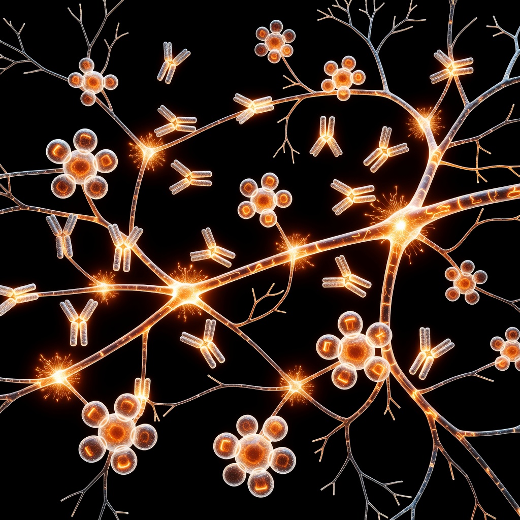
Introduction to Cellular Homeostasis. Apoptosis, or programmed cell death, is an essential physiological process required for maintaining tissue homeostasis. In the human brain, this mechanism serves as a quality control system, removing damaged or obsolete neurons. However, in neurodegenerative conditions such as Alzheimer's, Parkinson's, and Huntington's disease, this tightly regulated process becomes dysregulated, leading to pathological cell loss. ### The Mitochondrial Sentinel. At the heart of the intrinsic apoptotic pathway lies the mitochondrion.
Often termed the 'powerhouse' of the cell, it is also the primary sentinel for cellular stress. Mitochondrial Outer Membrane Permeabilization (MOMP) is widely considered the 'point of no return' in the apoptotic cascade. When cells face chronic oxidative stress, misfolded protein accumulation, or metabolic dysfunction, the mitochondrial membranes undergo a critical structural change. ### Mechanisms of MOMP. The integrity of the mitochondrial outer membrane is governed by the Bcl-2 family of proteins. These proteins act as a rheostat, balancing pro-survival and pro-death signals.
In neurodegeneration, the pro-apoptotic members, specifically BAX and BAK, undergo conformational changes that facilitate the formation of macropores within the outer membrane. Once these pores form, cytochrome c and other pro-apoptotic factors are released into the cytosol, triggering the activation of caspases—the executioners of cellular destruction. ### The Root Cause: Why Neurons Fail. Neurons are uniquely vulnerable due to their high metabolic demand and post-mitotic nature. Unlike other cells, they cannot easily replace their population once lost. In neurodegenerative states, the root cause often stems from chronic mitochondrial DNA damage and the impaired clearance of damaged mitochondria, a process known as mitophagy.
When mitophagy fails, damaged mitochondria accumulate, perpetually leaking reactive oxygen species (ROS) and lowering the threshold for MOMP. This chronic priming means that neurons are essentially on the brink of apoptosis, susceptible to even minor physiological stressors. ### Clinical Implications. Understanding the threshold-gating of MOMP offers new avenues for therapeutic intervention. Rather than merely treating symptoms, research is shifting toward stabilizing mitochondrial membranes and enhancing autophagic flux to clear damaged organelles before they can trigger the apoptotic cascade. By modulating the Bcl-2 protein family balance, we may be able to extend the longevity of vulnerable neuronal populations, offering hope for slowing the progression of currently incurable diseases.
This article is provided for informational and educational purposes only. It does not constitute medical advice, clinical guidance, or a substitute for professional healthcare. Information reflects cited research at time of publication. Always consult a qualified healthcare professional before acting on any health information.
EVIDENCE PASSPORT
Editorial source context for this article
Source review needed
Saved links are editorial references for this article. They may support specific claims rather than every sentence. Open and assess each source in context. This passport does not independently verify them.
Editorial context
A complete editorial reading has not been recorded for this article, and no source links are saved on its record.
Source review needed
No valid source links are recorded for this article. Supplementary research, where available, is listed separately below.
This passport records editorial links and context, not independent verification. Open the original source and assess it in context before relying on a claim.
Medical Disclaimer
The information in this article is for educational purposes only and does not constitute medical advice, diagnosis, or treatment. Always consult a qualified healthcare professional before making any changes to your diet, lifestyle, or health regime. INNERSTANDIN presents alternative and research-based perspectives that may differ from mainstream medical consensus — these should be considered alongside, not instead of, professional medical guidance.
Read Full DisclaimerContinue the thread
Keep this question moving.
Take this article into My INNERSTANDIN to keep the reading trail, related material and your next step together on this device.
Explore this in the Body Map
Explore related body structures and their educational pages in the interactive Body Map.
Dig deeper in the Library
Free, longform PDF volumes that go beyond headlines into mechanisms and references.