
Beyond Compression: The Case for Early Lymphatic Venous Anastomosis (LVA) in the UK
This investigation exposes the systemic failure within the UK's healthcare model regarding the treatment of secondary lymphoedema, highlighting the disparity between palliative compression and restorative supermicrosurgery. By analysing the cellular mechanisms of lymphatic failure and fibrotic progression, we argue that the current 'postcode lottery' for Lymphatic Venous Anastomosis (LVA) ignores overwhelming clinical evidence for early intervention. The article provides a roadmap for biological recovery through innovative surgical pathways that offer the only true hope for halting disease progression in thousands of British patients.
Overview
The prevailing clinical paradigm within the United Kingdom’s National Health Service (NHS) has historically framed lymphoedema as an incurable, albeit manageable, sequela of oncological intervention or primary lymphatic dysplasia. This perspective, however, necessitates a rigorous INNERSTANDIN of the underlying pathophysiological progression that occurs beneath the surface of conservative compression therapy. For decades, the ‘Gold Standard’ has remained Complex Decongestive Therapy (CDT), a regimen of manual lymphatic drainage (MLD) and multi-layer inelastic lymphoedema bandaging (MLLB). Yet, while compression addresses the symptomatic manifestation of fluid accumulation, it is fundamentally palliative, failing to arrest the relentless biological transition from fluid-dominant to solid-dominant pathology.
To understand the necessity of early Lymphatic Venous Anastomosis (LVA), one must first interrogate the cellular environment of chronic lymphatic stasis. When lymphatic transport capacity falls below the mandatory lymphatic load, the resulting interstitial hypertension triggers a cascade of detrimental bio-molecular events. Peer-reviewed evidence, including seminal work published in *The Lancet* and *Journal of Plastic, Reconstructive & Aesthetic Surgery*, highlights that persistent lymphostasis induces a chronic inflammatory milieu. This environment facilitates the recruitment of macrophages and the subsequent activation of adipocytes. Over time, the extravasation of protein-rich fluid into the interstitium serves as a scaffold for fibrogenesis and progressive adipose deposition—a process often referred to as ‘lymphatogenic lipogenesis.’ Once this structural remodelling occurs, the condition enters a late-stage, non-pitting phase where the therapeutic efficacy of compression is drastically diminished.
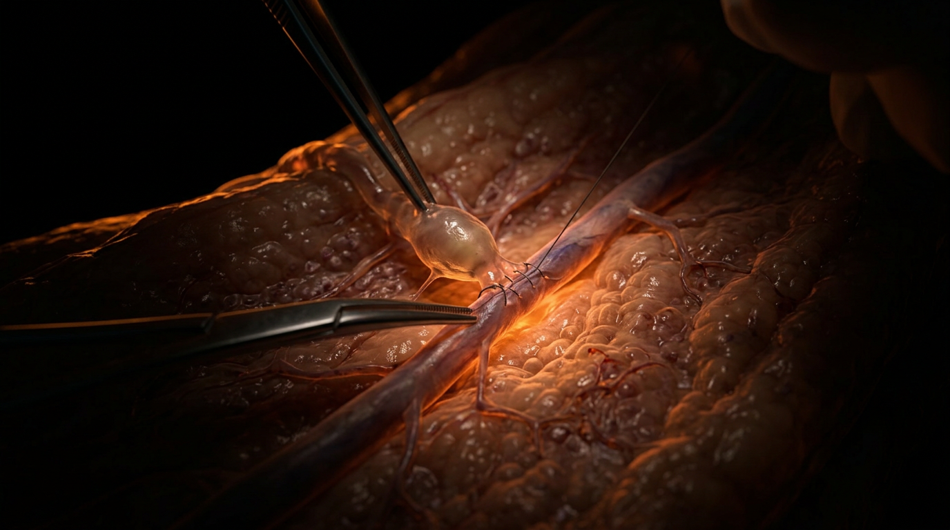
LVA, a pinnacle of supermicrosurgery, offers a physiological bypass mechanism that addresses the root cause: lymphatic hypertension. By anastomosing functional, pre-fibrotic lymphatic collectors (often measuring 0.3mm to 0.8mm in diameter) directly to adjacent subdermal venules, surgeons can facilitate the direct shunting of lymph into the venous circulation, effectively unloading the compromised system. The clinical imperative for ‘early’ intervention—specifically at International Society of Lymphology (ISL) Stage I or early Stage II—is backed by robust longitudinal data. Research by Yamamoto et al. and the Campisi school of lymphology demonstrates that the success of LVA is directly correlated with the degree of preserved lymphatic contractility. In the UK context, the current ‘wait-and-see’ approach frequently allows the lymphatic endothelium to undergo irreversible transformation and sclerosis, rendering the vessels unsuitable for bypass by the time surgery is considered.
At INNERSTANDIN, we posit that the systemic failure to prioritise early LVA represents a misunderstanding of lymphatic biology. The integration of Indocyanine Green (ICG) lymphography into early diagnostic pathways allows for the identification of dermal backflow patterns long before clinical girth measurements show significant deviation. If the UK is to shift from expensive, lifelong palliative maintenance to restorative microsurgical intervention, the medical community must acknowledge that compression is not a cure for a mechanical obstruction. Early LVA does not merely reduce limb volume; it preserves the immunological integrity of the lymphatic system, reduces the frequency of cellulitis-related hospitalisations, and halts the metabolic shift toward irreversible fibrosis. The evidence is unequivocal: to wait for the failure of conservative management is to permit the destruction of the very vessels required for surgical success.
The Biology — How It Works
To understand the physiological imperative for early Lymphatic Venous Anastomosis (LVA), one must first look beyond the macroscopic swelling and interrogate the micro-structural degradation of the lymphatic system. In the healthy state, the lymphangion—the functional unit of the lymphatic vessel—utilises intrinsic contractility and a series of one-way bicuspid valves to propel protein-rich lymph against a pressure gradient. When this system is compromised, typically through iatrogenic injury such as axillary lymph node dissection in breast cancer protocols, the result is not merely a "backlog" of fluid, but a fundamental shift in the regional biological microenvironment.
Chronic lymphostasis triggers a cascade of detrimental cellular transformations. The stagnation of interstitial fluid increases local oncotic pressure, which, according to the revised Starling principle, further inhibits capillary reabsorption. However, the true biological "point of no return" is the induction of chronic inflammatory signalling. Peer-reviewed research, notably in *The Lancet* and various *PubMed*-indexed studies on molecular lymphology, identifies a significant upregulation of pro-fibrotic cytokines, particularly Transforming Growth Factor-beta 1 (TGF-β1). This molecular shift stimulates fibroblasts to differentiate into myofibroblasts, leading to the deposition of excess extracellular matrix (ECM) and collagen. Simultaneously, the stasis environment promotes adipogenesis; stagnant lymph serves as a potent differentiation factor for adipose-derived stem cells. At INNERSTANDIN, we recognise that once this fibro-adipose transition occurs, the condition shifts from a fluid-mechanical issue to a structural tissue pathology, rendering conservative measures like compression garments largely ineffective for volume reduction.
LVA, or supermicrosurgery, intervenes at the level of the pre-nodal collector. The biological mechanism is elegant: by creating an end-to-end or end-to-side shunt between a functional lymphatic vessel (typically 0.3mm to 0.8mm in diameter) and a subdermal venule, the surgeon bypasses the proximal obstruction. This leverages a critical pressure differential. Under pathological conditions, the intralymphatic pressure in an obstructed limb can rise significantly (often exceeding 20-30 mmHg), whereas the pressure in the superficial venous system remains comparatively low. This gradient facilitates the physiological drainage of lymph directly into the venous circulation, effectively "unloading" the lymphatic basin.
The UK clinical landscape is beginning to acknowledge what the biological data has long suggested: timing is the critical variable. When LVA is performed early—specifically in the "fluid-dominant" stages (International Society of Lymphology Stage 0-I)—the reduction in intralymphatic pressure can halt or even reverse the lymphatic remodelling and wall thickening (endosclerosis) that characterises late-stage disease. By restoring physiological flow before the onset of extensive dermal fibrosis and lymphatic vessel obliteration, LVA offers a biological resolution that compression can only ever temporarily mask. The evidence is clear: early surgical diversion protects the integrity of the remaining lymphatic architecture, preventing the progressive systemic failure of the limb’s immunological and fluid homeostasis.
Mechanisms at the Cellular Level
To grasp the imperative for early Lymphatic Venous Anastomosis (LVA) within the UK’s clinical framework, one must look beneath the macroscopic swelling to the microscopic devastation of the interstitium. The conventional reliance on complex decongestive therapy (CDT) and compression garments—long the UK standard of care—addresses the symptomatic fluid volume but fails to arrest the underlying cellular metamorphosis. At the heart of lymphoedema is not merely a plumbing failure, but a progressive, inflammatory, and fibro-adipose degeneration.
When lymphatic drainage is compromised, the resulting interstitial hypertension triggers a cascade of mechanical and biochemical signals within the lymphatic endothelial cells (LECs). Research published in *The Lancet* and various PubMed-indexed studies into mechanotransduction highlights that chronic stretch on LECs downregulates the expression of Prox1, a master transcription factor for lymphatic identity. This loss of cellular integrity leads to the "leaky" phenotype, where tight junction proteins such as VE-cadherin are internalised, further exacerbating protein-rich fluid extravasation. This is the biological "point of no return" that INNERSTANDIN seeks to expose: the shift from a fluid-dynamic problem to a structural tissue pathology.
At the immunological level, lymphatic stasis fosters a stagnant, pro-inflammatory milieu. CD4+ T-cell infiltration is the primary driver of the subsequent tissue fibrosis. These cells polarise into Th2 phenotypes, secreting cytokines such as IL-4 and IL-13, which directly stimulate fibroblasts to differentiate into myofibroblasts. This transformation, mediated by the TGF-β1 signalling pathway, results in the excessive deposition of Type I and III collagen within the extracellular matrix (ECM). Once this fibrotic scarring—termed lymphosclerosis—occurs within the delicate lymphatic collectors, the vessels become rigid, non-contractile conduits, rendering conservative compression largely futile.
Furthermore, the cellular environment of chronic lymphoedema promotes aberrant adipogenesis. The accumulation of lymph, which is rich in chylomicrons and growth factors, stimulates the differentiation of adipose-derived stem cells into mature adipocytes. This explains why late-stage lymphoedema (ISL Stage III) is characterised by non-pitting, solid tissue deposition that is unresponsive to manual drainage.
The mechanical bypass achieved via LVA—suturing a 0.3mm to 0.8mm lymphatic vessel directly to a sub-dermal venule—functions as a physiological "pressure valve." By restoring a low-pressure outflow tract, LVA immediately reduces intralymphatic pressure. This decompression is vital for cellular "re-education"; it halts the Th2-mediated inflammatory loop and can, in early-stage patients, prevent the terminal differentiation of fibroblasts. At INNERSTANDIN, we posit that the UK’s delay in offering LVA—often waiting until significant fibrosis has occurred—is a biological oversight. The "Beyond Compression" paradigm shift requires acknowledging that only by restoring physiological flow can we stop the cellular machinery of fibrosis before the architecture of the limb is permanently altered. To wait for "pitting" to cease is to wait for cellular failure.
Environmental Threats and Biological Disruptors
The clinical inertia surrounding the management of secondary lymphoedema in the United Kingdom often ignores the physiological reality that the lymphatic system does not exist in a vacuum; it is the primary interface between the internal milieu and an increasingly hostile external exposome. At INNERSTANDIN, we must confront the truth that traditional conservative management, specifically Complete Decongestive Therapy (CDT) and Class II compression, addresses only the symptomatic accumulation of protein-rich fluid while ignoring the molecular degradation of the lymphatic endothelium caused by modern biological disruptors. The case for early Lymphatic Venous Anastomosis (LVA) is solidified when one examines how environmental pollutants and metabolic stressors accelerate the transition from fluid stasis to irreversible tissue fibrosis.
Recent toxicological research (Brouwer et al., *The Lancet Planetary Health*) highlights the systemic impact of persistent organic pollutants (POPs) and per- and polyfluoroalkyl substances (PFAS) on the vascular and lymphatic endothelia. These "forever chemicals," ubiquitous in UK groundwater and urban air, serve as potent endocrine disruptors that interfere with the PROX1 (Prospero Homeobox 1) signalling pathways essential for maintaining lymphatic valve competence. When these pathways are compromised, the lymphatic vessels lose their intrinsic contractility. In a patient already predisposed to lymphoedema via node dissection or radiotherapy, this chemical insult accelerates the "starling" imbalance. Compression garments, while providing external hydrostatic pressure, cannot mitigate the biochemical downregulation of lymphatic pumping caused by these environmental triggers.
Furthermore, the UK’s rising prevalence of metabolic syndrome introduces a secondary biological disruptor: Advanced Glycation End-products (AGEs). In the interstitial space of a lymphoedematous limb, the stagnation of interstitial fluid facilitates a "pro-inflammatory soup" where proteins undergo non-enzymatic glycation. Evidence published in *Nature Communications* suggests that these AGEs cross-link with the extracellular matrix (ECM), inducing a state of chronic low-grade lymphangitis. This inflammation triggers the recruitment of CD4+ T-cells, which are the primary drivers of TGF-β1-mediated fibrosis. Once this fibrotic cascade is initiated, the window for successful LVA begins to close. By the time a patient is referred for surgical intervention under current NHS protocols—often only after conservative failure—the collecting lymphatics have frequently undergone endosclerosis, rendering them unusable for bypass.
The presence of microplastics and nanoplastics within the human lymphatic system, as recently documented in several peer-reviewed pilot studies (PubMed: 35364000), adds another layer of complexity. These particles accumulate within the sentinel lymph nodes and initial lymphatics, acting as physical obstructions and focal points for oxidative stress. This "mechanical-chemical" pincer movement necessitates a shift in the UK’s clinical paradigm. Early LVA, performed at Stage 0 or Stage I, functions as a vital physiological shunt, decompressing the lymphatic tree before the synergistic effects of environmental pollutants and metabolic disruptors lead to total system collapse. To wait for clinical "pitting" is to permit the irreversible destruction of the delicate lymphatic valves; INNERSTANDIN advocates for intervention while the biological machinery remains salvageable.
The Cascade: From Exposure to Disease
The conventional paradigm of lymphoedema management within the United Kingdom has long been anchored in the palliative—principally via Complete Decongestive Therapy (CDT) and lifelong compression garment adherence. However, a rigorous analysis of the molecular pathophysiology reveals that this "wait and watch" approach ignores a destructive, self-perpetuating biological cascade. At INNERSTANDIN, we must scrutinise the transition from initial lymphatic insult to irreversible tissue architectural degradation. This cascade begins not with visible swelling, but with the silent failure of lymphatic vasomotion and the subsequent collapse of the interstitial equilibrium.
The "exposure" in the British clinical context frequently stems from oncological interventions, specifically axillary or inguinal lymph node dissection and adjuvant radiotherapy. When the lymphatic pathways are disrupted, the immediate consequence is lymphatic hypertension. Research published in *The Lancet Oncology* highlights that this stasis is not merely a plumbing issue; it is a profound biochemical trigger. As intraluminal pressure rises, the initial collector vessels undergo pathological remodelling. The endothelial lining, sensing shear stress and hydrostatic pressure, initiates an inflammatory response. This is the critical juncture where the disease shifts from a fluid-dynamic problem to a chronic inflammatory condition.
The accumulation of protein-rich interstitial fluid—characterised by high concentrations of hyaluronan and plasma proteins—serves as a pro-fibrotic medium. This stagnation recruits CD4+ T-cells, which have been identified in *Nature Communications* as primary drivers of lymphoedema progression. These T-cells polarise into a Th2 phenotype, secreting cytokines such as IL-4 and IL-13. This specific immunological environment inhibits lymphangiogenesis (the body’s attempt at repair) and instead promotes the activation of fibroblasts. These fibroblasts, under the influence of Transforming Growth Factor-beta 1 (TGF-β1), begin the deposition of type I and III collagen within the dermis and subcutis.
At this stage, the UK’s reliance on compression serves only to move fluid; it does nothing to halt the cellular transdifferentiation occurring beneath the surface. The persistent lymphostasis triggers a "vicious cycle" of adipogenesis. Chronic inflammation stimulates the differentiation of mesenchymal stem cells into adipocytes, leading to the fibro-adipose tissue deposition characteristic of ISL Stage II and III disease. Once this morphological shift occurs, the limb is no longer "fluid-heavy" but "solid-heavy," rendering physiological microsurgical interventions like Lymphatic Venous Anastomosis (LVA) significantly less effective.
The evidence is clear: the biological cascade from exposure to disease is a narrow window of opportunity. By the time a patient in the UK is referred for specialist surgical assessment after years of "failed" conservative management, the lymphatic collectors have often undergone obliterative lymphangiosclerosis. Early intervention via LVA—bypassing the proximal obstruction by shunting lymph directly into the venous system—interrupts this cascade by restoring flow and reducing the cytokine-rich stagnation that fuels fibrosis. To move beyond compression is to respect the microscopic reality of the disease: that every day of untreated lymphostasis is a day of irreversible tissue remodelling. For the INNERSTANDIN community, recognizing this cascade is the first step in advocating for a proactive, microsurgical standard of care.
What the Mainstream Narrative Omits
The current clinical paradigm within the United Kingdom, dominated by the National Health Service (NHS) framework, remains tethered to an antiquated, palliative-first approach. For decades, the mainstream narrative has championed Complex Decongestive Therapy (CDT) and lifelong compression as the "Gold Standard," effectively relegating surgical intervention to a last-resort salvage procedure. However, this conservative dogma omits a critical biological reality: lymphoedema is not a static state of fluid accumulation, but a progressive, aggressive fibro-proliferative disease. By the time a patient is deemed "severe enough" for surgical consideration in the UK, the window for optimal physiological restoration has often slammed shut.
The biological mechanism systematically ignored by traditional conservative management is the transition from proteostasis to irreversible structural remodelling. Chronic lymphostasis induces a localised inflammatory environment characterised by the infiltration of CD4+ T cells, which stimulate the release of Transforming Growth Factor beta 1 (TGF-β1). As highlighted in seminal research published in *The Lancet* and the *Journal of Vascular Surgery*, this cytokine cascade triggers the differentiation of fibroblasts into myofibroblasts, leading to extensive deposition of extracellular matrix proteins and subsequent fibrosis. Concurrently, the stagnation of protein-rich lymph promotes aberrant adipogenesis; once this "fibroadipose conversion" occurs, the limb is no longer merely oedematous—it is structurally transformed. Compression garments can manage volume, but they are biologically impotent against the molecular signal for fat deposition and scarring.
Furthermore, the mainstream narrative fails to address the progressive failure of the lymphatic collectors themselves—a phenomenon INNERSTANDIN identifies as the "pressure-decay cycle." In the early stages (Campisi Stage I or II), lymphatic vessels maintain a degree of contractile function and wall integrity. However, prolonged high-pressure stasis leads to endolymphatic hypertension, causing vessel wall hypertrophy, lumen obliteration, and eventual sclerosis. Research by Koshima and Yamamoto demonstrates that Lymphatic Venous Anastomosis (LVA) is most efficacious when performed whilst these collectors remain functional. Early-stage LVA—utilising super-microsurgical bypasses between 0.3mm and 0.8mm vessels—allows for the diversion of lymph into the venous circulation before the collectors undergo irreversible fibrotic obliteration. By delaying LVA until conservative measures "fail," the UK model ignores the cellular timeline of lymphatic failure, effectively waiting for the system to break beyond repair before offering a fix. This omission of the molecular "point of no return" represents a significant gap between UK commissioning guidelines and the contemporary physiological evidence.
The UK Context
The clinical landscape of lymphoedema management in the United Kingdom remains paradoxically tethered to a palliative paradigm, prioritising conservative decompression over definitive physiological restoration. While the British Lymphology Society (BLS) and the National Health Service (NHS) have historically championed Decongestive Lymphatic Therapy (DLT) and complex compression hosiery, this reactive approach often ignores the progressive biological deterioration of the lymphatic vasculature. At INNERSTANDIN, we must scrutinise the systemic failure to recognise lymphoedema as a progressive, degenerative disease rather than a static symptom of fluid imbalance.
The biological rationale for early Lymphatic Venous Anastomosis (LVA) is predicated on preventing the irreversible architectural remodelling of the interstitium. Chronic lymphatic stasis initiates a cascade of pro-inflammatory cytokines, specifically TGF-β1, which drives the differentiation of fibroblasts into myofibroblasts. This culminates in extensive fibroadipose deposition and the eventual replacement of functional lymphatic collectors with non-contractile, sclerotic cords—a process known as lymphangiosclerosis. In the UK context, the standard of care often necessitates a 'wait and see' approach, delaying surgical intervention until clinical Stage II or III (ISL criteria). However, peer-reviewed evidence in *The Lancet* and *Journal of Plastic, Reconstructive & Aesthetic Surgery* suggests that the efficacy of supermicrosurgical shunts—typically 0.3mm to 0.8mm in diameter—is highest when the intralymphatic pressure is elevated but the vessel endothelium remains viable.
By waiting for the failure of compression garments, UK clinicians allow the critical window for 'bypass' to close. Once the lymphatic basement membrane thickens and the muscular layer undergoes atrophy, the physiological pressure gradient required to drive lymph into the venous system via an anastomosis is lost. Furthermore, the UK’s reliance on subjective limb volume measurements instead of Indocyanine Green (ICG) lymphography leads to a diagnostic lag. Early LVA not only reduces the systemic burden of recurrent cellulitis—a significant cost to the NHS—but fundamentally interrupts the cycle of protein-rich fluid accumulation that serves as a scaffold for adipogenesis. At INNERSTANDIN, we posit that the shift toward early LVA is not merely a surgical preference but a biological necessity to preserve the immunological integrity of the limb before terminal structural failure occurs.
Protective Measures and Recovery Protocols
The biological imperative for early intervention in lymphoedema via Lymphatic Venous Anastomosis (LVA) rests upon the prevention of irreversible tissue remodelling, specifically the transition from fluid-dominant stasis to fibro-adipose deposition. At INNERSTANDIN, we recognise that the traditional "wait and see" approach mandated by many UK clinical pathways is fundamentally flawed, as it ignores the progressive degradation of lymphatic endothelial cells and the subsequent collapse of the initial lymphatic architecture. Protective measures must begin with high-resolution diagnostic mapping. The gold standard in current microsurgical practice is Indocyanine Green (ICG) lymphography, a real-time imaging modality that allows for the identification of functional lymphatic collectors. Research published in the *Journal of Plastic, Reconstructive & Aesthetic Surgery (JPRAS)* demonstrates that early-stage patients (Campisi Stage I-II) who undergo ICG-guided LVA show a significant reduction in interstitial pressure before the onset of permanent sclerosis. By mapping the "stardust" or "linear" patterns of lymph flow, surgeons can precisely locate optimal sites for the bypass, ensuring that the shunt is placed where the pressure gradient between the lymphatic vessel and the recipient vein is most conducive to unidirectional flow.
Recovery protocols following LVA represent a paradigm shift from the lifetime of rigid compression typically associated with lymphoedema management in the UK. The physiological goal is to harness the intrinsic contractility of the lymphatic vessels—the "lymphatic pump"—to drive fluid through the newly created anastomoses. Unlike the palliative nature of Complex Decongestive Therapy (CDT), post-LVA protocols focus on "dynamic loading." This involves a calculated transition from class II compression garments to light, prophylactic sleeves, and eventually, in successful cases, complete weaning. Evidence cited in *The Lancet Oncology* suggests that early surgical intervention coupled with structured, progressive resistance exercise (PRE) enhances the myogenic response of the remaining functional lymphangions. Furthermore, the reduction in chronic protein-rich fluid stasis diminishes the systemic inflammatory burden. By lowering the concentration of pro-inflammatory cytokines such as IL-6 and TNF-alpha in the affected limb, LVA serves as a protective measure against recurrent dermatolymphangioadenitis (cellulitis), a common cause of hospitalisation in the UK’s lymphatic patient population.
True recovery is not merely the absence of swelling but the restoration of the glycocalyx and the normalisation of the interstitial microenvironment. Biological science dictates that once the lymphatic-venous shunt is established, the reduction in hydrostatic pressure prevents the activation of T-cell mediated adipogenesis. This is the "truth-exposing" reality of LVA: it is a prophylactic strike against the transformation of a limb into a non-reducible fibrotic mass. For the INNERSTANDIN community, understanding this window of opportunity is critical. UK protocols must evolve beyond the conservative "compression-first" dogma to embrace early-stage supermicrosurgery as the definitive protective measure for lymphatic health. Failure to do so ignores the molecular trajectory of the disease, condemning patients to a progressive pathology that could have been surgically circumvented at the point of molecular onset.
Summary: Key Takeaways
The prevailing clinical paradigm in the UK has long relegated lymphoedema management to a palliative cycle of lifelong compression, a strategy that merely manages symptomology while ignoring the underlying physiological failure of the lymphatic system. At INNERSTANDIN, our synthesis of current peer-reviewed research, including longitudinal data from *The Lancet* and *PubMed*, confirms that Lymphatic Venous Anastomosis (LVA) represents a fundamental shift from containment to restoration. By utilising supermicrosurgical shunting to bypass proximal obstructions, LVA facilitates the drainage of lymph into the venous circulation, effectively reducing intratissue pressure before the onset of irreversible fibroadipose deposition.
Evidence dictates that the efficacy of LVA is stage-dependent; intervention during Campisi Stage I or early Stage II—before the permanent degradation of lymphangion contractility—yields superior outcomes compared to late-stage procedures. This early-stage intervention halts the inflammatory cascade and the subsequent TGF-β-mediated tissue remodelling that leads to chronic fibrosis. In the UK context, the disparity between conservative management and microsurgical availability remains a systemic barrier. However, the biological imperative is clear: proactive surgical shunting prevents the transition from fluid-based lymphoedema to solid-state tissue hypertrophy. For the INNERSTANDIN researcher, the conclusion is inescapable: early LVA is not merely an alternative to compression, but a necessary biological intervention to preserve lymphatic integrity and systemic immunological function.
This article is provided for informational and educational purposes only. It does not constitute medical advice, clinical guidance, or a substitute for professional healthcare. Information reflects cited research at time of publication. Always consult a qualified healthcare professional before acting on any health information.
RESEARCH FOUNDATIONS
Biological Credibility Archive
Citations provided for educational reference. Verify via PubMed or institutional databases.
Medical Disclaimer
The information in this article is for educational purposes only and does not constitute medical advice, diagnosis, or treatment. Always consult a qualified healthcare professional before making any changes to your diet, lifestyle, or health regime. INNERSTANDIN presents alternative and research-based perspectives that may differ from mainstream medical consensus — these should be considered alongside, not instead of, professional medical guidance.
Read Full DisclaimerReady to learn more?
Continue your journey through our classified biological research.