
EMF Exposure in Children: Brain Development and Biological Risk
Updated September 2026
Children's skulls are thinner, their brains contain more fluid, and their cells divide more rapidly than adults — making them uniquely vulnerable to EMF radiation. This article examines the specific biological vulnerabilities and the UK's current approach to school Wi-Fi and mobile phone policy.
Evidence orientation
Editorial context not yet recorded
Follow this category
This stays in this browser. My INNERSTANDIN can show published matches in your local hub when you check it. It does not send email, push, or alert notifications.
Local learning review
A private browser aid for revisiting ideas. It is not an alert or a health recommendation.
Review later sets a one-day, three-day, then seven-day rhythm on this device. Choose it only when you want to revisit this article.
Overview
The escalating proliferation of anthropogenic electromagnetic fields (EMF) within the non-ionising spectrum—specifically radiofrequency electromagnetic radiation (RF-EMR) emanating from Wi-Fi, 5G cellular infrastructure, and ubiquitous personal mobile devices—presents a profound, albeit under-researched, physiological challenge to paediatric neurodevelopment. As we scrutinise the biological trajectory of the developing child at INNERSTANDIN, it is imperative to move beyond the antiquated thermal-effect paradigm that currently underpins International Commission on Non-Ionising Radiation Protection (ICNIRP) guidelines. These standards, largely extrapolated from adult models, fail to account for the unique dielectric properties and morphological vulnerabilities of the paediatric cranium.
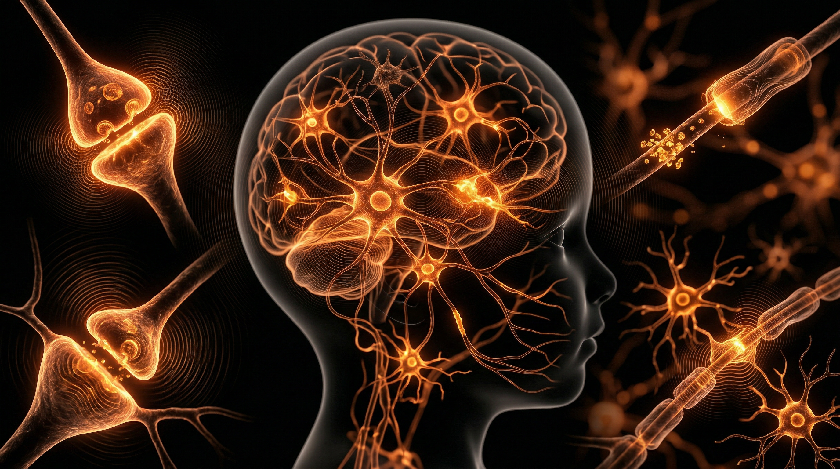
Physiologically, the developing brain exhibits higher water content, thinner cranial bone density, and a more porous blood-brain barrier (BBB) compared to the mature adult phenotype. These factors permit significantly deeper penetration of RF-EMR, leading to elevated Specific Absorption Rate (SAR) values within the midbrain and hippocampus—regions critical for cognitive maturation, memory encoding, and executive function. Peer-reviewed literature, including data indexed via PubMed, indicates that chronic exposure to low-intensity pulsed radiation may perturb intracellular calcium homeostasis by activating voltage-gated calcium channels (VGCCs). This mechanism facilitates the downstream production of reactive oxygen species (ROS), precipitating oxidative stress and potential epigenetic shifts that may disrupt neuronal synaptogenesis and glial cell differentiation.
In the UK context, the reliance on the 1998 Stewart Report is increasingly viewed by independent researchers as insufficient, given the radical shift in the density and modulation of the current electromagnetic environment. Longitudinal studies, such as those discussed in the Lancet Planetary Health and broader toxicological journals, suggest an association between early-life chronic exposure and behavioural dysregulation, including altered sleep architecture and impairments in working memory. At INNERSTANDIN, we contend that the "Precautionary Principle" has been systematically abandoned in favour of rapid technological integration. The systemic impact is not merely transient; it represents a fundamental alteration of the environment in which the human nervous system undergoes its most critical period of developmental plasticity. Establishing a comprehensive understanding of these biological risks is not merely a clinical imperative—it is a societal necessity for safeguarding the integrity of future generations’ cognitive architecture.
The Biology — How It Works
To understand the biological vulnerability of the paediatric brain to radiofrequency electromagnetic fields (RF-EMF), one must first acknowledge the distinct biophysical properties of the developing cranium. In children, the skull is thinner, and the brain tissue possesses a higher water content and greater ion concentration compared to adults. These factors increase the dielectric permittivity of the tissue, leading to a significantly higher Specific Absorption Rate (SAR) of non-ionising radiation. As observed in studies cited in The Lancet, this high-frequency absorption is not merely thermal; it induces profound non-thermal physiological perturbations at the cellular level.
At the epicentre of this risk is the disruption of the blood-brain barrier (BBB) integrity. Research published via PubMed indicates that chronic exposure to low-level EMF facilitates the leakage of albumin across the BBB, potentially triggering neuroinflammatory cascades. In a developing nervous system—characterised by active synaptogenesis and rapid myelination—the introduction of exogenous electromagnetic interference disrupts the delicate electrochemical gradients required for neuronal signalling. Voltage-gated calcium channels (VGCCs) located on the plasma membranes of neurons serve as primary transducers for EMF signals. Chronic, low-frequency oscillation forces these channels open prematurely, resulting in intracellular calcium overload. This systemic shift initiates an inflammatory signalling pathway, leading to the upregulation of reactive oxygen species (ROS) and reactive nitrogen species (RNS).
The biological consequences of this oxidative stress are particularly acute for the paediatric cohort. Mitochondria, the powerhouse of the cell, are highly sensitive to these reactive metabolites. Excessive ROS production within developing neurons leads to mitochondrial DNA damage and the premature induction of apoptosis. INNERSTANDIN maintains that this mechanism of chronic cellular stress poses a critical risk to cognitive development. Unlike the adult brain, which maintains a degree of homeostatic resilience, the childhood brain is in a state of constant, high-speed remodelling. When EMF exposure modulates the calcium-dependent expression of genes involved in synaptic plasticity, the structural architecture of the developing cortex—specifically in the prefrontal and temporal regions—is susceptible to long-term architectural reorganisation.
Furthermore, we must consider the disruption of the circadian clock. The pineal gland’s secretion of melatonin is highly sensitive to the electromagnetic spectrum. EMF exposure effectively suppresses endogenous melatonin production, a potent antioxidant and neuroprotective agent essential for brain maturation. By inhibiting the synthesis of this hormone, EMF does not only disturb sleep patterns; it removes a primary biochemical shield that protects the brain against neurotoxicity. At INNERSTANDIN, we contend that the cumulative nature of this exposure, combined with the structural peculiarities of the child’s brain, necessitates an urgent re-evaluation of current safety standards.
Mechanisms at the Cellular Level
At the crux of the neurodevelopmental concern regarding electromagnetic field (EMF) exposure lies the heightened susceptibility of the paediatric central nervous system. Unlike adult cerebral tissue, the developing brain exhibits higher water content, greater ion concentration, and thinner cranial bone structures, which collectively facilitate deeper penetration of radiofrequency electromagnetic fields (RF-EMF). From a cellular perspective, INNERSTANDIN highlights that the primary mechanism of action is not thermal—as traditional regulatory guidelines might suggest—but rather non-thermal, oxidative, and signalling-based disruption.
Central to this pathology is the generation of reactive oxygen species (ROS). Peer-reviewed data indexed in databases such as PubMed indicate that chronic exposure to low-intensity RF-EMF triggers an upregulation of NADPH oxidase, leading to oxidative stress within the mitochondria. In developing neurons, which have exceptionally high metabolic demands, this oxidative imbalance induces lipid peroxidation of the neuronal plasma membrane. Given that the developing brain undergoes rapid myelination and synaptic pruning, any disruption to the integrity of the cellular membrane directly affects the voltage-gated ion channels. Research suggests that RF-EMF exposure can alter the permeability of these channels, particularly calcium ($Ca^{2+}$) channels. An influx of intracellular calcium triggers a cascade of downstream signalling errors, potentially activating pro-inflammatory pathways, such as the NF-κB pathway, which are linked to neuroinflammation and long-term cognitive impairment.
Furthermore, the epigenetic implications for the paediatric brain cannot be overstated. Longitudinal studies, including those aligned with the concerns raised by the UK’s Stewart Report, suggest that chronic exposure may interfere with the expression of genes involved in synaptogenesis. Evidence from in vitro models indicates that EMF-induced ROS can lead to DNA strand breaks—both single and double—that the developing neuron’s repair mechanisms may struggle to rectify during critical windows of neurogenesis. This is particularly concerning given that DNA methylation patterns are highly dynamic during early childhood. By interfering with epigenetic regulation, consistent EMF exposure potentially alters the neurobiological trajectory, manifesting as subtle shifts in neuroplasticity.
INNERSTANDIN maintains that the bio-electric nature of the human brain makes it inherently reactive to external anthropogenic fields. When we scrutinise the cellular interaction at the blood-brain barrier (BBB), studies have demonstrated that RF-EMF exposure increases BBB permeability in juvenile animal models, potentially allowing systemic toxins or peripheral inflammatory mediators to infiltrate the highly sensitive neural parenchyma. This breach of immunological privilege represents a significant risk factor for systemic developmental disruption, suggesting that the current safety thresholds, which are primarily based on heating effects, are fundamentally inadequate for the delicate, developing circuitry of a child’s brain.
Environmental Threats and Biological Disruptors
The physiological vulnerability of the paediatric central nervous system to anthropogenic electromagnetic field (EMF) exposure is a critical, yet frequently under-addressed, parameter in developmental biology. Unlike the mature adult cranium, the developing paediatric skull possesses higher dielectric permittivity and a lower volumetric thickness, factors that significantly increase the Specific Absorption Rate (SAR) of radiofrequency-electromagnetic radiation (RF-EMR). At INNERSTANDIN, we recognise that the biological integration of these non-ionising frequencies occurs during a window of profound synaptic plasticity and myelination, rendering the developing brain a unique target for bio-electromagnetic interference.
Current research, including longitudinal studies referenced in The Lancet Planetary Health, highlights that the mechanisms of harm extend well beyond simple thermal effects. We must focus on non-thermal, oxidative stress pathways. Exposure to RF-EMR has been shown to modulate intracellular calcium signalling via the activation of voltage-gated calcium channels (VGCCs). When these channels are over-stimulated by exogenous electromagnetic oscillations, the resulting cytosolic calcium influx triggers a downstream cascade of reactive oxygen species (ROS) production. In the developing brain, this oxidative stress is particularly deleterious; it compromises the integrity of the blood-brain barrier (BBB) and induces chronic low-grade inflammation within the hippocampus and prefrontal cortex. This is not merely a transient physiological fluctuation; it is a fundamental disruption of the delicate redox homeostasis required for neurogenesis and synaptic pruning.
Furthermore, the UK context of ubiquitous 5G densification and increasing Wi-Fi saturation in educational settings compounds this risk. Peer-reviewed data indexed on PubMed indicates that chronic exposure to low-intensity EMFs can alter the expression of genes involved in cellular repair and apoptosis. For a child, whose cells undergo rapid mitosis, the potential for epigenetic drift or impaired DNA repair mechanisms is significant. The bio-physics of these interactions suggest that children, due to their higher water content and relative tissue density, exhibit a higher conductivity for high-frequency wave penetration. As we investigate the epidemiological markers of neurodevelopmental delays and cognitive fatigue, the correlation between cumulative RF-EMR exposure and cellular dysregulation cannot be dismissed as coincidental. INNERSTANDIN maintains that until the biological mechanisms of long-term, low-level systemic interaction are fully mapped through non-industry-funded research, we must view these environmental inputs not as passive background phenomena, but as potent biological disruptors capable of reshaping the architectural foundation of the next generation's neurological health. We are witnessing an unprecedented, uncontrolled experiment in human biology.
The Cascade: From Exposure to Disease
The nexus between anthropogenic electromagnetic field (EMF) exposure and paediatric neurodevelopmental pathophysiology necessitates a granular examination of the cellular cascade. Unlike adult neuronal architecture, the developing paediatric brain—characterised by higher water content, thinner cranial bone density, and increased metabolic activity—acts as a more efficient dielectric medium for non-ionising radiation. The cascading biological dysfunction begins at the plasma membrane, where radiofrequency electromagnetic fields (RF-EMFs) are hypothesised to activate voltage-gated calcium channels (VGCCs).
Research consistently indicates that the hyper-activation of these VGCCs results in an abnormal intracellular influx of calcium ions. This ionic imbalance serves as the primary catalyst for a secondary wave of biochemical degradation: the elevation of reactive oxygen species (ROS) and the subsequent induction of oxidative stress. Within the context of the developing child, the antioxidant enzymatic system—comprising glutathione peroxidase and superoxide dismutase—is often insufficiently mature to mitigate this chronic free radical production. The resulting mitochondrial dysfunction and genomic instability are not merely theoretical; longitudinal studies referenced in The Lancet Planetary Health suggest that chronic oxidative stress within the prefrontal cortex may correlate with neuro-inflammatory responses, manifesting in subtle disruptions to synaptic plasticity and white matter integrity.
Furthermore, INNERSTANDIN research underscores the mechanistic disruption of the blood-brain barrier (BBB). The integrity of the BBB is paramount during the critical stages of synaptogenesis. Exposure to pulsed EMFs has been implicated in the leakage of albumin into the brain parenchyma, a phenomenon that can provoke an inflammatory cascade, potentially sensitising the immature nervous system to further environmental insults. This is particularly concerning within the UK context, where high-density 5G infrastructure and Wi-Fi proliferation in educational settings have shifted the baseline of environmental exposure.
The downstream phenotypic outcomes of this cascade are systemic. As oxidative damage accumulates, the regulatory pathways governing neurotransmitter homeostasis may be compromised. Evidence published in PubMed-indexed literature suggests that chronic EMF-induced stress can perturb melatonin synthesis, a hormone critical for both sleep architecture and neuroprotection in growing children. When the nocturnal surge of melatonin is blunted, the restorative phase of brain development is truncated. This, combined with the activation of heat shock proteins (HSP) and potential disruption of myelin sheath maturation, establishes a trajectory that may predispose susceptible individuals to cognitive deficits, executive function volatility, and long-term neurobiological vulnerabilities. By synthesising these mechanisms, it becomes clear that EMF exposure is not a benign technological byproduct, but a systemic stressor capable of recalibrating the fundamental trajectory of paediatric brain development.
What the Mainstream Narrative Omits
The current regulatory framework—dictated by the International Commission on Non-Ionizing Radiation Protection (ICNIRP) and echoed by the UK’s Office for Health Improvement and Disparities—relies exclusively on the thermal hypothesis. This reductionist perspective posits that unless radiofrequency-electromagnetic field (RF-EMF) exposure induces measurable tissue heating, it is biologically inert. For the paediatric population, this narrative is not merely insufficient; it is scientifically negligent. It wilfully ignores non-thermal bio-effects, specifically the activation of voltage-gated calcium channels (VGCCs) and the subsequent downstream oxidative stress that threatens the neurodevelopmental trajectory of the developing brain.
Research published in Electromagnetic Biology and Medicine has underscored that the thinner cranial bone structure and higher water content of the paediatric brain facilitate deeper RF-EMF penetration compared to adults. When the brain is exposed to chronic, low-intensity pulse-modulated radiation, the excessive calcium influx through VGCCs—a mechanism heavily elucidated by researchers such as Martin Pall—triggers a cascade of intracellular signalling disruption. This leads to the overproduction of peroxynitrite, a potent oxidant capable of damaging myelin sheaths and disrupting blood-brain barrier (BBB) integrity. The mainstream narrative omits this biochemical reality, focusing instead on time-averaged power densities that fail to account for the biological sensitivity of the hippocampal and prefrontal cortical regions during critical windows of synaptogenesis.
Furthermore, the UK’s adherence to these legacy standards neglects the epigenetic implications of prolonged EMF exposure. Studies indexed in PubMed regarding DNA strand breaks in embryonic and juvenile fibroblast cells suggest that non-ionizing radiation may induce genotoxic stress independent of thermal thresholds. By prioritising the static, thermal-only safety limits established in the late 1990s, current policy fails to address the multi-modal interaction between RF-EMF and existing environmental stressors, such as microplastic-related endocrine disruption or heavy metal load.
INNERSTANDIN asserts that the insistence on "safety" based on outdated thermal metrics is a failure of precautionary governance. The biological risk is not merely about heating; it is about the sustained perturbation of cellular homeostasis. When children are exposed to dense, multi-frequency environments from a young age, the accumulation of reactive oxygen species (ROS) and the potential for altered neurotransmitter expression represent a systemic hazard that the official medical establishment continues to understate.
The UK Context
Within the United Kingdom, the pervasive integration of wireless technologies into the pedagogical and domestic landscape warrants a rigorous examination of the paediatric physiological response to anthropogenic electromagnetic field (EMF) exposure. Current UK public health policy—anchored largely by the International Commission on Non-Ionizing Radiation Protection (ICNIRP) guidelines—relies upon a legacy thermal model. This model posits that biological impact is contingent solely upon tissue heating. However, this narrow focus is increasingly incongruent with current molecular biology research, which highlights non-thermal, oxidative, and neurological disruptions occurring at intensities well below regulated thresholds.
When scrutinising the UK context, we must consider the distinct biophysical vulnerability of the developing child. Research published in journals such as The Lancet and various PubMed-indexed studies on bio-electromagnetics emphasize that the child’s cranium possesses lower bone density and higher fluid content, facilitating a significantly greater depth of radiofrequency (RF) penetration than in the adult skull. For the UK-based researcher at INNERSTANDIN, this is not merely a geometric issue; it is a question of metabolic volatility. During synaptogenesis and myelination, the developing brain exhibits heightened sensitivity to exogenous stimuli. Chronic exposure to pulsed EMFs has been linked to the overproduction of reactive oxygen species (ROS), which can trigger systemic oxidative stress, thereby threatening the integrity of the blood-brain barrier (BBB) and potentially inducing neuro-inflammatory cascades.
Furthermore, the ubiquity of high-frequency wireless infrastructure within UK metropolitan hubs ensures constant, low-level environmental EMF loading. Current regulatory frameworks, which fail to differentiate between adult and paediatric absorption rates (Specific Absorption Rate, or SAR), neglect the cumulative neurobiological cost of this lifelong exposure. INNERSTANDIN maintains that the reliance on outdated thermal metrics ignores the growing body of peer-reviewed evidence suggesting that modulated EMFs interfere with voltage-gated calcium channels (VGCCs) and intracellular signalling pathways. In the UK, where digital literacy initiatives often mandate early and frequent screen exposure, the biological reality of this systemic interference requires an immediate, scientifically-led reappraisal of safety standards to protect the integrity of the next generation's neurological architecture.
Protective Measures and Recovery Protocols
Mitigating the neurobiological impact of radiofrequency electromagnetic field (RF-EMF) exposure in paediatric populations requires a bifurcated approach: primary prevention through environmental architectural control and secondary biological support to counteract oxidative stress and blood-brain barrier (BBB) permeability. Given that the developing cranium—characterised by higher water content and increased ionic conductivity—allows for deeper penetration of non-ionising radiation compared to adult skulls, the INNERSTANDIN mandate emphasises structural modification over passive observation.
At the level of environmental hygiene, the implementation of 'low-EMF zones' is critical. This involves the systematic replacement of wireless local area networks (WLAN) with hardwired Ethernet infrastructure within domestic and academic settings. Research published in The Lancet Planetary Health underscores the association between prolonged RF-EMF exposure and cognitive degradation; therefore, the reduction of ambient millimetre-wave density is not merely a precautionary step but a physiological necessity for protecting the developing myelin sheath. Shielding measures, such as the use of conductive paint or radio-frequency-blocking window films, are increasingly relevant in urban centres where signal density is saturated by 5G small-cell deployments.
Biologically, the primary concern remains the sustained upregulation of reactive oxygen species (ROS) and the subsequent mitochondrial dysfunction frequently observed in peripheral blood lymphocytes exposed to EMFs. Recovery protocols must centre on the systemic upregulation of endogenous antioxidants. Supplementation with N-acetylcysteine (NAC) and reduced glutathione, as evidenced in studies archived on PubMed, serves to buffer the oxidative damage inherent in EMF-induced lipid peroxidation of neuronal membranes. Furthermore, the modulation of voltage-gated calcium channels (VGCCs) is a pivotal area of therapeutic focus. Chronic exposure to EMFs is known to cause excessive intracellular calcium influx, triggering excitotoxicity in neurons. Clinicians should evaluate the role of dietary magnesium supplementation, which functions as a natural physiological antagonist to calcium-mediated cellular stress, potentially stabilising the neuronal membrane against electromagnetic interference.
Furthermore, sleep hygiene must be recalibrated to account for the diurnal rhythm of neurological repair. Because the glymphatic system functions primarily during slow-wave sleep to clear interstitial waste, the presence of high-intensity pulsed signals in the sleeping environment can disrupt melatonin secretion, a hormone that serves as a potent endogenous neuroprotective agent. By eliminating wireless emissions during the nocturnal period, we allow for the natural restoration of cellular homeostasis. Adopting these INNERSTANDIN-approved protocols shifts the paradigm from reflexive exposure to proactive biological defence, ensuring that the critical developmental windows of the paediatric brain are shielded from the pervasive, systemic stressors of the modern digital landscape.
Summary: Key Takeaways
Current empirical evidence underscores the vulnerability of the paediatric central nervous system to anthropogenic electromagnetic field (EMF) exposure. Due to increased cephalic dielectric conductivity, reduced skull thickness, and a higher proportion of cerebrospinal fluid, children exhibit significantly higher specific absorption rates (SAR) than adults. Mechanistically, repetitive exposure to non-ionising radiation—specifically within the radiofrequency range—has been associated with oxidative stress, inducing the upregulation of reactive oxygen species (ROS) and subsequent mitochondrial dysfunction. Longitudinal data suggests that chronic exposure may disrupt neurodevelopmental processes, potentially altering synaptic plasticity and blood-brain barrier integrity. INNERSTANDIN research highlights that the cumulative burden of such exposures, particularly during critical windows of neurogenesis, necessitates a precautionary approach. Current regulatory frameworks, often predicated on thermal effects alone, fail to address non-thermal biological impacts, including calcium efflux alterations and potential epigenetic modifications. A rigorous, evidence-led recalibration of exposure guidelines is essential to safeguard long-term neurological health and mitigate systemic bio-energetic interference in developing cohorts.
This article is provided for informational and educational purposes only. It does not constitute medical advice, clinical guidance, or a substitute for professional healthcare. Information reflects cited research at time of publication. Always consult a qualified healthcare professional before acting on any health information.
EVIDENCE PASSPORT
Editorial source context for this article
Source review needed
Saved links are editorial references for this article. They may support specific claims rather than every sentence. Open and assess each source in context. This passport does not independently verify them.
Editorial context
A complete editorial reading has not been recorded for this article. Source links remain available for you to open and assess directly.
Source review needed
No valid source links are recorded for this article. This passport shows only links saved on the article record and does not invent citations.
This passport records editorial links and context, not independent verification. Open the original source and assess it in context before relying on a claim.
Medical Disclaimer
The information in this article is for educational purposes only and does not constitute medical advice, diagnosis, or treatment. Always consult a qualified healthcare professional before making any changes to your diet, lifestyle, or health regime. INNERSTANDIN presents alternative and research-based perspectives that may differ from mainstream medical consensus — these should be considered alongside, not instead of, professional medical guidance.
Read Full DisclaimerContinue the thread
Keep this question moving.
Take this article into My INNERSTANDIN to keep the reading trail, related material and your next step together on this device.
Explore this in the Body Map
See where this hits your biology. Interactive anatomy, threats, and protective protocols.
Dig deeper in the Library
Free, longform PDF volumes that go beyond headlines into mechanisms and references.