Hypoxia-Inducible Factor Signaling and Its Role in Impairing Neurovascular Unit Barriers
Discuss this resourceUpdated August 2026
An exploration of how cellular oxygen sensing mechanisms, specifically Hypoxia-Inducible Factor (HIF) pathways, contribute to the degradation of the blood-brain barrier and broader neurovascular unit dysfunction.
Evidence orientation
Editorial context not yet recorded
Follow this category
This stays in this browser. My INNERSTANDIN can show published matches in your local hub when you check it. It does not send email, push, or alert notifications.
Local learning review
A private browser aid for revisiting ideas. It is not an alert or a health recommendation.
Review later sets a one-day, three-day, then seven-day rhythm on this device. Choose it only when you want to revisit this article.
Introduction to the Neurovascular Unit
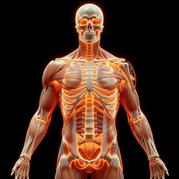
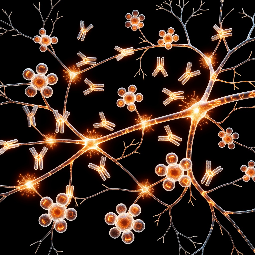
The neurovascular unit (NVU) is the functional foundation of brain health, comprising endothelial cells, astrocytes, pericytes, and neurons. Its primary role is to maintain the Blood-Brain Barrier (BBB), a selective gatekeeper that regulates ion homeostasis and prevents neurotoxic influx. When the NVU becomes compromised, the resulting vascular leakage leads to neuroinflammation and neurodegeneration.
The HIF Signaling Pathway: A Molecular Oxygen Sensor
Hypoxia-Inducible Factor (HIF) serves as a primary transcription factor responding to low oxygen availability. Under normoxic conditions, HIF-alpha subunits are hydroxylated and targeted for proteasomal degradation. However, during states of chronic hypoperfusion or localized metabolic stress, oxygen levels drop, stabilizing HIF-1alpha. This stabilized subunit translocates to the nucleus to trigger the expression of over 100 genes, including Vascular Endothelial Growth Factor (VEGF).
Mechanisms of BBB Impairment
The transition from acute protective response to chronic pathology is where HIF-1 signaling impairs the NVU.
- VEGF Overexpression and Tight Junction Disruption: HIF-1-driven VEGF upregulation promotes endothelial permeability. Persistent VEGF signaling leads to the downregulation of junctional proteins like Claudin-5 and Occludin, physically dismantling the barrier.
- Matrix Metalloproteinase (MMP) Activation: HIF-1 stimulates the release of MMPs, particularly MMP-2 and MMP-9. These enzymes digest the basal lamina, effectively tearing the structural scaffold that keeps brain microvessels intact.
- Pericyte Detachment: Pericytes are essential for maintaining capillary stability. HIF-1 signaling disrupts the signaling cross-talk between endothelial cells and pericytes (specifically PDGF-B/PDGFR-beta signaling), leading to pericyte loss and subsequent microvascular collapse.
Root Causes: Why Does This Happen?
The chronic activation of HIF-1 in the brain is often rooted in systemic metabolic dysregulation. Chronic inflammation, advanced glycation end-products (AGEs) from poor dietary choices, and mitochondrial dysfunction all create a micro-environment of perceived cellular hypoxia, even when systemic oxygen levels appear normal. Furthermore, sleep apnea and chronic stress-induced hypertension reduce the efficiency of cerebral blood flow, fueling the HIF-1 fire.
Clinical Implications and Future Directions
Targeting the HIF-1 signaling pathway represents a therapeutic frontier for neurological disorders such as vascular dementia, stroke recovery, and even Alzheimer’s disease. Current research is focusing on stabilizing the basal lamina and modulating HIF-1 transcriptional activity without compromising its baseline protective roles. For patients, maintaining metabolic health through anti-inflammatory protocols remains the most viable preventative strategy to keep the NVU barrier intact.
This article is provided for informational and educational purposes only. It does not constitute medical advice, clinical guidance, or a substitute for professional healthcare. Information reflects cited research at time of publication. Always consult a qualified healthcare professional before acting on any health information.
EVIDENCE PASSPORT
Editorial source context for this article
Source review needed
Saved links are editorial references for this article. They may support specific claims rather than every sentence. Open and assess each source in context. This passport does not independently verify them.
Editorial context
A complete editorial reading has not been recorded for this article, and no source links are saved on its record.
Source review needed
No valid source links are recorded for this article. Supplementary research, where available, is listed separately below.
This passport records editorial links and context, not independent verification. Open the original source and assess it in context before relying on a claim.
Medical Disclaimer
The information in this article is for educational purposes only and does not constitute medical advice, diagnosis, or treatment. Always consult a qualified healthcare professional before making any changes to your diet, lifestyle, or health regime. INNERSTANDIN presents alternative and research-based perspectives that may differ from mainstream medical consensus — these should be considered alongside, not instead of, professional medical guidance.
Read Full DisclaimerContinue the thread
Keep this question moving.
Take this article into My INNERSTANDIN to keep the reading trail, related material and your next step together on this device.
Explore this in the Body Map
Explore related body structures and their educational pages in the interactive Body Map.
Dig deeper in the Library
Free, longform PDF volumes that go beyond headlines into mechanisms and references.
RABBIT HOLE
Follow the biological thread deeper