
Metabolic Pathway Interference: How Lead Inhibits Heme Biosynthesis and Iron Homeostasis
Discuss this resourceUpdated August 2026
An in-depth exploration of how lead toxicity disrupts the heme biosynthetic pathway and iron metabolism, leading to cellular dysfunction and systemic health impacts.
Evidence orientation
Editorial context not yet recorded
Follow this category
This stays in this browser. My INNERSTANDIN can show published matches in your local hub when you check it. It does not send email, push, or alert notifications.
Local learning review
A private browser aid for revisiting ideas. It is not an alert or a health recommendation.
Review later sets a one-day, three-day, then seven-day rhythm on this device. Choose it only when you want to revisit this article.
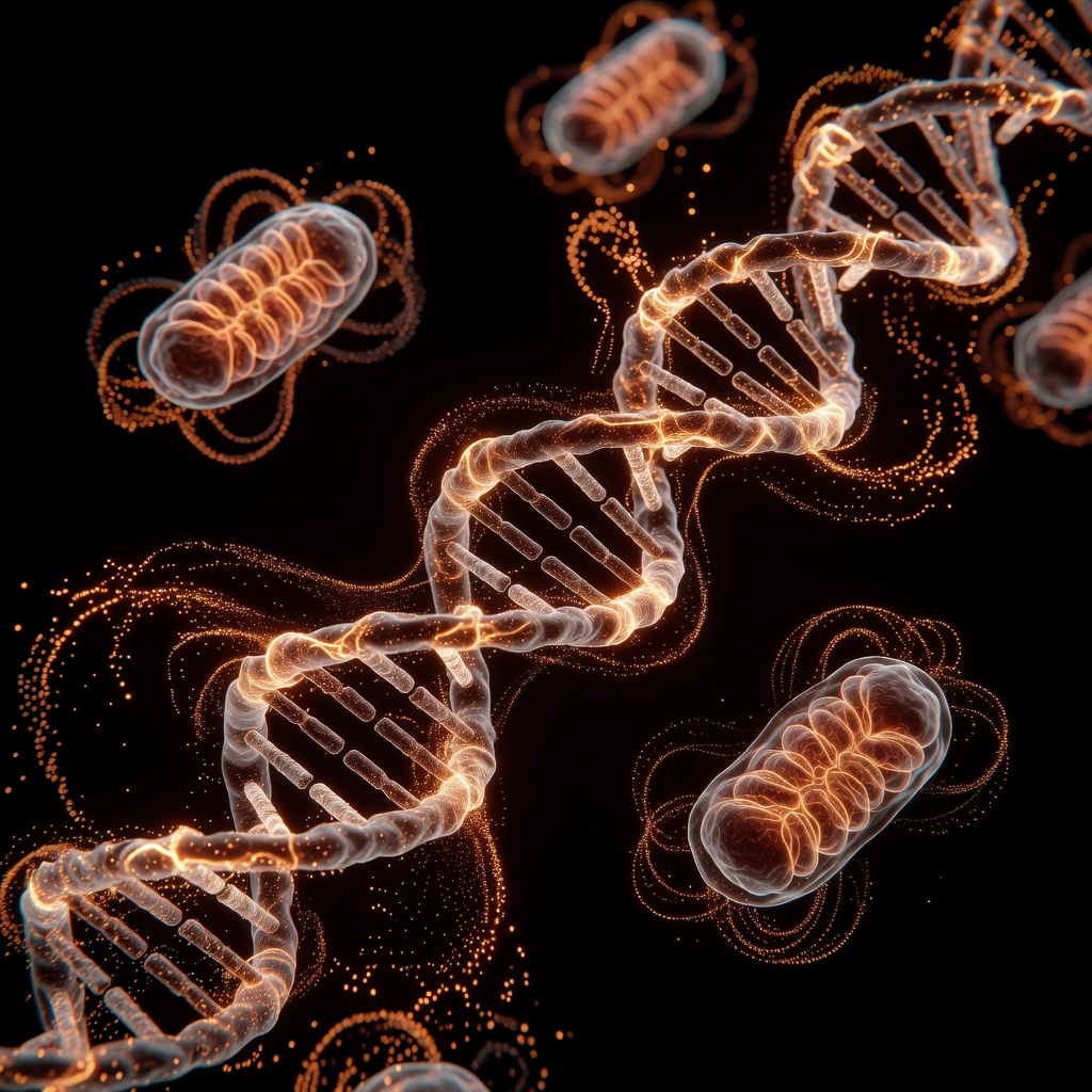
Introduction: The Invisible Saboteur. Lead, a potent heavy metal, does not merely circulate; it actively sabotages the molecular architecture of human physiology. By mimicking essential minerals, lead disrupts critical metabolic pathways, most notably heme biosynthesis. This article explores the root-cause mechanisms of lead-induced enzymatic inhibition and iron dysregulation. ### The Heme Biosynthesis Pathway. Heme is the vital prosthetic group of hemoglobin, myoglobin, and the cytochrome P450 enzymes.
Its production requires a highly coordinated sequence of eight enzymatic steps. Lead interferes with at least three of these enzymes, most significantly Delta-aminolevulinic acid dehydratase (ALAD) and Ferrochelatase. By substituting for zinc or competing with iron, lead halts the conversion of porphobilinogen into heme, resulting in a backlog of precursors and a catastrophic drop in oxygen transport capacity. ### Ferrochelatase Inhibition. Ferrochelatase is the final gatekeeper of the heme pathway, responsible for inserting the iron ion into the protoporphyrin IX ring. Lead’s high affinity for the enzyme’s sulfhydryl groups renders it inactive.
When ferrochelatase is inhibited, zinc often takes the place of iron, creating zinc-protoporphyrin (ZnPP), a clinical hallmark of lead exposure. This process effectively starves cells of functional hemoglobin, leading to microcytic anemia. ### Iron Homeostasis and Divalent Metal Transporters. Lead is a master of molecular mimicry. Because its ionic radius is similar to that of calcium and iron, it exploits the Divalent Metal Transporter 1 (DMT1) to enter cells. Once inside, lead disrupts the iron-sulfur cluster proteins that regulate the Iron Regulatory Proteins (IRPs).
This creates a 'false' signal of iron deficiency within the cell, triggering the body to absorb more iron, which inadvertently facilitates further lead uptake. It is a vicious cycle of metal dysregulation that depletes the body’s reserves of essential nutrients while facilitating heavy metal bioaccumulation. ### Cellular Consequences. The cumulative effect of these disruptions is oxidative stress and mitochondrial dysfunction. Without adequate heme, the electron transport chain falters, diminishing ATP production. Simultaneously, the buildup of intermediate metabolites like delta-aminolevulinic acid generates reactive oxygen species, further damaging cell membranes and DNA. ### Conclusion.
Lead toxicity is not just about direct tissue damage; it is a metabolic disruption that recalibrates how the body manages essential nutrients. Addressing lead exposure requires more than chelation; it necessitates a focus on restoring enzymatic function and optimizing nutritional status to counteract the metal's persistent interference.
This article is provided for informational and educational purposes only. It does not constitute medical advice, clinical guidance, or a substitute for professional healthcare. Information reflects cited research at time of publication. Always consult a qualified healthcare professional before acting on any health information.
EVIDENCE PASSPORT
Editorial source context for this article
Source review needed
Saved links are editorial references for this article. They may support specific claims rather than every sentence. Open and assess each source in context. This passport does not independently verify them.
Editorial context
A complete editorial reading has not been recorded for this article, and no source links are saved on its record.
Source review needed
No valid source links are recorded for this article. Supplementary research, where available, is listed separately below.
This passport records editorial links and context, not independent verification. Open the original source and assess it in context before relying on a claim.
Medical Disclaimer
The information in this article is for educational purposes only and does not constitute medical advice, diagnosis, or treatment. Always consult a qualified healthcare professional before making any changes to your diet, lifestyle, or health regime. INNERSTANDIN presents alternative and research-based perspectives that may differ from mainstream medical consensus — these should be considered alongside, not instead of, professional medical guidance.
Read Full DisclaimerContinue the thread
Keep this question moving.
Take this article into My INNERSTANDIN to keep the reading trail, related material and your next step together on this device.
Explore this in the Body Map
Explore related body structures and their educational pages in the interactive Body Map.
Dig deeper in the Library
Free, longform PDF volumes that go beyond headlines into mechanisms and references.