
Microvascular Permeability and Lymphatic Dysfunction: A Mechanistic Analysis of Lipoedema Progression
Updated August 2026
This article explores the intricate physiological breakdown of the microvasculature and lymphatic system that drives the chronic progression of lipoedema, focusing on capillary fragility and extracellular matrix remodeling.
Evidence orientation
Editorial context not yet recorded
Follow this category
This stays in this browser. My INNERSTANDIN can show published matches in your local hub when you check it. It does not send email, push, or alert notifications.
Local learning review
A private browser aid for revisiting ideas. It is not an alert or a health recommendation.
Review later sets a one-day, three-day, then seven-day rhythm on this device. Choose it only when you want to revisit this article.
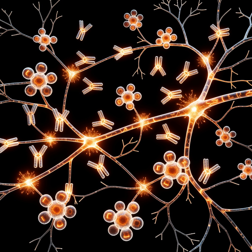
Introduction to Lipoedema Pathophysiology. Lipoedema is a chronic, progressive adipose tissue disorder that goes far beyond simple fat accumulation. At its core, the condition represents a systemic dysfunction of the microvascular and lymphatic environments. Understanding the progression of lipoedema requires a deep dive into the 'double hit' of microvascular permeability and subsequent lymphatic insufficiency. ## The Vascular Component: Increased Permeability. In the healthy human body, the microvasculature maintains a strict barrier, regulated by endothelial cells and the glycocalyx.
In patients with lipoedema, research indicates a compromised capillary barrier. This increased permeability allows for the excessive leakage of plasma proteins and fluids into the interstitial space. This influx creates an oncotic pull, drawing further fluid into the extracellular matrix, which manifests as the characteristic heaviness and swelling associated with the condition. ## Lymphatic Overload and Stasis. The lymphatic system serves as the body’s primary drainage mechanism for interstitial fluid and leaked proteins. As the microvasculature leaks fluid into the tissues, the lymphatic system is forced into a state of hyper-filtration.
Initially, lymphatic vessels may compensate with increased contraction rates. However, over time, the constant overload leads to lymphangiospasm and eventually structural damage to the lymphatic collectors. This creates a vicious cycle: as lymphatic drainage capacity drops, fluid retention increases, creating a hypoxic environment for adipocytes. ## Adipocyte Hypertrophy and Inflammation. The chronic accumulation of protein-rich fluid in the extracellular matrix creates a pro-inflammatory environment. Adipocytes, sensitive to their surroundings, undergo hypertrophy and hyperplasia.
This rapid growth leads to mechanical stress and local tissue hypoxia, which further triggers the release of inflammatory cytokines. This chemical signaling encourages the degradation of the collagen matrix, leading to the fibrosis and nodular texture palpable under the skin of lipoedema patients. ## The Role of Mechanical Stress. The progression of lipoedema is not merely chemical; it is mechanical. As the tissue becomes saturated with fluid and enlarged fat cells, the interstitial pressure rises. This pressure compresses local microvessels, further impeding venous return and lymphatic flow.
This 'mechanical entrapment' is why simple lifestyle interventions often fail to address the underlying physiological distortion; the structural integrity of the tissue must be supported to reverse or slow the damage. ## Conclusion. The progression of lipoedema is a multi-factorial event rooted in a breakdown of the microcirculatory system. By addressing microvascular permeability and supporting lymphatic transit, we move closer to a root-cause management strategy that prioritizes cellular health over cosmetic symptom control. Understanding these mechanisms is the first step toward effective patient advocacy and long-term management.
This article is provided for informational and educational purposes only. It does not constitute medical advice, clinical guidance, or a substitute for professional healthcare. Information reflects cited research at time of publication. Always consult a qualified healthcare professional before acting on any health information.
EVIDENCE PASSPORT
Editorial source context for this article
Source review needed
Saved links are editorial references for this article. They may support specific claims rather than every sentence. Open and assess each source in context. This passport does not independently verify them.
Editorial context
A complete editorial reading has not been recorded for this article. Source links remain available for you to open and assess directly.
Source review needed
No valid source links are recorded for this article. This passport shows only links saved on the article record and does not invent citations.
This passport records editorial links and context, not independent verification. Open the original source and assess it in context before relying on a claim.
Medical Disclaimer
The information in this article is for educational purposes only and does not constitute medical advice, diagnosis, or treatment. Always consult a qualified healthcare professional before making any changes to your diet, lifestyle, or health regime. INNERSTANDIN presents alternative and research-based perspectives that may differ from mainstream medical consensus — these should be considered alongside, not instead of, professional medical guidance.
Read Full DisclaimerContinue the thread
Keep this question moving.
Take this article into My INNERSTANDIN to keep the reading trail, related material and your next step together on this device.
Explore this in the Body Map
See where this hits your biology. Interactive anatomy, threats, and protective protocols.
Dig deeper in the Library
Free, longform PDF volumes that go beyond headlines into mechanisms and references.