
Peripheral Sensitization and Nociceptor Hyperexcitability in Vulvodynia: A Neuropathological Overview
Discuss this resourceUpdated August 2026
An in-depth exploration of the neurobiological drivers behind vulvodynia, focusing on how peripheral nerves become hyper-reactive and the mechanisms of chronic pelvic pain.
Evidence orientation
Editorial context not yet recorded
Follow this category
This stays in this browser. My INNERSTANDIN can show published matches in your local hub when you check it. It does not send email, push, or alert notifications.
Local learning review
A private browser aid for revisiting ideas. It is not an alert or a health recommendation.
Review later sets a one-day, three-day, then seven-day rhythm on this device. Choose it only when you want to revisit this article.
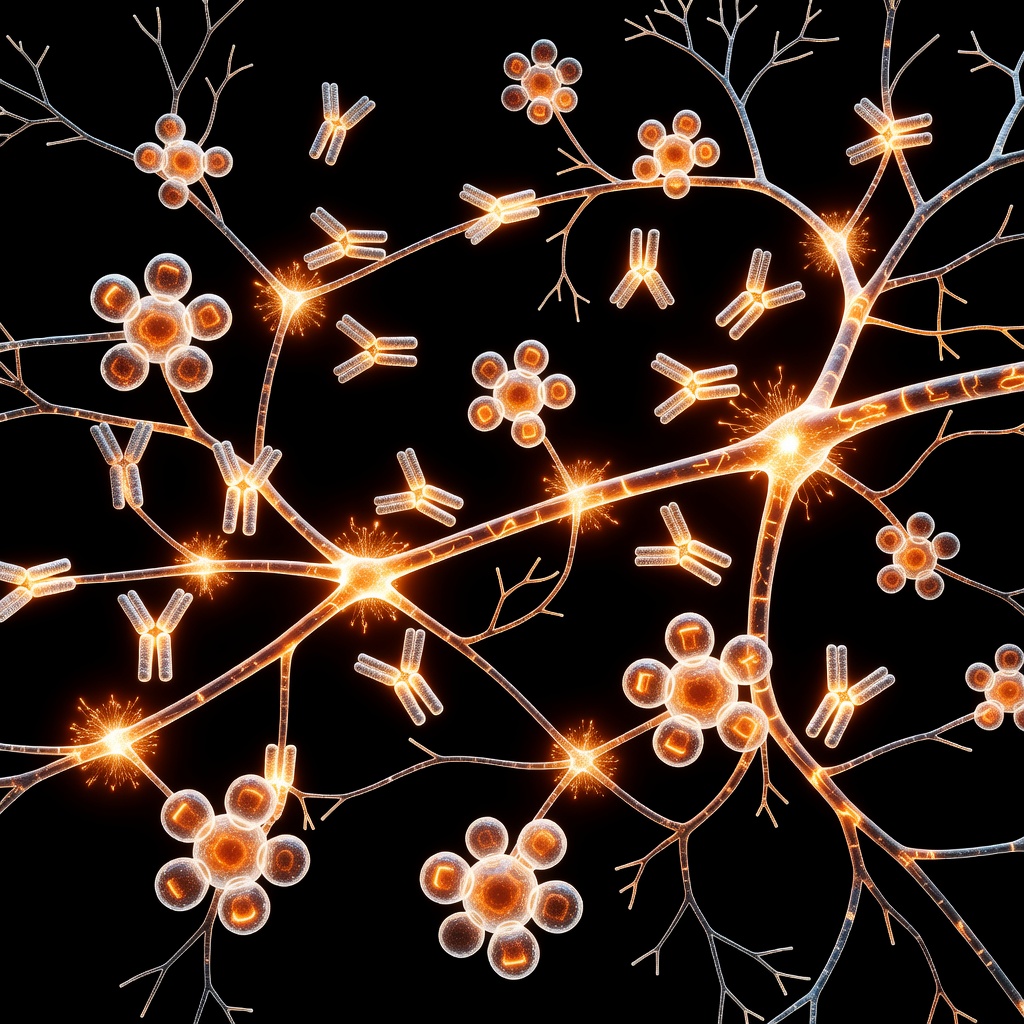
Vulvodynia is defined as chronic vulvar discomfort, most often described as burning, stinging, irritation, or rawness, occurring in the absence of an identifiable infection or skin pathology. For many, this condition is not merely 'pain' but a complex neurological recalibration. At the heart of this experience lies the phenomenon of peripheral sensitization.
The Anatomy of the Nociceptive System
To understand vulvodynia, we must first look at nociceptors—specialized sensory receptors responsible for detecting noxious stimuli such as heat, pressure, or chemical irritation. Under normal conditions, these nerves maintain a high threshold for activation. However, in patients with vulvodynia, this system undergoes a process known as 'hyperexcitability.'
Peripheral Sensitization: The Root Cause
Peripheral sensitization occurs when nociceptors become sensitized due to persistent inflammatory signaling, micro-trauma, or neurogenic inflammation. This process involves a reduction in the threshold required for these nerves to fire and an increase in the frequency of their firing. In the vulvar vestibule, research indicates a pathological increase in the density of nerve fibers (hyperinnervation), which exacerbates the transmission of pain signals to the dorsal horn of the spinal cord.
Molecular Drivers of Hyperexcitability
Several molecular pathways contribute to this shift in nerve sensitivity:
- Ion Channel Alterations: The upregulation of sodium channels, particularly Nav1.7 and Nav1.8, lowers the activation threshold, making nerves 'hyper-ready' to signal pain.
- Nerve Growth Factor (NGF): Elevated levels of NGF in the vulvar tissue promote the sprouting of new nerve fibers, creating a denser web of pain-responsive receptors.
- Neurogenic Inflammation: The release of substance P and calcitonin gene-related peptide (CGRP) from nerve endings triggers further inflammation, creating a self-sustaining feedback loop of excitation.
From Peripheral to Central: The Clinical Bridge
When peripheral nociceptors remain chronically activated, the central nervous system undergoes plastic changes. This is the bridge between localized peripheral sensitization and the systemic experience of pelvic pain. As signals bombard the spinal cord, central neurons become more responsive, leading to allodynia—the experience of pain from stimuli that should not be painful, such as the touch of clothing or light palpation.
Conclusion: Toward Targeted Therapies
Understanding that vulvodynia is driven by neuropathological changes in the peripheral nervous system shifts the focus from 'psychological' or 'undefined' pain to a physiological reality. By targeting these specific ion channels and nerve growth pathways, we can begin to offer more robust, evidence-based interventions for those navigating chronic pelvic discomfort.
This article is provided for informational and educational purposes only. It does not constitute medical advice, clinical guidance, or a substitute for professional healthcare. Information reflects cited research at time of publication. Always consult a qualified healthcare professional before acting on any health information.
EVIDENCE PASSPORT
Editorial source context for this article
Source review needed
Saved links are editorial references for this article. They may support specific claims rather than every sentence. Open and assess each source in context. This passport does not independently verify them.
Editorial context
A complete editorial reading has not been recorded for this article, and no source links are saved on its record.
Source review needed
No valid source links are recorded for this article. Supplementary research, where available, is listed separately below.
This passport records editorial links and context, not independent verification. Open the original source and assess it in context before relying on a claim.
Medical Disclaimer
The information in this article is for educational purposes only and does not constitute medical advice, diagnosis, or treatment. Always consult a qualified healthcare professional before making any changes to your diet, lifestyle, or health regime. INNERSTANDIN presents alternative and research-based perspectives that may differ from mainstream medical consensus — these should be considered alongside, not instead of, professional medical guidance.
Read Full DisclaimerContinue the thread
Keep this question moving.
Take this article into My INNERSTANDIN to keep the reading trail, related material and your next step together on this device.
Explore this in the Body Map
Explore related body structures and their educational pages in the interactive Body Map.
Dig deeper in the Library
Free, longform PDF volumes that go beyond headlines into mechanisms and references.