
The Role of Lead-Induced Calcium Signaling Disruption in Neuronal Apoptosis
Discuss this resourceUpdated August 2026
An in-depth exploration of how lead exposure interferes with intracellular calcium homeostasis, triggering neurotoxicity and programmed cell death in the central nervous system.
Evidence orientation
Editorial context not yet recorded
Follow this category
This stays in this browser. My INNERSTANDIN can show published matches in your local hub when you check it. It does not send email, push, or alert notifications.
Local learning review
A private browser aid for revisiting ideas. It is not an alert or a health recommendation.
Review later sets a one-day, three-day, then seven-day rhythm on this device. Choose it only when you want to revisit this article.
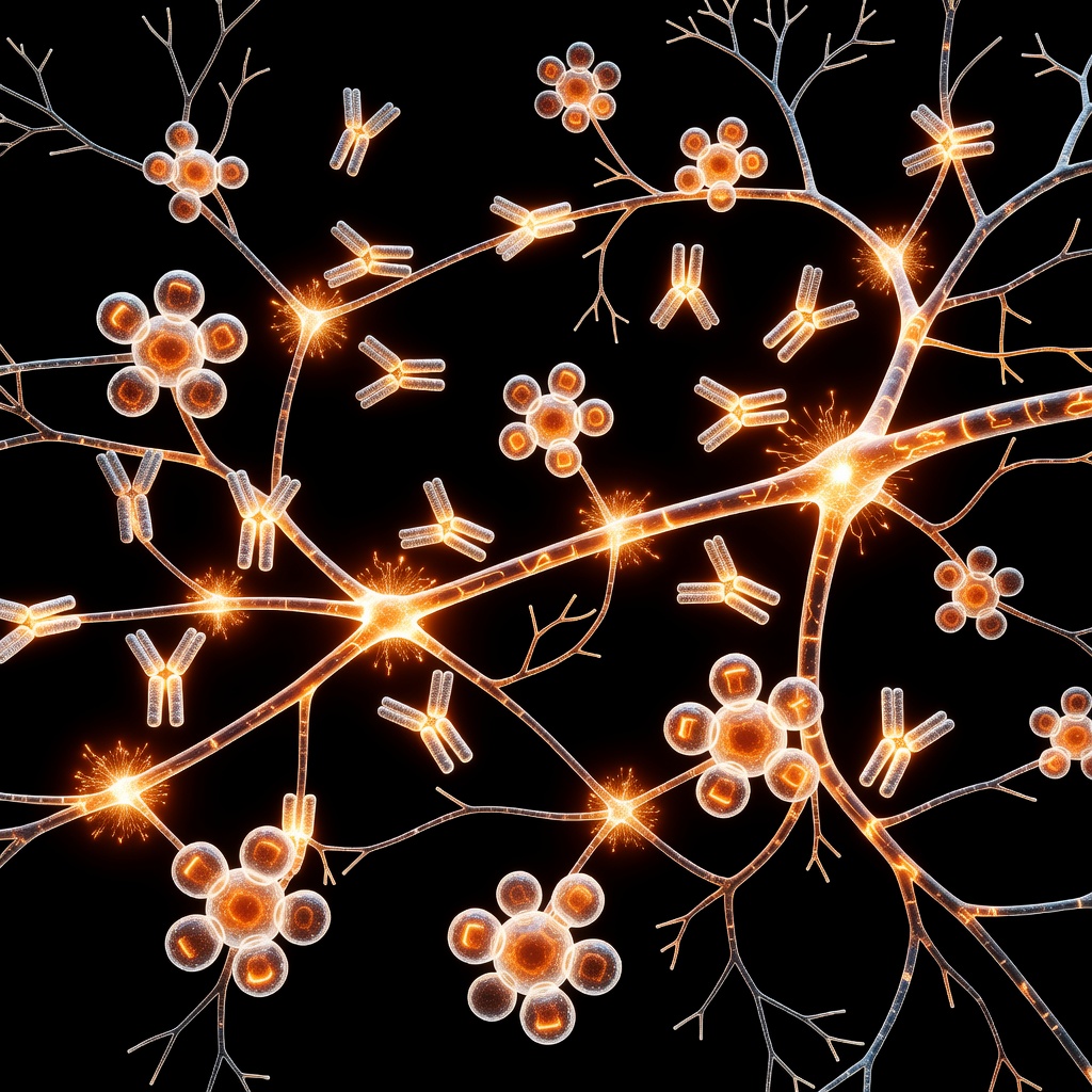
The Silent Neurotoxin: Lead and the Calcium Paradox. Lead (Pb) remains one of the most potent environmental neurotoxins, capable of causing profound cognitive and neurological impairment. At the cellular level, the mechanism of this toxicity is rooted in the disruption of intricate signaling pathways. Central to this is the relationship between lead and calcium (Ca2+). Because lead acts as a molecular mimic of calcium, it effectively hijacks the cell's signaling machinery. ### The Mimicry Mechanism.
Calcium is the universal secondary messenger in the human body, vital for neurotransmitter release, synaptic plasticity, and gene expression. Lead possesses a similar ionic radius and charge density to calcium, allowing it to bypass cellular defense mechanisms. It enters neurons through voltage-gated calcium channels, effectively masquerading as a vital nutrient. Once inside, lead binds to calcium-binding proteins like calmodulin and protein kinase C (PKC) with significantly higher affinity than calcium itself. This aberrant binding locks these proteins in an 'always-on' state or inhibits them entirely, leading to catastrophic signaling failures. ### Disruption of Homeostasis.
Inside the neuron, lead disrupts the sequestration of calcium within the endoplasmic reticulum and mitochondria. By interfering with the sarco/endoplasmic reticulum Ca2+-ATPase (SERCA) pumps, lead prevents the cell from clearing excess calcium from the cytosol. This creates a state of chronic intracellular calcium overload. When cytoplasmic calcium levels remain abnormally high, the cell activates calcium-dependent proteases and lipases, which begin to degrade the structural integrity of the neuron from the inside out. ### The Path to Apoptosis. The culmination of this signaling chaos is programmed cell death, or apoptosis.
Mitochondria, overwhelmed by lead-induced calcium influx, undergo a transition known as the Mitochondrial Permeability Transition (MPT). This causes the release of Cytochrome c into the cytosol. Once in the cytosol, Cytochrome c triggers the activation of the caspase cascade—the 'executioner' enzymes that dismantle the cell's DNA and cytoskeleton. In the developing brain, this process is particularly devastating because neurons cannot easily regenerate. Lead-induced apoptosis essentially 'prunes' healthy brain tissue, leading to the well-documented developmental delays and behavioral deficits associated with chronic exposure. ### Therapeutic Perspectives.
Understanding that lead toxicity is a problem of ionic disruption opens new doors for potential remediation. While chelation therapy remains the clinical standard for high-level acute exposure, research into dietary calcium supplementation and antioxidants suggests a potential protective role in mitigating lead-induced damage. By maintaining robust calcium homeostasis through nutritional support, we can theoretically harden the cellular defense against the insidious entry of heavy metal mimics. As we continue to study the long-term impacts of environmental toxins, focusing on the root-cause mechanics of cellular signaling is essential for developing effective public health interventions.
This article is provided for informational and educational purposes only. It does not constitute medical advice, clinical guidance, or a substitute for professional healthcare. Information reflects cited research at time of publication. Always consult a qualified healthcare professional before acting on any health information.
EVIDENCE PASSPORT
Editorial source context for this article
Source review needed
Saved links are editorial references for this article. They may support specific claims rather than every sentence. Open and assess each source in context. This passport does not independently verify them.
Editorial context
A complete editorial reading has not been recorded for this article, and no source links are saved on its record.
Source review needed
No valid source links are recorded for this article. Supplementary research, where available, is listed separately below.
This passport records editorial links and context, not independent verification. Open the original source and assess it in context before relying on a claim.
Medical Disclaimer
The information in this article is for educational purposes only and does not constitute medical advice, diagnosis, or treatment. Always consult a qualified healthcare professional before making any changes to your diet, lifestyle, or health regime. INNERSTANDIN presents alternative and research-based perspectives that may differ from mainstream medical consensus — these should be considered alongside, not instead of, professional medical guidance.
Read Full DisclaimerContinue the thread
Keep this question moving.
Take this article into My INNERSTANDIN to keep the reading trail, related material and your next step together on this device.
Explore this in the Body Map
Explore related body structures and their educational pages in the interactive Body Map.
Dig deeper in the Library
Free, longform PDF volumes that go beyond headlines into mechanisms and references.