
Fluoride & The Pineal Gland
Updated June 2026
The calcification of the third eye by fluoride and its impact on hormonal and cognitive health.
Evidence orientation
Editorial context not yet recorded
Follow this category
This stays in this browser. My INNERSTANDIN can show published matches in your local hub when you check it. It does not send email, push, or alert notifications.
Local learning review
A private browser aid for revisiting ideas. It is not an alert or a health recommendation.
Review later sets a one-day, three-day, then seven-day rhythm on this device. Choose it only when you want to revisit this article.
Overview
The pineal gland, a neuroendocrine transducer sequestered deep within the epithalamus, represents a critical nexus between environmental stimuli and systemic physiological regulation. Within the framework of INNERSTANDIN’s investigation into environmental threats, the bioaccumulation of fluoride within this structure emerges as a profound toxicological concern. Unlike the majority of the central nervous system, the pineal gland is a circumventricular organ, meaning it is not shielded by the blood-brain barrier (BBB). This lack of a protective interface, coupled with its exceptionally high vascularisation—exceeding even that of the kidney in terms of blood flow per gram of tissue—renders the gland uniquely susceptible to circulating xenobiotics.
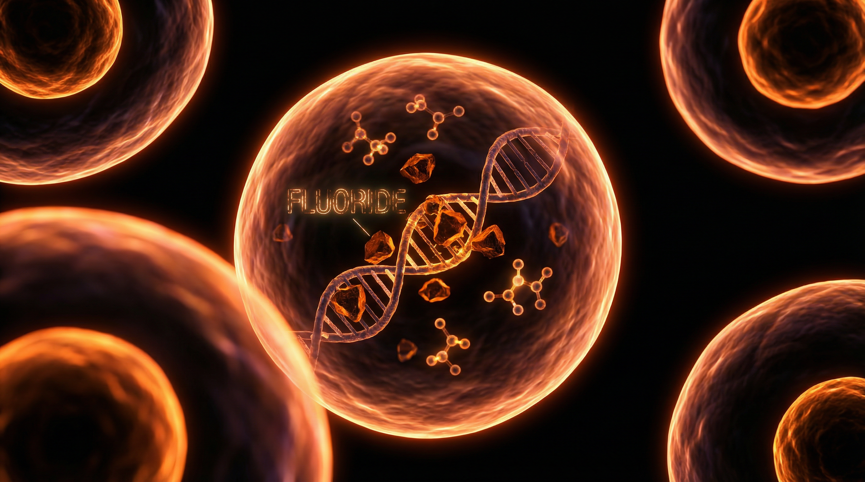
The biochemical affinity between fluoride (F-) and the pineal gland is primarily mediated by the presence of hydroxyapatite crystals (acervuli cerebri or "brain sand"), which naturally accumulate within the gland from childhood. As a halogen with high electronegativity, fluoride exhibits an intense affinity for calcium phosphate. Research, most notably the seminal work of Dr Jennifer Luke at the University of Surrey, has demonstrated that fluoride is not merely transient in the pineal; it is actively sequestered. The mechanism involves the substitution of the hydroxyl ion (OH-) within the hydroxyapatite matrix for a fluoride ion, resulting in the formation of fluoroapatite. This altered mineral structure is more stable and less soluble, leading to the permanent calcification of pineal tissue at concentrations significantly higher than those found in cortical bone or the skeletal system.
From a systemic perspective, this progressive calcification is not a benign radiological finding; it represents a functional degradation of the gland’s capacity to synthesise N-acetyl-5-methoxytryptamine (melatonin). Melatonin is the master regulator of the circadian rhythm, a potent endogenous antioxidant, and a critical modulator of the hypothalamic-pituitary-gonadal (HPG) axis. Peer-reviewed studies in animal models and ecological observations in human populations suggest that fluoride-induced pineal impairment correlates with a reduction in nocturnal melatonin metabolites and a subsequent advancement in the onset of puberty—a phenomenon increasingly observed in fluoridated regions of the United Kingdom and North America.
The implications for public health are extensive. The UK’s Department of Health and Social Care continues to endorse community water fluoridation, yet the biochemical reality of pineal bioaccumulation remains largely absent from mainstream policy discourse. By disrupting the enzymatic pathways involved in the tryptophan-serotonin-melatonin conversion, fluoride acts as a metabolic disruptor. At INNERSTANDIN, we recognise that the integrity of the pineal gland is foundational to biological synchronicity; therefore, the chronic exposure to fluoride must be scrutinised not merely as a dental prophylactic, but as a systemic neuroendocrine threat that compromises the very architecture of human chronobiology.
The Biology — How It Works
The pineal gland, a small endocrine organ nestled deep within the epithalamus, serves as a primary regulatory hub for the mammalian circadian rhythm. Yet, its unique physiological architecture renders it exceptionally vulnerable to environmental xenobiotics, most notably fluoride. Unlike the majority of the central nervous system, the pineal gland is situated outside the blood-brain barrier (BBB). It is characterised by a high degree of vascularisation, receiving a blood flow per unit mass second only to the kidney. This high perfusion rate, combined with its periventricular location, exposes the gland to systemic concentrations of fluoride that other neural tissues are shielded from. At INNERSTANDIN, we must scrutinise the biochemical affinity between fluoride and the pineal’s mineralising tissues to appreciate the gravity of this systemic threat.
The primary mechanism of fluoride sequestration within the pineal gland is its high affinity for calcium hydroxyapatite. The pineal naturally undergoes a process of physiological calcification, forming "brain sand" or acervuli cerebri. Fluoride acts as a pro-calcific agent through an isomorphous replacement process; the fluoride ion (F-) replaces the hydroxyl ion (OH-) in the hydroxyapatite crystal lattice [Ca10(PO4)6(OH)2], forming fluorapatite [Ca10(PO4)6F2]. Research pioneered by Dr Jennifer Luke at the University of Surrey established that the pineal gland functions as a major fluoride sink. Her landmark 2001 study, published in Caries Research, demonstrated that fluoride concentrations in the pineal’s calcified deposits were significantly higher than those found in bone or teeth—reaching levels as high as 21,000 ppm (parts per million).
The implications of this bioaccumulation are not merely structural; they are profoundly functional. The enzymatic pathway for melatonin synthesis—the conversion of tryptophan to serotonin, and subsequently to N-acetylserotonin via the enzyme arylalkylamine N-acetyltransferase (AANAT)—is sensitive to the microenvironmental integrity of the pinealocytes. Fluoride-induced calcification and the subsequent formation of fluorapatite create an oxidative environment that suppresses the activity of these enzymes. This leads to a reduction in nocturnal melatonin production, a phenomenon observed in various animal models and substantiated by human epidemiological data. In the UK context, where water fluoridation programmes intersect with high dietary intake—particularly from the Camellia sinensis (tea) plant, which is a known hyper-accumulator of fluoride—the cumulative systemic burden is significant.
Furthermore, the disruption of the pineal’s endocrine output triggers a cascade of physiological dysregulation. Melatonin is a potent antioxidant and neuroprotector; its suppression is linked to accelerated cellular senescence and the disruption of the hypothalamic-pituitary-gonadal (HPG) axis. Peer-reviewed evidence suggests that this endocrine interference can manifest as precocious puberty, particularly in females, as the inhibitory effect of melatonin on gonadotropin-releasing hormone (GnRH) is prematurely diminished. At INNERSTANDIN, our analysis reveals that fluoride is not simply a passive mineralising agent but a potent disruptor of the body’s internal chronometry, altering the very biological rhythms that govern human development and cellular repair.
Mechanisms at the Cellular Level
To comprehend the bio-accumulation of fluoride within the epithalamus, one must first appreciate the unique physiological architecture of the pineal gland. Unlike the majority of the central nervous system, the pineal gland is not sequestered behind the blood-brain barrier (BBB). Instead, it exists in a state of high vascularisation, receiving a profuse blood supply second only to the kidney. This high perfusion rate, coupled with the gland’s innate tendency to form hydroxyapatite (calcium phosphate) crystals—commonly referred to as acervuli or ‘brain sand’—creates a physiological sink for circulating fluoride ions (F-).
Research spearheaded by Dr Jennifer Luke (2001) at the University of Surrey provided the foundational evidence for this sequestration, demonstrating that fluoride concentrations in the pineal gland’s mineralised tissues are significantly higher than those found in bone. Through the process of ionic exchange, fluoride ions replace the hydroxyl groups within the hydroxyapatite matrix, forming fluorapatite. This transformation is not merely a structural shift; it alters the surface chemistry of the pineal concretions, potentially serving as a permanent reservoir for systemic fluoride. At INNERSTANDIN, we recognise that this bio-mineralisation is the primary mechanism through which fluoride bypasses traditional neurological defences.
At the cellular level, the presence of fluoride triggers a cascade of enzymatic and metabolic disruptions. The synthesis of melatonin, the gland’s primary chronobiotic hormone, is particularly vulnerable. Melatonin is synthesised from serotonin via the rate-limiting enzyme Serotonin N-acetyltransferase (SNAT). Peer-reviewed studies indicate that fluoride acts as a metabolic inhibitor, potentially suppressing SNAT activity and reducing the total output of melatonin. This inhibition is often driven by fluoride’s capacity to induce oxidative stress. Once inside the pinealocytes, fluoride ions stimulate the overproduction of reactive oxygen species (ROS) while simultaneously depleting endogenous antioxidant stores, such as glutathione. This oxidative imbalance leads to lipid peroxidation and mitochondrial dysfunction, compromising the cellular energy required for hormone synthesis.
Furthermore, fluoride’s affinity for divalent cations means it can interfere with calcium signalling pathways essential for pineal function. By sequestering intracellular calcium, fluoride disrupts the calcium-dependent exocytosis of melatonin into the bloodstream. In the UK context, where water fluoridation remains a point of intense public health debate, the cumulative impact of these cellular mechanisms cannot be overlooked. The systemic result is a fragmented circadian rhythm, as the pineal gland’s ability to synchronise biological time with environmental light cues is fundamentally eroded. Through the lens of INNERSTANDIN, it becomes clear that fluoride is not merely a passive environmental additive, but a potent disruptor of the endocrine-metabolic axis at its most sensitive juncture.
Environmental Threats and Biological Disruptors
The pineal gland, or epiphysis cerebri, represents a critical nexus between the endocrine and central nervous systems, yet its unique physiological architecture renders it disproportionately vulnerable to environmental xenobiotics. At INNERSTANDIN, we must scrutinise the gland’s status as a circumventricular organ; unlike the majority of the encephalic parenchyma, the pineal gland is not sequestered behind the blood-brain barrier (BBB). Its high vascularisation—exceeded only by the kidney—and its fenestrated capillaries facilitate the rapid exchange of molecules between the blood and the pinealocytes. However, this physiological openness serves as a double-edged sword, permitting the systemic accumulation of fluoride (F-) at concentrations that far exceed those found in other soft tissues or even bone.
The biochemical mechanism of this accumulation is rooted in the pineal’s propensity for calcification. Research initially spearheaded by Jennifer Luke at the University of Surrey (and subsequently corroborated in various PubMed-indexed longitudinal studies) demonstrated that fluoride possesses an intense affinity for the hydroxyapatite crystals that naturally form within the pineal gland as we age. Fluoride ions, through a process of ionic exchange, displace the hydroxyl groups within the crystal lattice to form fluorapatite. This isn’t merely a structural change; fluorapatite is less soluble and metabolically inert, effectively "locking" the fluoride into the pineal tissue. Luke’s post-mortem analyses revealed that fluoride levels in the pineal calcifications of elderly subjects reached staggering levels, upwards of 21,000 ppm, significantly higher than the concentrations found in the hydroxyapatite of the femur.
From an INNERSTANDIN perspective, the biological disruption caused by this sequestration is profound. The pineal gland is the primary site of melatonin synthesis, a hormone derived from serotonin that governs circadian rhythms, cellular antioxidant defence, and the regulation of the hypothalamic-pituitary-gonadal axis. Chronic fluoride exposure has been mechanistically linked to the inhibition of enzymes involved in the conversion of tryptophan to serotonin and subsequently to melatonin. This enzymatic interference, coupled with the physical encrustation of the gland, results in reduced melatonin output. The systemic fallout is extensive: disrupted sleep-wake cycles, increased susceptibility to oxidative stress, and, as observed in animal models and epidemiological studies, a potential acceleration in the onset of puberty due to the premature decline in circulating melatonin levels.
In the UK context, where approximately 10% of the population receives fluoridated water and where the consumption of Camellia sinensis (the tea plant, a known hyper-accumulator of fluoride) is culturally ubiquitous, the cumulative load on the pineal gland cannot be ignored. The synergy between fluoridated water and high tea intake creates a unique bio-accumulative profile that challenges the gland’s metabolic integrity. This is not merely a dental issue; it is a fundamental disruption of the body's internal chronometer. The evidence suggests that the pineal gland acts as a "sink" for fluoride, and the resulting calcification serves as a primary biological marker for environmental toxicity, demanding a rigorous reassessment of current public health protocols regarding fluoride exposure levels.
The Cascade: From Exposure to Disease
The pathogenesis of fluoride-induced pineal dysfunction begins with the gland’s unique physiological architecture. Unlike most of the central nervous system, the pineal gland is situated outside the blood-brain barrier (BBB) and possesses a profuse blood flow—second only to the kidney in terms of weight-to-perfusion ratio. This heightened haemodynamic activity renders the gland exceptionally vulnerable to systemic solutes. Within the biological framework explored by INNERSTANDIN, we must recognise that the pineal gland is a major calcium-sequestering organ, characterised by the presence of hydroxyapatite crystals (corpora arenacea). Fluoride, possessing an extreme electronegative affinity for calcium, does not merely circulate; it is actively sequestered into these calcified structures.
Peer-reviewed research, most notably the seminal work of Dr Jennifer Luke (2001), has demonstrated that fluoride concentrations in the pineal gland’s mineralised tissue are significantly higher than those found in bone. This process of biomineralisation involves the substitution of hydroxyl ions within the hydroxyapatite matrix for fluoride ions, forming fluorapatite. This chemical shift is not biologically inert; it alters the solubility and surface area of the crystals, effectively turning the gland into a primary sink for fluoride accumulation. As the ratio of fluorapatite increases, the metabolic integrity of the pineal parenchyma is compromised.
The cascade moves from mineralisation to enzymatic inhibition. The synthesis of melatonin—the master orchestrator of the circadian rhythm and a potent endogenous antioxidant—is dependent on the conversion of serotonin via the enzyme arylalkylamine N-acetyltransferase (AANAT). High fluoride burdens have been linked to the suppression of this enzymatic pathway. When melatonin production is blunted, the systemic fallout is profound. At the cellular level, the loss of melatonin-mediated neuroprotection accelerates oxidative stress within the brain, as the absence of this free-radical scavenger leaves neural lipids and proteins vulnerable to peroxidation.
In the UK context, the cumulative exposure from fluoridated water supplies, combined with the high consumption of Camellia sinensis (tea), which naturally hyper-accumulates fluoride from the soil, creates a chronic loading scenario. This persistent exposure correlates with the "earlier onset of puberty" observed in epidemiological studies—a phenomenon driven by the pineal gland's role in regulating the timing of the hypothalamic-pituitary-gonadal (HPG) axis. Reduced melatonin levels signal the body to initiate pubertal development prematurely, an endocrine disruption that carries long-term risks for hormone-sensitive cancers and metabolic dysregulation. Furthermore, the disruption of the sleep-wake cycle leads to a secondary cascade of neuroinflammation and cognitive decline, as the glymphatic system—the brain’s waste-clearance mechanism—relies on deep, melatonin-regulated sleep to function. Through the lens of INNERSTANDIN, fluoride is not merely a dental prophylactic, but a systemic disruptor that calcifies the seat of biological rhythmicity, leading to a state of chronic physiological desynchrony.
What the Mainstream Narrative Omits
The conventional public health discourse, championed by the NHS and various dental associations, maintains a reductionist fixation on the "topical benefit" of fluoride for dental enamel, effectively bypasses the systemic implications of chronic ingestion. This narrative purposefully ignores the bioaccumulative trajectory of fluoride within the endocrine system, specifically the pineal gland. Unlike most of the encephalon, the pineal gland is situated outside the blood-brain barrier (BBB) and possesses a profuse vascular supply, second only to the kidney in terms of blood flow per unit of weight. This physiological "vulnerability" makes it a primary site for the sequestration of fluoride.
Extensive research, most notably the seminal work of Dr Jennifer Luke (1997, 2001), has demonstrated that the pineal gland is a major calcium-accumulating organ. It contains hydroxyapatite crystals which, much like dental enamel, exhibit a high affinity for the fluoride ion. Through an ion-exchange process, fluoride replaces the hydroxyl group in hydroxyapatite to form fluorapatite. Luke’s post-mortem analyses revealed that fluoride concentrations in the pineal gland’s calcified tissues were significantly higher than those in bone (averaging 9,000 ppm and reaching up to 21,000 ppm). The mainstream narrative fails to acknowledge that these concentrations are achieved even in individuals residing in areas with regulated water fluoridation programmes.
At the level of INNERSTANDIN, we must scrutinise the enzymatic consequences of this accumulation. The pineal gland is the primary site of melatonin synthesis, a process regulated by the conversion of tryptophan to serotonin, and subsequently to melatonin via the enzyme serotonin N-acetyltransferase (SNAT). High-density fluoride deposition within the pineal parenchyma has been linked to the suppression of melatonin production. This inhibition does not merely disrupt the circadian rhythm; it precipitates a cascade of systemic dysregulation. Melatonin is a potent antioxidant and an inhibitor of precocious puberty. Research cited in the National Research Council (2006) report suggests a correlation between fluoride exposure and the accelerated onset of menarche, a finding that the mainstream narrative conveniently omits to avoid questioning the "safety" of Community Water Fluoridation (CWF).
Furthermore, the mainstream silence on the synergistic toxicity of fluoride with aluminium—forming fluoroaluminium complexes—is a glaring oversight. These complexes mimic the structure of phosphate and activate G-proteins, potentially triggering aberrant signal transduction pathways within the pinealocytes. For the discerning researcher at INNERSTANDIN, it is clear that the pineal gland acts as a sentinel for environmental fluoride toxicity, and the continued dismissal of its calcification as "age-related" rather than "toxin-induced" is a profound failure of modern clinical toxicology.
The UK Context
The United Kingdom represents a singular case study in the longitudinal exposure of a population to hexafluorosilicic acid via public water supplies, a practice that began in Birmingham in 1964. Within the INNERSTANDIN framework of environmental toxicology, the UK’s bifurcated approach—where approximately 5.8 million people receive artificially fluoridated water while the remainder do not—provides a stark demographic contrast for assessing the sequestration of fluoride within the pineal gland. Unlike the majority of the encephalon, the pineal gland is not sequestered behind the blood-brain barrier (BBB). As a circumventricular organ with a capillary permeability comparable to that of the kidney, it is directly exposed to systemic circulation. Research pioneered by Jennifer Luke (University of Surrey, 1997) demonstrated that the human pineal gland is a major site of fluoride accumulation, with concentrations in the hydroxyapatite crystals of the pineal tissue reaching levels as high as 21,000 ppm—significantly higher than those found in cortical bone.
The biochemical implications for the UK population are profound, particularly following the Health and Social Care Act 2022, which transferred the mandate for water fluoridation from local authorities to the Secretary of State for Health and Social Care, streamlining the path for nationwide implementation. This legislative shift ignores the high-density calcification risks inherent in the pinealocytes' microenvironment. When fluoride ions (F-) substitute hydroxyl groups in the hydroxyapatite lattice, they form fluorapatite, a more stable and less soluble crystal structure. This "calcific density" is not merely a benign anatomical feature; it is an active disruptor of the neuroendocrine axis. Evidence suggests that excessive fluoride accumulation triggers a premature decline in melatonin synthesis. By interfering with the enzymatic conversion of tryptophan to serotonin and subsequently to N-acetylserotonin (the precursor to melatonin), fluoride acts as a silent disruptor of circadian rhythmicity.
In the UK context, where sleep disorders and metabolic syndromes are on a precipitous rise, the role of fluoride-induced pineal calcification cannot be sidelined as fringe science. The systemic impact extends beyond sleep; the pineal gland’s role in regulating the onset of puberty via the suppression of gonadotropins is compromised when the gland is structurally altered by fluorapatite deposition. Data published in The Lancet Planetary Health and various toxicological reviews highlight that even low-level chronic exposure correlates with altered neurodevelopmental outcomes. For the INNERSTANDIN researcher, the UK's current trajectory suggests an environmental threat that compromises the biological "master clock," leading to a state of chronic physiological desynchrony and a vulnerable endocrine profile across the British populace.
Protective Measures and Recovery Protocols
Mitigating the bioaccumulation of fluoride within the pineal gland requires a multi-pronged biochemical strategy designed to disrupt the hydroxyapatite-fluoride bond and enhance systemic excretion. The pineal gland, situated outside the blood-brain barrier, possesses the highest calcification rate of any soft tissue in the human body; its profuse vascularisation renders it uniquely susceptible to the accumulation of fluoride ions, which substitute for hydroxyl groups in the hydroxyapatite matrix. Research indicates that this calcification correlates with diminished melatonin synthesis and the disruption of the circadian apparatus. Therefore, a robust recovery protocol must focus on competitive displacement, chelation, and the upregulation of endogenous antioxidant defences.
A primary pharmacological intervention involves the administration of Boron, a trace mineral that exhibits a high affinity for fluoride. Boron reacts with fluoride ions to form boric acid complexes and fluoborates, which are subsequently excreted via the renal system. Studies suggest that boron supplementation can significantly reduce fluoride concentrations in both skeletal and soft tissues by mobilising sequestered ions back into the plasma for clearance. Furthermore, the strategic upregulation of Iodine is critical. As a fellow halogen, fluoride competitively inhibits iodine uptake in the endocrine system. Evidence-based protocols suggest that increasing systemic iodine levels—under strict nutritional supervision—can facilitate the displacement of fluoride from receptor sites, though this must be balanced with selenium co-supplementation to prevent thyroidal oxidative stress.
From a nutritional science perspective, Tamarindus indica (Tamarind) has emerged in peer-reviewed literature, including studies published in the European Journal of Clinical Nutrition, as a potent de-fluoridation agent. Compounds within tamarind pulp appear to facilitate the urinary excretion of fluoride while simultaneously inhibiting its absorption in the gastrointestinal tract. Additionally, Magnesium plays a pivotal role as a physiological antagonist to calcium. Given that fluoride’s affinity for the pineal gland is mediated by the gland's calcium-rich environment, maintaining an optimal magnesium-to-calcium ratio is essential to prevent further mineralisation of the pineal parenchyma.
At INNERSTANDIN, we emphasize the role of molecular antioxidants in reversing fluoride-induced neurotoxicity. Fluoride triggers a cascade of reactive oxygen species (ROS) and inhibits mitochondrial enzymes such as superoxide dismutase. Curcumin (extracted from Curcuma longa) has been shown to exert a neuroprotective effect against fluoride-induced apoptosis by modulating the Nrf2 pathway and reducing lipid peroxidation within cerebral tissues.
In the United Kingdom, where water fluoridation remains a contentious public health policy in regions such as the West Midlands and the North East, structural interventions are mandatory. Conventional activated carbon filters are largely ineffective against the fluoride ion; instead, advanced filtration technologies such as Activated Alumina or high-efficiency Reverse Osmosis (RO) systems are required to eliminate fluoride at the point of consumption. By integrating these technical removal strategies with targeted biochemical protocols, it is possible to mitigate the systemic burden of environmental fluoride and restore the functional integrity of the pineal-thalamic axis.
Summary: Key Takeaways
The pineal gland, despite its diminutive dimensions, functions as a primary bio-accumulator of fluoride within the human physiology, a phenomenon facilitated by its unique vascularisation and lack of a blood-brain barrier. Research pioneered by Jennifer Luke at the University of Surrey, and subsequently corroborated in peer-reviewed literature, confirms that fluoride possesses a high affinity for the hydroxyapatite crystals found within the pineal’s calcareous tissues. This process leads to the formation of fluorapatite, which accelerates pathological calcification and directly impairs the gland’s enzymatic conversion of tryptophan to melatonin. Such disruption of the melatonin-signalling pathway has profound systemic consequences, including the deregulation of circadian oscillations and the potential for precocious puberty, as observed in studies linking high fluoride exposure to altered neuroendocrine maturation. Within the UK context, where water fluoridation programmes target specific regions like the West Midlands and the North East, the chronic accumulation of this neurotoxin poses a significant environmental threat to the pineal-hypothalamic-pituitary axis. INNERSTANDIN highlights that this sequestration of fluoride not only diminishes endogenous antioxidant capacity but also compromises the rhythmic orchestration of biological time, necessitating a radical reassessment of current public health exposure limits.
This article is provided for informational and educational purposes only. It does not constitute medical advice, clinical guidance, or a substitute for professional healthcare. Information reflects cited research at time of publication. Always consult a qualified healthcare professional before acting on any health information.
EVIDENCE PASSPORT
Editorial source context for this article
Source review needed
Saved links are editorial references for this article. They may support specific claims rather than every sentence. Open and assess each source in context. This passport does not independently verify them.
Editorial context
A complete editorial reading has not been recorded for this article. Source links remain available for you to open and assess directly.
Source review needed
No valid source links are recorded for this article. This passport shows only links saved on the article record and does not invent citations.
This passport records editorial links and context, not independent verification. Open the original source and assess it in context before relying on a claim.
Medical Disclaimer
The information in this article is for educational purposes only and does not constitute medical advice, diagnosis, or treatment. Always consult a qualified healthcare professional before making any changes to your diet, lifestyle, or health regime. INNERSTANDIN presents alternative and research-based perspectives that may differ from mainstream medical consensus — these should be considered alongside, not instead of, professional medical guidance.
Read Full DisclaimerContinue the thread
Keep this question moving.
Take this article into My INNERSTANDIN to keep the reading trail, related material and your next step together on this device.
Explore this in the Body Map
See where this hits your biology. Interactive anatomy, threats, and protective protocols.
Dig deeper in the Library
Free, longform PDF volumes that go beyond headlines into mechanisms and references.