
The Hidden Epidemic of Subclinical Hypothyroidism and Why UK Labs Miss It
Updated September 2026
Millions of people suffer from thyroid symptoms while being told their blood tests are 'normal.' This article explains the flaws in standard thyroid testing and how to identify true thyroid dysfunction.
Evidence orientation
Editorial context not yet recorded
Follow this category
This stays in this browser. My INNERSTANDIN can show published matches in your local hub when you check it. It does not send email, push, or alert notifications.
Local learning review
A private browser aid for revisiting ideas. It is not an alert or a health recommendation.
Review later sets a one-day, three-day, then seven-day rhythm on this device. Choose it only when you want to revisit this article.
Overview
The prevailing diagnostic paradigm within the National Health Service (NHS) regarding thyroid function operates upon a rigid, algorithmic adherence to the thyroid-stimulating hormone (TSH) reference range. INNERSTANDIN posits that this narrow statistical interpretation masks a pervasive, subclinical crisis. In the UK, the standard diagnostic threshold typically permits TSH levels up to 4.5 or 5.0 mIU/L before a clinical diagnosis of hypothyroidism is conferred. However, this ‘one-size-fits-all’ quantitative approach disregards the nuanced physiological requirements of the peripheral tissues, where the metabolic conversion of thyroxine (T4) to the biologically active triiodothyronine (T3) dictates cellular respiration and homeostatic stability.
Evidence published in The Lancet and various endocrinological journals suggests that the ‘healthy’ reference range for TSH is significantly narrower than current laboratory practice implies, with optimal metabolic function often clustering between 0.5 and 2.5 mIU/L. By adopting an overly permissive upper limit, UK clinical laboratories effectively normalise a state of tissue-level hypothyroidism. Patients exhibiting TSH levels between 3.0 and 5.0 mIU/L—often referred to as ‘high-normal’—frequently present with classic symptomatic profiles: metabolic slowing, cognitive fatigue, thermoregulatory dysregulation, and altered lipid metabolism. Yet, these individuals are routinely discharged from the diagnostic pipeline, dismissed as ‘euthyroid’ despite the systemic indicators of hypothalamic-pituitary-thyroid (HPT) axis dysfunction.
The disconnect between laboratory ‘normality’ and physiological reality is further exacerbated by the failure to screen for peripheral conversion efficiency or thyroid peroxidase (TPO) antibodies in asymptomatic patients. This oversight ignores the emerging data on the impact of chronic low-grade inflammation and micronutrient deficiencies (such as selenium and ferritin status) on enzymatic deiodination. When INNERSTANDIN scrutinises the current UK testing protocols, we identify a systemic reliance on a solitary biomarker—TSH—which is inherently lagging and fails to reflect the nuanced tissue-specific bioavailability of T3. Consequently, the UK patient population is subjected to a ‘wait and watch’ strategy, effectively allowing a manageable metabolic drift to progress into established endocrine pathology. This administrative failure in diagnostics not only degrades the quality of life for millions but represents a fundamental misunderstanding of the systemic, multi-organ impact of compromised thyroid hormone signalling on cardiometabolic health and neuro-endocrine resilience.
The Biology — How It Works
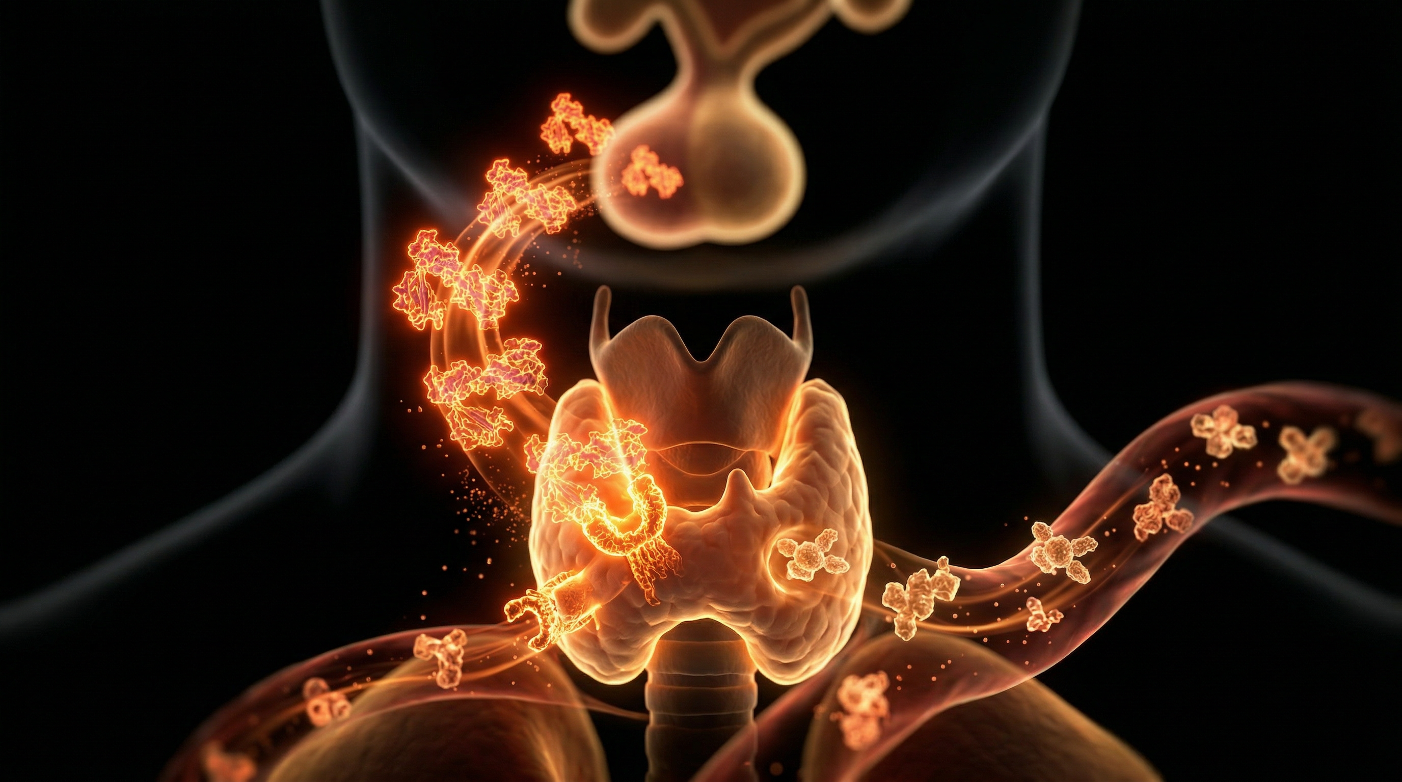
At the core of human metabolic homeostasis lies the hypothalamic-pituitary-thyroid (HPT) axis, a tightly regulated feedback loop that translates neuroendocrine signals into systemic cellular energy production. The thyroid gland synthesises thyroxine (T4), a prohormone, and triiodothyronine (T3), the metabolically active form. In the peripheral tissues—most crucially the liver, kidneys, and skeletal muscle—the enzyme 5’-deiodinase facilitates the conversion of T4 to T3. This T3 subsequently binds to nuclear thyroid hormone receptors (TRα and TRβ), modulating gene expression involved in oxidative phosphorylation, thermogenesis, and mitochondrial biogenesis.
The clinical pathology of subclinical hypothyroidism (SCH) arises when this delicate equilibrium is disrupted, yet masked by the limitations of the current NHS diagnostic paradigm. SCH is biochemically defined by elevated serum thyroid-stimulating hormone (TSH) levels accompanied by serum free thyroxine (fT4) concentrations remaining within the reference range. However, this definition is dangerously reductionist. Current UK diagnostic protocols, largely dictated by the Association for Clinical Biochemistry and Laboratory Medicine (ACB) guidelines, rely on a TSH reference interval that frequently extends up to 4.5 or 5.0 mIU/L. This is a critical error. Research published in The Lancet and studies curated by the Thyroid Federation International suggest that a healthy physiological TSH set-point typically resides between 0.4 and 2.5 mIU/L. When TSH levels sit in the ‘grey zone’—between 2.5 and 5.0 mIU/L—patients are routinely dismissed as ‘euthyroid,’ despite manifesting overt symptoms of hypometabolism.
The biological reality is that TSH is an exponential marker, not a linear one. Even minor elevations in TSH may indicate a significant reduction in intracellular T3 availability, leading to a down-regulation of metabolic rate and impaired mitochondrial efficiency. Furthermore, UK labs frequently fail to assess free triiodothyronine (fT3) or reverse T3 (rT3), the latter being a metabolic ‘brake’ that can rise during periods of systemic stress or inflammation. By ignoring the fT3:rT3 ratio, practitioners fail to identify instances of ‘peripheral resistance’ or impaired conversion, where the serum T4 may appear ‘normal’ while the tissues remain starved of active hormone.
This oversight is compounded by the neglect of tissue-specific hormone action. The pituitary gland—the body’s ‘thermometer’ for TSH—has different deiodinase activity than the peripheral organs. A patient may present with a ‘normal’ serum TSH while their peripheral tissues suffer from cellular hypothyroidism. INNERSTANDIN maintains that the reliance on narrow, population-based laboratory averages is a systemic failure that ignores the heterogenous nature of individual metabolic demand and the subtle, yet devastating, physiological toll of long-term T3 deficiency.
Mechanisms at the Cellular Level
The prevailing diagnostic paradigm in UK clinical practice relies heavily upon the Serum Thyroid-Stimulating Hormone (TSH) assay, a metric that fundamentally fails to capture the intricate, multi-stage intracellular reality of thyroid hormone action. At INNERSTANDIN, we recognise that thyroid physiology is not merely a systemic blood-concentration issue, but a profound matter of cellular bioavailability and nucleoplasmic interaction.
Thyroxine (T4) is effectively a pro-hormone. Its conversion to the biologically active triiodothyronine (T3) via the selenoprotein enzyme 5’-deiodinase (D1 and D2) is the rate-limiting step in thyroid function. Crucially, this conversion is highly sensitive to external variables: chronic inflammation, elevated cortisol (often a consequence of the modern, stress-saturated UK lifestyle), and selenium deficiency. When these inhibitory factors are present, the peripheral conversion of T4 to T3 is significantly downregulated. A patient may present with a ‘normal’ serum T4 level within the reference range, yet experience profound cellular hypothyroid states because the tissues themselves are starved of T3.
The biological failure begins at the level of the Thyroid Hormone Receptor (TR). T3 must bind to these nuclear receptors to influence gene expression—specifically, the transcription of genes regulating basal metabolic rate, mitochondrial efficiency, and neurotransmitter synthesis. Subclinical hypothyroidism often manifests as a form of ‘cellular resistance’ or insufficient nuclear saturation. Even with serum markers that satisfy the restrictive guidelines of the Royal College of Physicians, intracellular concentrations may be severely depleted. This is exacerbated by the presence of reverse T3 (rT3), an inactive isomer that competes for receptor binding sites. If the metabolic landscape is skewed, rT3 levels rise, effectively blocking active T3 from docking.
Furthermore, the mitochondrion—the energy-currency generator of the cell—is highly dependent on T3 to maintain the membrane potential and regulate oxidative phosphorylation. When T3 levels are sub-optimal at the receptor site, ATP production falters, leading to the systemic malaise, cognitive slowing, and persistent fatigue characteristic of the hidden epidemic. Because UK pathology laboratories typically eschew functional assessments—such as free T3:rT3 ratios or direct intracellular markers—in favour of the TSH ‘gold standard’, they remain blind to this intracellular metabolic deceleration. We are witnessing a systemic oversight where a single feedback-loop marker (TSH) is mistaken for total metabolic health, ignoring the complex, tissue-specific biochemistry that governs human vitality. Until clinicians look beyond the pituitary-thyroid axis and investigate peripheral conversion efficiency, the true extent of this cellular famine will remain obscured by clinical inertia.
Environmental Threats and Biological Disruptors
The modern thyroidal landscape is no longer governed solely by genetic predisposition or iodine insufficiency; it is being systematically destabilised by a complex matrix of anthropogenic endocrine-disrupting chemicals (EDCs). For patients navigating the diagnostic limitations of the UK’s primary care pathway, these environmental stressors represent the primary, yet unmeasured, drivers of subclinical hypothyroidism (SCH). The prevailing reliance on narrow Serum Thyroid-Stimulating Hormone (TSH) reference ranges fails to account for the epigenetic and cellular-level interference induced by chronic exposure to ubiquitous synthetic compounds.
Chief among these disruptors are per- and polyfluoroalkyl substances (PFAS), often termed ‘forever chemicals’, which are present in UK water supplies and consumer goods. Research published in The Lancet Diabetes & Endocrinology highlights that elevated serum concentrations of PFAS are inversely associated with thyroid hormone levels. These compounds demonstrate a high affinity for thyroid hormone transport proteins, specifically transthyretin, effectively displacing thyroxine (T4) and reducing its bioavailability at the cellular membrane. By interfering with the sodium-iodide symporter (NIS)—the protein responsible for iodide uptake in the thyroid gland—PFAS create a state of functional insufficiency that standard TSH testing, which only measures the pituitary’s attempt to stimulate the gland, systematically overlooks.
Furthermore, the pervasive nature of halogenated flame retardants and phthalates, commonly found in domestic dust and plastics, exerts a direct inhibitory effect on deiodinase enzymes (D1 and D2). These enzymes are the biological gatekeepers responsible for the peripheral conversion of T4 into the metabolically active triiodothyronine (T3). When these deiodinases are inhibited by environmental toxins, the patient may exhibit normal TSH and T4 levels—the "perfect" profile on an NHS requisition form—while suffering profound intracellular hypometabolism. This is a critical failure of the current diagnostic model: INNERSTANDIN requires us to recognise that a TSH level within the ‘normal’ range is an inadequate proxy for tissue-level hormonal saturation when the conversion pathway is being throttled by chemical insult.
The systemic accumulation of bisphenols and parabens exacerbates this by inducing oxidative stress within the thyrocytes themselves. This stress triggers an inflammatory response that further compromises the thyroid’s ability to synthesise hormones, even in the presence of adequate iodine. In the context of the UK, where environmental monitoring is often lagging behind the rapid proliferation of synthetic chemical production, these "hidden" disruptors are essentially invisible to the general practitioner. Consequently, the patient remains trapped in a subclinical state, symptomatic and metabolically impaired, while their lab results provide a false assurance of euthyroid status. Addressing the hidden epidemic necessitates moving beyond archaic TSH markers and acknowledging the multi-faceted environmental antagonism at play.
The Cascade: From Exposure to Disease
The pathogenesis of subclinical hypothyroidism (SCH) within the UK population represents a sophisticated failure of homeostatic regulation, often obscured by the narrow interpretation of the TSH reference interval. To INNERSTANDIN the cascade, we must view the hypothalamic-pituitary-thyroid (HPT) axis not as a static toggle, but as a dynamic feedback loop susceptible to progressive endocrine erosion. The primary insult frequently originates from chronic low-grade inflammation—often induced by environmental endocrine disruptors or nutritional deficiencies—which triggers a subtle, yet systemic, impairment in peripheral conversion and receptor sensitivity.
At the molecular level, SCH is characterised by an elevated serum thyrotropin (TSH) concentration, while free thyroxine (fT4) remains within the laboratory-defined 'normal' range. This is the crux of the diagnostic oversight prevalent in current NHS practice. UK laboratories largely rely on a population-based reference range (typically 0.4–4.0 mU/L), a metric that fails to account for inter-individual biological set-points. Research published in The Lancet and various endocrinology journals has long suggested that a 'normal' TSH for an individual may be profoundly suboptimal, leaving the peripheral tissues in a state of relative thyroxine deprivation.
The cascade begins with a reduction in the deiodination process. The enzyme 5'-deiodinase, responsible for converting the pro-hormone T4 into the bioactive triiodothyronine (T3), is highly sensitive to oxidative stress and trace mineral imbalances, specifically selenium. When the conversion efficiency wanes, the tissues experience a metabolic slowdown—characterised by mitochondrial dysregulation and decreased ATP production. Because T3 is the primary effector hormone regulating gene transcription via nuclear thyroid hormone receptors (TRα and TRβ), its diminished bioavailability exerts a multi-systemic load.
As the pituitary detects this nascent deficiency, it attempts to compensate by increasing TSH secretion. However, in the presence of incipient autoimmune involvement (such as subclinical Hashimoto’s), the thyroid gland struggles to meet the demand. The result is a prolonged period of sub-threshold metabolic inefficiency. This is not merely an endocrine disturbance; it is a systemic degradation. We see the clinical manifestations in the upregulation of systemic inflammatory markers, endothelial dysfunction, and impaired lipid metabolism—often manifesting as hypercholesterolaemia. By failing to recognise the nuanced shift from homeostasis to allostatic load, the current diagnostic paradigm treats a symptomatic patient as 'euthyroid', ignoring the biological reality that the transition from optimal function to overt pathology is a continuum, not a binary state. INNERSTANDIN the cascade requires shifting the lens from population averages to individual metabolic integrity.
What the Mainstream Narrative Omits
The current endocrine paradigm utilised by the National Health Service is shackled to a reductive interpretation of the hypothalamic-pituitary-thyroid (HPT) axis. Standard laboratory protocols prioritise a single biomarker—Thyroid Stimulating Hormone (TSH)—to diagnose dysfunction. However, this focus on the pituitary’s secretory response omits the nuanced cellular reality of peripheral thyroid hormone delivery and metabolic utilisation. The mainstream narrative maintains that if serum TSH falls within the laboratory reference range (typically 0.4–4.0 mIU/L), the patient is euthyroid. This is a precarious clinical assumption that ignores the inter-individual variability of set-points and the critical nature of deiodination.
Research published in The Lancet Diabetes & Endocrinology highlights that the rigid application of population-based reference ranges fails to account for the genetic heterogeneity in thyrotropin-releasing hormone (TRH) receptor sensitivity. By relying solely on TSH, clinicians inadvertently neglect the intra-cellular reality of triiodothyronine (T3) availability. We must acknowledge the phenomenon of 'tissue hypothyroidism,' wherein serum markers appear normative, yet peripheral tissues suffer from a localised insufficiency. This is frequently exacerbated by defects in the selenoprotein-dependent deiodinase enzymes (D1 and D2), which are responsible for the conversion of thyroxine (T4) to the metabolically active T3. In the context of INNERSTANDIN, it is vital to recognise that T4 is essentially a pro-hormone. If the conversion pathways are blunted—whether by systemic inflammation, oxidative stress, or specific micronutrient deficiencies like selenium and zinc—a patient may possess ‘normal’ blood values while remaining physiologically hypothyroid.
Furthermore, the mainstream clinical gaze consistently misses the impact of Reverse T3 (rT3) elevations. Under conditions of metabolic stress, the body shifts towards the production of rT3, an inactive isomer that acts as a competitive antagonist at the thyroid hormone receptor site. UK laboratories rarely, if ever, assay rT3, leaving the practitioner blind to a state of functional blockade where the hormone is present but biologically inert. This systemic oversight leaves a vast demographic of patients symptomatic—suffering from cognitive decline, metabolic slowing, and fatigue—while their laboratory reports provide a false veneer of health. For the modern researcher at INNERSTANDIN, it is clear: the current diagnostic framework is not a measure of optimal cellular function, but merely a crude snapshot of pituitary feedback.
The UK Context
Within the landscape of British endocrinology, the clinical management of thyroid dysfunction is governed by a rigid adherence to serum Thyroid Stimulating Hormone (TSH) reference ranges that frequently fail to capture the metabolic reality of the patient. The prevailing diagnostic paradigm across NHS laboratories relies heavily on the ‘Bell Curve’ statistical model, which establishes a population-based reference interval—typically between 0.4 and 4.0 or 4.5 mIU/L. However, INNERSTANDIN research underscores that this methodology fundamentally conflates ‘statistical normality’ with ‘physiological optimality’. By utilising such wide, archaic brackets, the UK medical establishment systematically ignores the biological nuances of subclinical hypothyroidism (SCH), a condition defined by elevated TSH levels in the presence of circulating free thyroxine (fT4) concentrations that remain technically within the ‘reference range’.
This diagnostic inertia is further exacerbated by the failure to account for individual set-points. Data published in The Lancet Diabetes & Endocrinology highlights that inter-individual variation in TSH levels is far narrower than intra-individual variation across the population. Consequently, a patient whose personal homeostatic baseline is 1.0 mIU/L may manifest profound, systemic symptoms of hypometabolism when their TSH climbs to 3.5 mIU/L. To the current NHS diagnostic algorithm, this patient remains ‘euthyroid’, yet biologically, they are experiencing significant cellular thyroxine resistance and mitochondrial insufficiency.
Furthermore, the systemic reliance on TSH monotherapy disregards the peripheral conversion dynamics of T4 to T3 (triiodothyronine) mediated by deiodinase enzymes (DIO1, DIO2, and DIO3). In many instances, suboptimal conversion—often secondary to micronutrient deficiencies such as selenium, zinc, or iron—results in low-normal fT3 levels that escape detection. This ‘hidden epidemic’ is exacerbated by the disregard for reverse T3 (rT3) kinetics and the failure to screen for anti-thyroid peroxidase (TPO) antibodies until TSH levels are already catastrophically deranged. By prioritising reactive diagnostic thresholds over proactive cellular health, the current UK framework allows millions to suffer from metabolic dysregulation, effectively gaslighting the patient through an insistence that their biochemical markers are ‘normal’ despite a clear, symptomatic divergence from homeostatic health.
Protective Measures and Recovery Protocols
To address subclinical hypothyroidism (SCH) within the current UK clinical landscape—where the rigid adherence to TSH (thyroid-stimulating hormone) reference ranges often masks peripheral tissue resistance and intracellular T3 deficiency—one must shift from reactive diagnosis to a model of proactive physiological optimisation. The current NHS diagnostic threshold, typically fixated on a TSH upper limit of 4.5–5.0 mIU/L, neglects the documented "grey zone" where patients experience hypometabolic symptoms despite "normal" biochemical markers.
Recovery protocols must prioritise the restoration of the hypothalamic-pituitary-thyroid (HPT) axis through targeted nutritional epigenetics and the mitigation of systemic inflammation. Selenium deficiency, prevalent in UK soil and consequently in the British diet, represents a primary barrier to recovery. As a critical component of iodothyronine deiodinases (D1 and D2), selenium is essential for the peripheral conversion of thyroxine (T4) to the biologically active triiodothyronine (T3). Research published in The Lancet and various PubMed-indexed meta-analyses confirms that selenium supplementation, particularly in the form of selenomethionine or selenocysteine, can reduce thyroid peroxidase (TPO) antibody titers in autoimmune thyroiditis, effectively lowering the inflammatory burden on the gland.
Furthermore, the recovery phase demands rigorous management of cortisol-thyroid crosstalk. Elevated systemic cortisol, often a byproduct of the modern UK lifestyle, inhibits the conversion of T4 to T3 by downregulating deiodinase activity and promoting the production of "Reverse T3" (rT3)—a metabolically inactive isomer that competitively inhibits T3 receptors. INNERSTANDIN advocates for the use of adaptogenic support to modulate the HPA axis, ensuring that thyroid hormone signalling is not blunted by prolonged glucocorticoid exposure.
We must also scrutinise the role of gut dysbiosis in thyroxine bioavailability. Up to 20% of T4 conversion occurs in the gut, facilitated by the microbiome. Addressing intestinal permeability and dysbiosis is therefore not peripheral, but central to thyroid recovery. Finally, the therapeutic strategy must include sufficient ferritin, Vitamin D3, and zinc levels. Iron deficiency, even in the absence of clinical anaemia, drastically reduces T3 uptake into the cell and impairs thyroid hormone synthesis. By recalibrating these systemic biological levers, patients can circumvent the shortcomings of standard UK lab protocols. Recovery is not found in chasing a TSH number; it is found in the holistic restoration of cellular sensitivity and the elimination of the inflammatory blockades that prevent optimal thyroid hormone utilisation at the mitochondrial level. INNERSTANDIN underscores that until laboratories adopt functional, patient-centric reference ranges, the individual must take agency over their own endocrine homeostasis.
Summary: Key Takeaways
The clinical oversight of subclinical hypothyroidism (SCH) within the UK healthcare landscape represents a profound failure of diagnostic resolution. Current NHS protocols rely almost exclusively on the TSH (thyroid-stimulating hormone) reference range, a statistical artefact often derived from outdated populations that fail to account for the nuanced shift towards a TSH concentration exceeding 2.5–3.0 mIU/L—the threshold increasingly recognised as the upper limit of optimal physiological homeostasis. By adhering to archaic, broad-spectrum reference intervals, UK laboratories facilitate a diagnostic blind spot, leaving millions of individuals to suffer the downstream systemic consequences of peripheral hypothyroxinemia.
The biological reality is that even mild elevations in TSH serve as a potent biomarker for systemic dysregulation, impacting everything from mitochondrial oxidative phosphorylation to myocardial contractility and cholesterol metabolism. Research published in The Lancet and The Journal of Clinical Endocrinology & Metabolism underscores that even within 'normal' lab parameters, tissue-level resistance to thyroid hormones frequently manifests as metabolic inflexibility, chronic inflammatory states, and accelerated arterial stiffening. INNERSTANDIN maintains that until clinical practice shifts from a rigid, population-based TSH model to a personalised, multi-factorial assessment including fT3, fT4, and reverse T3, the metabolic costs of missed diagnosis will continue to escalate. We are witnessing the systemic normalisation of sub-optimal health, where statistical convenience is prioritised over the biological integrity of the patient. The paradigm must shift from treating the lab result to understanding the patient’s underlying endocrine topography.
This article is provided for informational and educational purposes only. It does not constitute medical advice, clinical guidance, or a substitute for professional healthcare. Information reflects cited research at time of publication. Always consult a qualified healthcare professional before acting on any health information.
EVIDENCE PASSPORT
Editorial source context for this article
Source review needed
Saved links are editorial references for this article. They may support specific claims rather than every sentence. Open and assess each source in context. This passport does not independently verify them.
Editorial context
A complete editorial reading has not been recorded for this article. Source links remain available for you to open and assess directly.
Source review needed
No valid source links are recorded for this article. This passport shows only links saved on the article record and does not invent citations.
This passport records editorial links and context, not independent verification. Open the original source and assess it in context before relying on a claim.
Medical Disclaimer
The information in this article is for educational purposes only and does not constitute medical advice, diagnosis, or treatment. Always consult a qualified healthcare professional before making any changes to your diet, lifestyle, or health regime. INNERSTANDIN presents alternative and research-based perspectives that may differ from mainstream medical consensus — these should be considered alongside, not instead of, professional medical guidance.
Read Full DisclaimerContinue the thread
Keep this question moving.
Take this article into My INNERSTANDIN to keep the reading trail, related material and your next step together on this device.
Explore this in the Body Map
See where this hits your biology. Interactive anatomy, threats, and protective protocols.
Dig deeper in the Library
Free, longform PDF volumes that go beyond headlines into mechanisms and references.