
Mechanisms of Fibrotic Remodeling in the Pericardial Sac Following Inflammatory Insults
Discuss this resourceUpdated August 2026
An in-depth exploration of the pathological pathways leading to pericardial fibrosis, focusing on the transition from acute inflammation to chronic restrictive states.
Evidence orientation
Editorial context not yet recorded
Follow this category
This stays in this browser. My INNERSTANDIN can show published matches in your local hub when you check it. It does not send email, push, or alert notifications.
Local learning review
A private browser aid for revisiting ideas. It is not an alert or a health recommendation.
Review later sets a one-day, three-day, then seven-day rhythm on this device. Choose it only when you want to revisit this article.
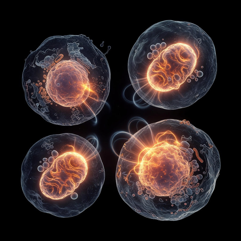
The pericardium is a complex, fibroelastic sac that encapsulates the heart, providing critical lubrication and structural stabilization. When this delicate membrane suffers an inflammatory insult—whether infectious, autoimmune, or traumatic—the body initiates a repair response that, if dysregulated, leads to fibrotic remodeling. Understanding this transition is essential for addressing cardiac constraints at their root.
The Inflammatory Trigger
Inflammation of the pericardium, or pericarditis, serves as the initial catalyst. The recruitment of neutrophils and macrophages to the epicardial and pericardial surfaces is intended as a defensive mechanism. However, when the insult persists or the immune response becomes chronic, the signaling environment changes. Pro-inflammatory cytokines, specifically IL-1β, IL-6, and TNF-α, shift the local cellular landscape, activating quiescent fibroblasts.
The Fibroblast-to-Myofibroblast Transition
At the heart of fibrotic remodeling is the transformation of resident fibroblasts into myofibroblasts. This process is driven primarily by Transforming Growth Factor-beta 1 (TGF-β1). Once activated, myofibroblasts exhibit enhanced contractility and massive production of extracellular matrix (ECM) components, including collagen types I and III.
In a healthy state, ECM turnover is balanced by matrix metalloproteinases (MMPs). During fibrotic remodeling, this balance is lost. The upregulation of Tissue Inhibitors of Metalloproteinases (TIMPs) creates an environment where collagen deposition significantly outpaces degradation, leading to the thickening and stiffening of the pericardial layers.
Chronic Remodeling and Restrictive Sequelae
The accumulation of dense, fibrous tissue bridges the visceral and parietal layers of the pericardium. Over time, these adhesions can coalesce into a calcified, rigid shell—a condition known as constrictive pericarditis. This "encasement" physically limits the heart's ability to fill during diastole, resulting in impaired stroke volume and systemic congestion.
Root-Cause Considerations
Addressing fibrotic remodeling requires moving beyond symptom management. Integrative health approaches look at the systemic drivers of inflammation, such as metabolic dysfunction, chronic low-grade oxidative stress, and the resolution phase of the inflammatory cascade. By supporting the body's ability to resolve inflammation through proper nutrient signaling and cellular homeostasis, we can potentially interrupt the progression of the fibrotic cascade before it becomes structural.
Conclusion
Fibrotic remodeling of the pericardial sac is a testament to the heart's vulnerability to chronic inflammatory cycles. By focusing on the molecular pathways of the myofibroblast and the extracellular matrix, we gain a clearer understanding of how to protect cardiac membrane health and preserve long-term cardiovascular resilience.
This article is provided for informational and educational purposes only. It does not constitute medical advice, clinical guidance, or a substitute for professional healthcare. Information reflects cited research at time of publication. Always consult a qualified healthcare professional before acting on any health information.
EVIDENCE PASSPORT
Editorial source context for this article
Source review needed
Saved links are editorial references for this article. They may support specific claims rather than every sentence. Open and assess each source in context. This passport does not independently verify them.
Editorial context
A complete editorial reading has not been recorded for this article, and no source links are saved on its record.
Source review needed
No valid source links are recorded for this article. Supplementary research, where available, is listed separately below.
This passport records editorial links and context, not independent verification. Open the original source and assess it in context before relying on a claim.
Medical Disclaimer
The information in this article is for educational purposes only and does not constitute medical advice, diagnosis, or treatment. Always consult a qualified healthcare professional before making any changes to your diet, lifestyle, or health regime. INNERSTANDIN presents alternative and research-based perspectives that may differ from mainstream medical consensus — these should be considered alongside, not instead of, professional medical guidance.
Read Full DisclaimerContinue the thread
Keep this question moving.
Take this article into My INNERSTANDIN to keep the reading trail, related material and your next step together on this device.
Explore this in the Body Map
Explore related body structures and their educational pages in the interactive Body Map.