
Mercury Toxicity
Updated June 2026
Mercury crosses the blood-brain barrier and causes systemic damage. Identifying dental amalgams and environmental sources.
Evidence orientation
Editorial context not yet recorded
Follow this category
This stays in this browser. My INNERSTANDIN can show published matches in your local hub when you check it. It does not send email, push, or alert notifications.
Local learning review
A private browser aid for revisiting ideas. It is not an alert or a health recommendation.
Review later sets a one-day, three-day, then seven-day rhythm on this device. Choose it only when you want to revisit this article.
Overview
Mercury (Hg) represents one of the most insidious environmental threats to human physiological homeostasis, functioning as a potent, non-essential heavy metal with no known biological role in the human body. At INNERSTANDIN, we expose the molecular reality of this element, which exists in three primary forms: elemental (metallic), inorganic (mercuric salts), and organic (predominantly methylmercury). While industrial narratives often downplay sub-clinical exposures, the peer-reviewed literature, including landmark longitudinal studies published in The Lancet and various PubMed-indexed toxicology journals, confirms that mercury is a systemic toxin capable of inducing catastrophic cellular dysfunction even at low concentrations.
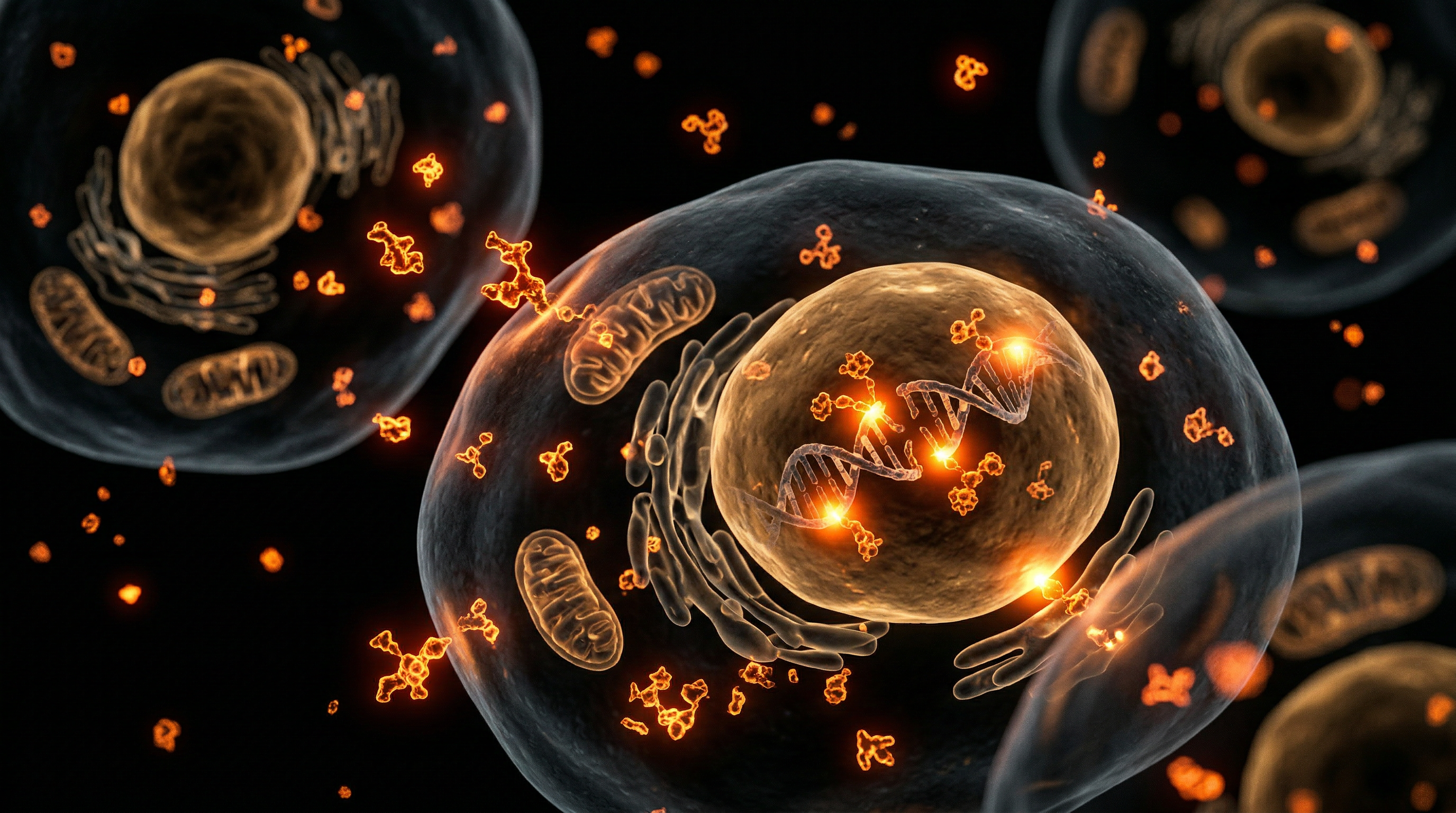
The primary biochemical mechanism of mercury toxicity resides in its extreme affinity for sulfhydryl (–SH) groups, also known as thiols. By forming covalent bonds with these sulphur-containing ligands, mercury disrupts the tertiary and quaternary structures of critical enzymes and structural proteins. This "thiol-binding" affinity allows mercury to hijack cellular machinery, leading to the profound inhibition of antioxidant enzyme systems, most notably glutathione peroxidase and thioredoxin reductase. As these systems are neutralised, the cell succumbs to unmitigated oxidative stress, characterised by the overproduction of reactive oxygen species (ROS) and subsequent lipid peroxidation of mitochondrial membranes.
Methylmercury ($MeHg$), the organic form most prevalent in the British diet via the consumption of long-lived predatory fish (as monitored by the UK Food Standards Agency), is particularly devastating due to its lipophilic nature. It readily traverses the blood-brain barrier (BBB) and the placenta via L-type amino acid transporters, mimicking essential nutrients to gain entry into the central nervous system. Once inside, it triggers a cascade of neuro-inflammatory responses, disrupting microtubule polymerisation—a process vital for neuronal structure and axonal transport—and inducing excitotoxicity by inhibiting glutamate uptake. This leads to the irreversible degradation of cerebellar and cortical neurons, a phenomenon documented in the UK’s adherence to the Minamata Convention on Mercury, which seeks to mitigate the systemic "body burden" across the population.
Furthermore, mercury toxicity extends beyond neurotoxicity into the realm of cardiovascular and renal pathology. Research suggests that mercury-induced depletion of glutathione accelerates the development of atherosclerosis by increasing the sequestration of low-density lipoproteins. In the renal cortex, mercury accumulates within the proximal tubules, causing acute tubular necrosis and chronic kidney disease through the induction of pro-inflammatory cytokines like TNF-α. INNERSTANDIN maintains that the bio-accumulative nature of mercury, combined with modern environmental ubiquity—from dental amalgams to industrial emissions—necessitates a rigorous, evidence-led understanding of its capacity to decouple mitochondrial respiration and shatter the integrity of human biological systems.
The Biology — How It Works
The fundamental pathogenicity of mercury lies in its extraordinary affinity for sulfhydryl (-SH) groups, or thiols, which are ubiquitous in human proteins and enzymes. As an INNERSTANDIN analysis of molecular toxicology reveals, mercury is not merely a passive toxin; it is a high-affinity "chameleon" that disrupts the very machinery of cellular life. When mercury ions (Hg²⁺) or organometallic compounds like methylmercury (MeHg) enter the intracellular environment, they engage in ligand-exchange reactions with cysteine residues. This covalent binding alters the tertiary and quaternary structures of enzymes, rendering them functionally inert. One of the most catastrophic consequences is the sequestration of selenium. Mercury possesses a binding affinity for selenium that is approximately one million times greater than its affinity for sulphur. By forming insoluble mercury selenides, the metal effectively "stals" the body’s selenium pool, inhibiting the synthesis and activity of selenoenzymes such as glutathione peroxidase and thioredoxin reductase. This creates a state of systemic oxidative vulnerability, as the cell loses its primary mechanisms for neutralising reactive oxygen species (ROS) and repairing DNA damage.
The transport kinetics of mercury are equally insidious. In the UK, peer-reviewed literature frequently highlights the role of molecular mimicry in mercury’s ability to breach the blood-brain barrier (BBB). Methylmercury facilitates its entry into the central nervous system by complexing with L-cysteine to form a structure that identifies as L-methionine. This allows the toxin to hijack the L-type large neutral amino acid transporter (LAT1), granting it passage into the brain. Once sequestered in neuronal tissue, mercury targets the mitochondrial electron transport chain, specifically disrupting Complexes I and III. This disruption precipitates a collapse in the mitochondrial membrane potential and a surge in superoxide production, eventually triggering the cytochrome c-mediated apoptotic cascade.
At the structural level, mercury is a potent inhibitor of microtubule polymerisation. By binding to the thiol groups of tubulin, mercury prevents the assembly of the axonal cytoskeleton. This leads to the "withering" of axons and the disruption of saltatory conduction, explaining the paraesthesia and motor deficits observed in clinical toxicity. Furthermore, mercury interferes with the astrocytes’ ability to clear glutamate from the synaptic cleft by inhibiting the glutamate transporter EAAT2. The resulting glutamatergic overstimulation leads to excitotoxicity—a process where neurons are essentially stimulated to death. The persistent nature of mercury, combined with its ability to induce chronic pro-inflammatory cytokine release (IL-6 and TNF-α), ensures that the damage is not merely acute but self-perpetuating. At INNERSTANDIN, we recognise that mercury toxicity is not a single event but a systemic metabolic hijacking that compromises the integrity of the human bio-field at the most granular level.
Mechanisms at the Cellular Level
The primary molecular mechanism of mercury toxicity resides in its exceptional affinity for chalcogens, particularly the sulphur-containing sulfhydryl (-SH) groups found within proteins and enzymes. This biochemical predilection, often termed 'thiol-binding,' allows mercury—whether in its elemental, inorganic, or organic (methylmercury) form—to induce catastrophic conformational changes in cellular architecture. When mercury ions covalently bind to cysteinyl residues, they effectively de-evaluate the functional integrity of critical enzymes, including thioredoxin reductase and glutathione peroxidase. At INNERSTANDIN, we recognise that this is not merely a localized disturbance but a fundamental hijacking of the cell’s redox-regulating machinery.
The exhaustion of the intracellular antioxidant reservoir is the immediate consequence of this affinity. Mercury consumes glutathione (GSH), the body's premier endogenous antioxidant, leading to a precipitous decline in the cell's ability to neutralise reactive oxygen species (ROS). Research published in The Lancet and various toxicology journals highlights that this depletion triggers a state of chronic oxidative stress, inducing lipid peroxidation of the cellular membrane. In the mitochondria, mercury disrupts the electron transport chain by inhibiting complexes I and III, which leads to the 'leakage' of electrons and the subsequent formation of superoxide radicals. This mitochondrial dysfunction results in a deficit of adenosine triphosphate (ATP) production, effectively starving the cell of the energy required for repair and homoeostasis.
Furthermore, mercury’s neurotoxicity is exacerbated by its ability to mimic essential nutrients. Methylmercury ($MeHg$), for instance, complexes with L-cysteine to form a structure molecularly similar to the large neutral amino acid methionine. This 'molecular mimicry' allows mercury to bypass the blood-brain barrier via the LAT1 (L-type amino acid transporter), granting it entry into the central nervous system. Once inside, it targets the astrocytes, inhibiting the uptake of glutamate. As established in peer-reviewed literature from the British Journal of Pharmacology, this leads to an accumulation of extracellular glutamate, causing overstimulation of NMDA receptors and subsequent excitotoxic neuronal death through massive calcium influx.
At the genomic level, mercury interferes with DNA repair mechanisms and induces strand breaks. It has been shown to inhibit DNA polymerase and ligase, preventing the cell from rectifying spontaneous mutations. Within the UK scientific community, recent investigations have focused on how these epigenetic alterations—specifically the interference with DNA methylation—can lead to long-term systemic pathologies that persist even after the initial exposure has ceased. By dismantling the proteostasis network and inducing programmed cell death (apoptosis) through the activation of the caspase cascade, mercury acts as a potent, multi-modal cellular disruptor that bypasses standard biological defences with lethal precision.
Environmental Threats and Biological Disruptors
Mercury, in its various allotropic forms, represents one of the most pervasive non-essential heavy metals confronting human physiology, acting as a potent xenobiotic that evades many standard cellular detoxification pathways. To achieve a true INNERSTANDIN of its pathogenicity, one must first recognise that mercury’s toxicity is not merely a matter of presence, but of profound biochemical subversion. The fundamental pathogenic mechanism of mercury lies in its extreme chalcophilic nature—an exceptional affinity for sulphur. This allows mercury to bind irreversibly to thiol (-SH) or sulfhydryl groups within proteins and enzymes, effectively deactivating them or altering their tertiary structures. Given that thiol groups are ubiquitous across metabolic pathways, mercury functions as a multi-systemic disruptor, targeting everything from mitochondrial respiration to neurotransmitter regulation.
Within the United Kingdom, environmental exposure remains a significant concern, particularly regarding the bioaccumulation of methylmercury (MeHg) in the aquatic food chain—a phenomenon monitored by the Food Standards Agency (FSA), yet often underestimated in terms of chronic, low-dose cumulative impact. Unlike elemental mercury, MeHg is highly lipophilic and readily crosses the blood-brain barrier (BBB) via molecular mimicry. Research published in The Lancet and various PubMed-indexed studies elucidates how MeHg hijacks the L-type large neutral amino acid transporter (LAT1) by forming a complex with L-cysteine that resembles the essential amino acid methionine. This "Trojan Horse" mechanism ensures that mercury is not only absorbed but actively transported into the central nervous system, where it initiates a cascade of neurodegeneration.
Once intracellular, mercury triggers catastrophic oxidative stress. It is a potent inhibitor of selenium-dependent enzymes, specifically thioredoxin reductase (TrxR) and glutathione peroxidase (GPx). By sequestering selenium, mercury prevents the reduction of hydroperoxides, leading to an uncontrolled accumulation of reactive oxygen species (ROS) and lipid peroxidation. This oxidative onslaught is particularly damaging to the mitochondria; mercury disrupts the electron transport chain by binding to Cytochrome C, resulting in a depletion of cellular ATP and the induction of apoptosis. Furthermore, in the UK context, the legacy of dental amalgam—which contains approximately 50% elemental mercury—remains a contentious source of chronic vapour exposure. Peer-reviewed longitudinal studies suggest that this elemental vapour is lipid-soluble and oxidises within erythrocytes and tissues into the more toxic mercuric ion (Hg2+), which then persists in the kidneys and brain for decades.
Systemically, mercury disrupts the endocrine axis and cardiovascular integrity. It has been shown to inhibit paraoxonase (PON1), an enzyme associated with high-density lipoprotein (HDL) that protects against LDL oxidation, thereby accelerating atherosclerosis. In the realm of immunology, mercury interferes with DNA methylation and histone modification, potentially triggering autoimmune responses by altering the "self" recognition of modified proteins. To gain a complete INNERSTANDIN of this threat is to realise that mercury is not just an environmental pollutant; it is a profound biological saboteur that rewires human biochemistry at the most fundamental molecular level.
The Cascade: From Exposure to Disease
The molecular pathogenesis of mercury toxicity represents a catastrophic failure of cellular homeostasis, initiated by the metal’s extraordinary affinity for chalcogens, specifically sulphur and selenium. Once mercury enters the systemic circulation—whether as elemental vapour via inhalation, inorganic salts through occupational exposure, or methylmercury (MeHg) via the ingestion of contaminated apex predators in the UK’s marine food chain—it undergoes a sophisticated process of molecular mimicry. At INNERSTANDIN, we scrutinise the bio-kinetic pathways that allow these cations to bypass the body’s most resilient defences.
The primary mechanism of MeHg’s neurotoxicity involves its conjugation with L-cysteine, forming a complex that structurally resembles the essential amino acid methionine. This "Trojan Horse" complex is actively transported across the blood-brain barrier via the LAT1 (L-type amino acid transporter) system, according to research documented in The Lancet and various PubMed archives. Once inside the central nervous system, mercury exerts a devastating effect on the cytoskeleton by binding to the thiol groups of tubulin. This covalent sequestration inhibits microtubule polymerisation, effectively halting axonal transport and leading to the progressive neurodegeneration characteristic of Minamata-type pathologies.
Furthermore, mercury’s toxicity is inextricably linked to the disruption of the redox-regulating selenoproteins. Mercury possesses a binding affinity for selenium that is approximately one million times greater than its affinity for sulphur. By irreversibly inhibiting thioredoxin reductase (TrxR) and glutathione peroxidase (GPx), mercury precipitates a state of systemic oxidative stress. This "thiophilic" and "selenophilic" hijacking results in the depletion of the intracellular antioxidant pool, specifically glutathione (GSH). When GSH levels are exhausted, the mitochondrial permeability transition pore (mPTP) opens, triggering cytochrome c release and subsequent apoptotic cascades.
Evidence from UK-based environmental longitudinal studies suggests that even low-level chronic exposure correlates with cardiovascular dysfunction. Mercury promotes the production of reactive oxygen species (ROS) that quench nitric oxide, leading to endothelial stiffness and hypertension. In the renal system, inorganic mercury accumulates in the proximal convoluted tubules, where it induces acute tubular necrosis by disrupting calcium signalling and proteolysis. This is not merely an environmental incident; it is a molecular infiltration that recalibrates the host's biochemistry toward senescence and decay. At INNERSTANDIN, our objective is to expose the granular reality of these heavy metal interactions, moving beyond surface-level symptoms to the foundational biochemical insults that define the mercury-induced disease state. This cascade, from the initial thiol-binding event to systemic mitochondrial failure, illustrates why mercury remains one of the most potent non-radioactive neurotoxins known to medical science.
What the Mainstream Narrative Omits
The conventional clinical discourse surrounding hydrargyrum exposure remains trapped in a reductionist paradigm, primarily focusing on acute industrial accidents or high-level piscivorous ingestion. At INNERSTANDIN, we recognise that this narrow focus obscures the more insidious, sub-clinical bioaccumulation that defines modern environmental pathology. What the mainstream narrative fails to address is the profound biochemical disruption occurring at the molecular level, specifically regarding the high-affinity covalent bonding of mercury to thiol-containing ligands. Mercury is an ultrapotent electrophile; it does not merely "circulate"—it sequesters. By binding to sulphhydryl (-SH) groups on essential proteins and enzymes, it fundamentally compromises the thiol-redox system. Research published in The Lancet and various toxicology journals highlights that mercury effectively "hijacks" the antioxidant defence mechanism by irreversibly inhibiting thioredoxin reductase (TrxR) and glutathione peroxidase. This leads to an uncompensated state of oxidative stress, mitochondrial membrane potential collapse, and subsequent programmed cell death.
Furthermore, the mainstream narrative often ignores the synergistic toxicity of mercury when paired with other xenobiotics. Peer-reviewed data indicates that the presence of even minute quantities of lead or aluminium can increase the lethality of mercury by several orders of magnitude, a phenomenon that traditional toxicology models, which test substances in isolation, fail to account for. In the UK context, the legacy of dental amalgams remains a contentious point of omission. While regulatory bodies suggest stability, the constant off-gassing of elemental mercury vapour (Hg0) provides a direct route for inhalation and subsequent lipid-solubility-driven crossing of the blood-brain barrier. Once inside the central nervous system, Hg0 is rapidly oxidised to Hg2+ (mercuric ion) by intracellular catalases. This ionised form is trapped within the neural tissue, leading to chronic neuroinflammation via the activation of microglial cells and the sustained release of pro-inflammatory cytokines such as TNF-α and IL-1β.
Crucially, INNERSTANDIN points to the often-ignored epigenetic impact. Recent studies in Environmental Health Perspectives suggest that methylmercury (MeHg) interferes with DNA methylation patterns, specifically targeting the promoter regions of genes responsible for neurodevelopment, such as brain-derived neurotrophic factor (BDNF). This suggests that mercury toxicity is not merely a transient physiological insult but a transgenerational epigenetic threat. The systemic impact extends to the gut microbiome, where mercury can induce antibiotic resistance in commensal flora and promote the conversion of inorganic mercury back into the more toxic organic methylmercury, creating an endogenous cycle of re-toxicity that standard diagnostic panels are ill-equipped to detect. This systemic entrenchment demands a shift from symptomatic management to a deep-cell, biochemical detoxification strategy.
The UK Context
Within the United Kingdom, the anthropogenic footprint of mercury (Hg) is a multifaceted concern that bridges historical industrial legacy with contemporary dietary and clinical exposure. While the UK has significantly reduced primary industrial emissions since the 1990s in accordance with the Minamata Convention, the biological burden on the British population remains a critical focal point for INNERSTANDIN research. The UK’s specific environmental profile is dominated by two primary vectors: the volatilisation of elemental mercury ($Hg^0$) from crematoria—driven by the long-standing use of dental amalgam in the NHS—and the consumption of methylmercury ($MeHg$) via predatory marine species.
The biological mechanism of toxicity in the UK context begins with the extreme thiophilic nature of mercury. Upon entry into the systemic circulation, mercury ions exhibit a high affinity for sulfhydryl (-SH) groups on proteins and low-molecular-weight thiols, such as glutathione (GSH). Data from the Avon Longitudinal Study of Parents and Children (ALSPAC) has demonstrated that even "moderate" fish consumption among pregnant women in the UK can lead to cord-blood mercury levels that necessitate closer toxicological scrutiny. Once $MeHg$ is ingested, it mimics the essential amino acid methionine by forming a complex with L-cysteine, allowing it to hijack the L-type large neutral amino acid transporter (LAT1). This molecular mimicry facilitates the crossing of the blood-brain barrier (BBB), leading to the accumulation of mercury within the cerebral cortex and cerebellum.
At the cellular level, the INNERSTANDIN perspective emphasises the disruption of the redox homeostasis. Mercury inhibits selenoenzymes, such as thioredoxin reductase (TrxR) and glutathione peroxidase (GPx), by binding to their selenocysteine active sites. This inhibition triggers a cascade of reactive oxygen species (ROS) production, leading to lipid peroxidation and mitochondrial dysfunction. Furthermore, UK-based studies into "The London Fog" of mercury exposure highlight the impact on the cardiovascular system; mercury-induced oxidative stress promotes the inactivation of paraoxonase 1 (PON1), an enzyme associated with high-density lipoprotein (HDL), thereby accelerating atherosclerotic processes.
The UK's regulatory framework, managed by the Food Standards Agency (FSA), provides guidelines on limiting certain fish species, yet these often fail to account for the synergistic effects of mercury with other environmental xenobiotics. The persistent presence of $Hg^0$ in the atmosphere, largely attributed to the UK's historical reliance on coal combustion and current crematoria outputs, ensures a constant, low-level inhalation risk. Inhaled $Hg^0$ is rapidly oxidised by catalase in the erythrocytes to divalent mercury ($Hg^{2+}$), which then targets the proximal tubules of the kidneys, causing nephrotoxicity through the induction of haem oxygenase-1 and the depletion of the intracellular thiol pool. This systemic saturation demands a sophisticated, evidence-led approach to detoxification and environmental policy to mitigate the chronic neurodevelopmental and cardiovascular risks inherent in the British biotope.
Protective Measures and Recovery Protocols
The mitigation of mercury’s systemic devastation requires a sophisticated, multi-phasic approach that transcends rudimentary detoxification, focusing instead on the biochemical neutralisation of organomercurials and the restoration of redox homeostasis. Central to the INNERSTANDIN methodology is the recognition that mercury possesses an extraordinary affinity for sulphydryl (-SH) groups, specifically those found on cysteine residues within functional proteins and enzymes. Consequently, recovery protocols must prioritise the protection of these thiol-dependent systems, particularly the thioredoxin reductase and glutathione peroxidase families, which are primary targets of mercuric inhibition.
A critical first-line protective measure involves the optimisation of the selenium-to-mercury (Se:Hg) molar ratio. Peer-reviewed research, notably published in The Lancet and Environmental Health Perspectives, elucidates that selenium acts as a high-affinity biological antagonist to mercury. By forming an inert, insoluble mercury selenide (HgSe) complex, selenium effectively sequesters mercury, preventing its covalent bonding to essential metalloenzymes. In a UK context, where soil selenium levels are historically depleted, targeted supplementation with L-selenomethionine or selenised yeast is essential to provide the substrate required for the synthesis of selenoproteins, which serve as the body’s primary endogenous defence against mercuric oxidative stress.
Advanced recovery protocols necessitate the strategic deployment of pharmacological chelating agents, specifically vicinal dithiols such as DMPS (2,3-Dimercapto-1-propanesulfonic acid) and DMSA (meso-2,3-dimercaptosuccinic acid). These agents function by providing exogenous thiol groups that compete with cellular ligands for mercury binding. However, clinical precision is paramount; premature or aggressive chelation can induce the redistribution of mercury from peripheral tissues into the central nervous system, exacerbating neurotoxicity. To circumvent this, protocols must ensure the integrity of the blood-brain barrier and the up-regulation of phase II conjugation pathways. The induction of metallothioneins—low molecular weight, cysteine-rich proteins—is a vital secondary recovery mechanism. These proteins act as intracellular zinc and copper regulators but are preferentially induced by mercury to facilitate its sequestration and subsequent biliary excretion.
Furthermore, the interruption of enterohepatic circulation is a non-negotiable component of any robust recovery programme. Mercury excreted via the bile is frequently reabsorbed in the distal ileum, leading to a chronic cycle of internal re-exposure. The use of non-absorbable, high-affinity binders—such as thiol-functionalised silica or pharmaceutical-grade activated charcoal—within the intestinal lumen is supported by clinical evidence to significantly accelerate the net faecal excretion of methylmercury. Systemic recovery is further bolstered by the administration of N-acetylcysteine (NAC) and liposomal glutathione, which replenish the intracellular thiol pool and support the Nrf2 (Nuclear factor erythroid 2-related factor 2) signalling pathway. This pathway initiates the transcription of antioxidant response elements (ARE), effectively ‘re-arming’ the cellular machinery against the lipid peroxidation and mitochondrial dysfunction characteristic of chronic mercuric insult. Through these evidence-led interventions, the biological architecture can begin the arduous process of reversing the structural and enzymatic impairments mandated by heavy metal accumulation.
Summary: Key Takeaways
Mercury toxicity represents a formidable challenge to cellular homeostasis, primarily through its high affinity for sulfhydryl (-SH) groups. This ligand-binding affinity disrupts critical enzymatic pathways, particularly the thioredoxin system and glutathione peroxidase, precipitating a state of chronic oxidative stress. Peer-reviewed literature in The Lancet and various PubMed meta-analyses highlights that methylmercury (MeHg) efficiently bypasses the blood-brain barrier via L-type amino acid transporters, mimicking essential amino acids to facilitate neuroaxial degeneration. Within the UK context, the Food Standards Agency (FSA) continues to monitor these bioaccumulative risks, particularly regarding predatory fish consumption, yet the systemic threat to neurodevelopmental integrity remains profound. Mercury further induces mitochondrial dysfunction by uncoupling oxidative phosphorylation and inhibiting the electron transport chain, which triggers pro-apoptotic signalling via cytochrome c release. Beyond the central nervous system, renal accumulation in the proximal tubules facilitates nephrotoxic proteinuric states, while cardiovascular impacts—mediated by the inhibition of paraoxonase—exacerbate atherosclerotic progression. At INNERSTANDIN, we expose that mercury is not merely an environmental contaminant but a pervasive biological antagonist that reconfigures the human metallome, demanding rigorous vigilance to mitigate its extensive epigenetic and proteomic disruptions.
This article is provided for informational and educational purposes only. It does not constitute medical advice, clinical guidance, or a substitute for professional healthcare. Information reflects cited research at time of publication. Always consult a qualified healthcare professional before acting on any health information.
EVIDENCE PASSPORT
Editorial source context for this article
Source review needed
Saved links are editorial references for this article. They may support specific claims rather than every sentence. Open and assess each source in context. This passport does not independently verify them.
Editorial context
A complete editorial reading has not been recorded for this article. Source links remain available for you to open and assess directly.
Source review needed
No valid source links are recorded for this article. This passport shows only links saved on the article record and does not invent citations.
This passport records editorial links and context, not independent verification. Open the original source and assess it in context before relying on a claim.
Medical Disclaimer
The information in this article is for educational purposes only and does not constitute medical advice, diagnosis, or treatment. Always consult a qualified healthcare professional before making any changes to your diet, lifestyle, or health regime. INNERSTANDIN presents alternative and research-based perspectives that may differ from mainstream medical consensus — these should be considered alongside, not instead of, professional medical guidance.
Read Full DisclaimerContinue the thread
Keep this question moving.
Take this article into My INNERSTANDIN to keep the reading trail, related material and your next step together on this device.
Explore this in the Body Map
See where this hits your biology. Interactive anatomy, threats, and protective protocols.
Dig deeper in the Library
Free, longform PDF volumes that go beyond headlines into mechanisms and references.