
Molecular Mechanisms of Lead-Induced Oxidative Stress and Mitochondrial Dysfunction
Discuss this resourceUpdated August 2026
An in-depth exploration into how lead (Pb) exposure disrupts cellular respiration and triggers systemic oxidative damage at the mitochondrial level, detailing the biochemical pathways responsible for chronic toxicity.
Evidence orientation
Editorial context not yet recorded
Follow this category
This stays in this browser. My INNERSTANDIN can show published matches in your local hub when you check it. It does not send email, push, or alert notifications.
Local learning review
A private browser aid for revisiting ideas. It is not an alert or a health recommendation.
Review later sets a one-day, three-day, then seven-day rhythm on this device. Choose it only when you want to revisit this article.
Introduction to Lead Toxicity
Lead (Pb) remains a critical public health challenge despite regulatory efforts. As a potent neurotoxin and systemic poison, lead does not possess a known physiological function in the human body. Its toxicity is primarily driven by its ability to mimic essential divalent cations like calcium (Ca2+) and zinc (Zn2+), allowing it to infiltrate critical cellular processes. This article examines the molecular mechanisms underlying lead-induced oxidative stress and the subsequent collapse of mitochondrial integrity.
The Redox Paradox and Reactive Oxygen Species (ROS)
Lead is categorized as a non-redox-active metal, meaning it does not participate directly in Fenton-type reactions. However, it paradoxically acts as a powerful pro-oxidant. Lead induces oxidative stress by depleting the cell’s primary antioxidant reserves, particularly glutathione (GSH). By binding to the sulfhydryl (-SH) groups of antioxidant enzymes such as superoxide dismutase (SOD), catalase, and glutathione peroxidase, lead renders these defense systems impotent. This depletion creates a state of redox imbalance, leading to the accumulation of reactive oxygen species (ROS) that induce lipid peroxidation and DNA damage.
Mitochondrial Dysfunction: The Core of the Crisis
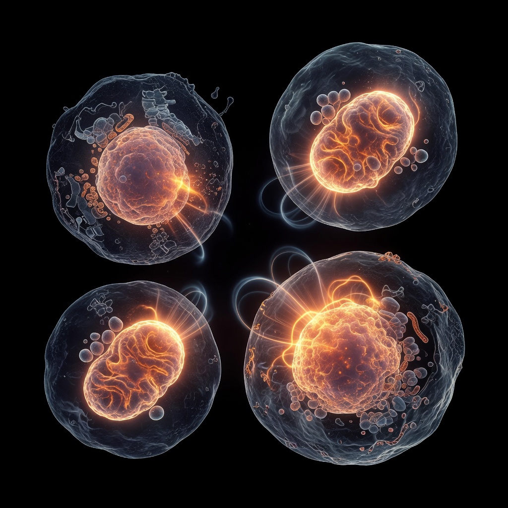
Mitochondria are the primary targets of lead toxicity due to their role in energy production and their high membrane sensitivity. Lead ions compete with calcium for entry into the mitochondrial matrix via the uniporter. Once inside, lead disrupts the Electron Transport Chain (ETC). Specifically, lead inhibits complexes I, II, and IV, resulting in the leakage of electrons that react with molecular oxygen to form superoxide radicals. This not only impairs ATP synthesis but also triggers the opening of the Mitochondrial Permeability Transition Pore (MPTP). The loss of mitochondrial membrane potential is a hallmark of lead-induced apoptosis, signaling the cell to undergo programmed death.
Molecular Mimicry and Calcium Signaling
Lead’s ability to replace calcium is perhaps its most insidious mechanism. Because lead has a higher affinity for calcium-binding proteins, such as calmodulin and protein kinase C (PKC), it dysregulates intracellular signaling pathways. In neurons, this leads to synaptic dysfunction and excitotoxicity. The chronic elevation of intracellular calcium, facilitated by lead, keeps the mitochondria in a state of hyper-metabolic stress, further accelerating the production of ROS and creating a vicious cycle of cellular degradation.
Clinical Implications and Future Directions
Understanding these pathways is essential for developing therapeutic chelating agents that move beyond simply removing metal ions and instead focus on antioxidant restoration. Future interventions must prioritize mitochondrial protection and the mitigation of lipid peroxidation to counteract the long-term systemic impact of lead exposure.
This article is provided for informational and educational purposes only. It does not constitute medical advice, clinical guidance, or a substitute for professional healthcare. Information reflects cited research at time of publication. Always consult a qualified healthcare professional before acting on any health information.
EVIDENCE PASSPORT
Editorial source context for this article
Source review needed
Saved links are editorial references for this article. They may support specific claims rather than every sentence. Open and assess each source in context. This passport does not independently verify them.
Editorial context
A complete editorial reading has not been recorded for this article, and no source links are saved on its record.
Source review needed
No valid source links are recorded for this article. Supplementary research, where available, is listed separately below.
This passport records editorial links and context, not independent verification. Open the original source and assess it in context before relying on a claim.
Medical Disclaimer
The information in this article is for educational purposes only and does not constitute medical advice, diagnosis, or treatment. Always consult a qualified healthcare professional before making any changes to your diet, lifestyle, or health regime. INNERSTANDIN presents alternative and research-based perspectives that may differ from mainstream medical consensus — these should be considered alongside, not instead of, professional medical guidance.
Read Full DisclaimerContinue the thread
Keep this question moving.
Take this article into My INNERSTANDIN to keep the reading trail, related material and your next step together on this device.
Explore this in the Body Map
Explore related body structures and their educational pages in the interactive Body Map.
Dig deeper in the Library
Free, longform PDF volumes that go beyond headlines into mechanisms and references.