Ocular Neurodegeneration: The Link Between Glaucoma, Alzheimer’s, and Parkinson’s
Scientific evidence suggests that the eye is an extension of the brain and shares similar neurodegenerative pathways. This article explores how early ocular changes can be used as biomarkers for cognitive decline.
# Ocular Neurodegeneration: The Link Between Glaucoma, Alzheimer’s, and Parkinson’s
Overview
For decades, the medical establishment has viewed the eye as an isolated organ, a self-contained camera-like device that merely transmits signals to the brain. If you had a problem with your vision, you saw an optician or an ophthalmologist. If you had a problem with your memory or motor skills, you saw a neurologist. This compartmentalised approach to medicine has failed millions of patients.
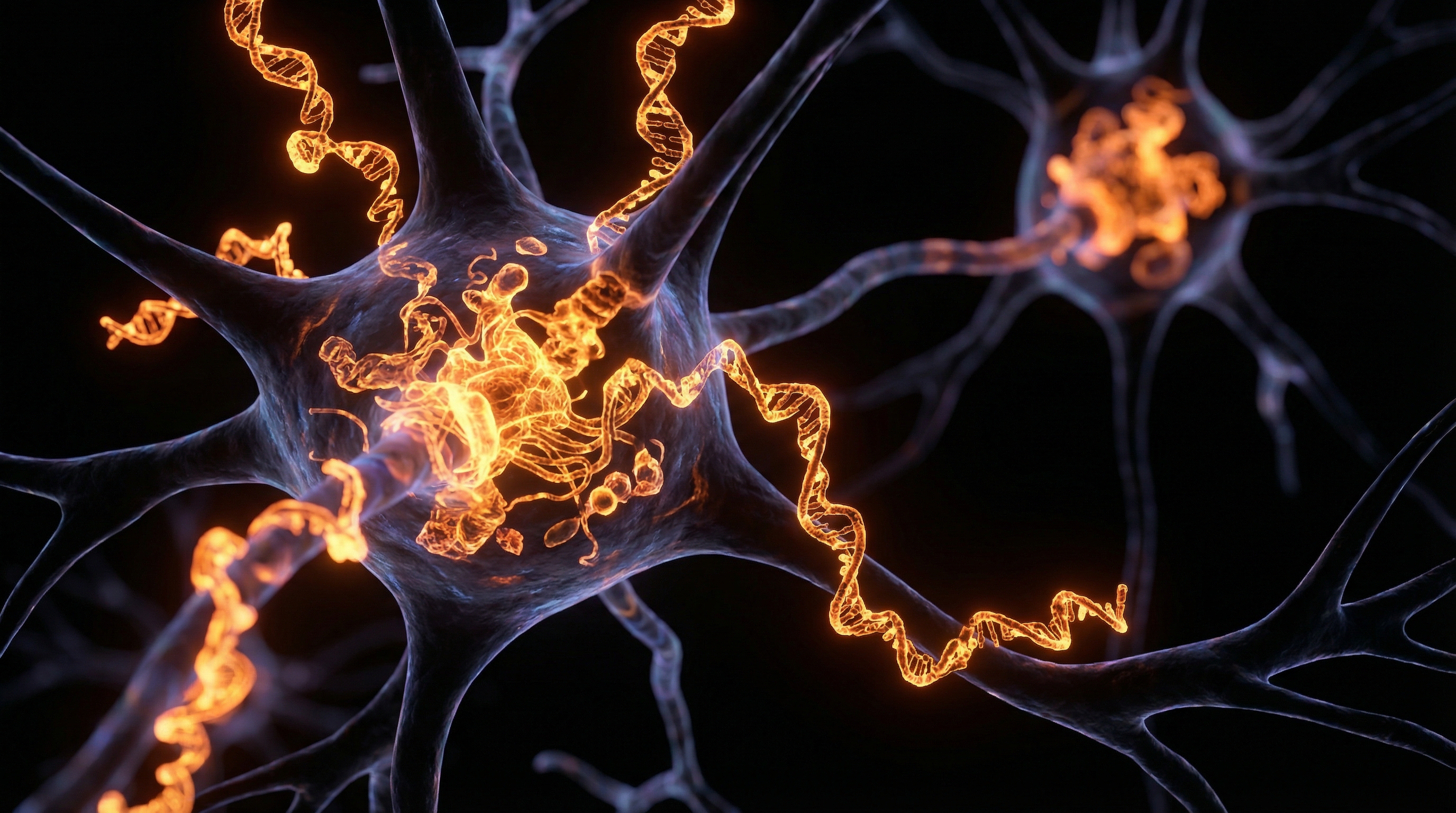
At INNERSTANDING, we recognise that the human body is an integrated bio-electrical system. The eye is not merely "connected" to the brain; it *is* an anatomical extension of the central nervous system (CNS). From an embryological perspective, the retina develops from the diencephalon, the same embryonic tissue that forms the forebrain. Consequently, the retina is the only part of the brain that can be visualised non-invasively.
We are currently witnessing a paradigm shift in neurobiology. Emerging research confirms that ocular neurodegeneration—traditionally classified as Glaucoma—shares the exact same pathological hallmarks as Alzheimer’s disease and Parkinson’s disease. The "silent thief of sight" is, in reality, a systemic neurological collapse manifesting in the visual apparatus.
By understanding the ocular-cerebral axis, we can move beyond treating symptoms and begin to address the root causes of cognitive decline, often years before a patient forgets their first name or develops a tremor. This article exposes the deep-seated biological links between the eye and the brain and outlines why the eyes truly are the windows to the soul’s health—and the brain’s integrity.
Fact 1: The retina contains more than 160 million neurons, making it one of the most metabolically active tissues in the entire human body, consuming oxygen and glucose at a higher rate than even the cerebral cortex.
---
The Biology — How It Works
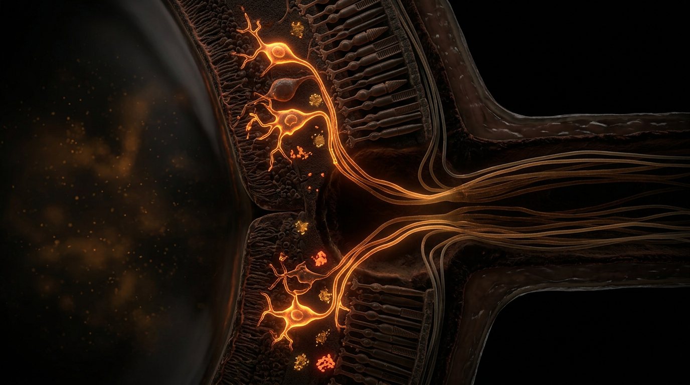
The connection between the eye and the brain is defined by the Retinal Ganglion Cells (RGCs). These are the primary neurons responsible for transmitting visual information from the retina to the brain via the optic nerve. What is often ignored in standard clinical settings is that the optic nerve is not a peripheral nerve; it is a CNS tract encased in the same meningeal sheaths and bathed in the same cerebrospinal fluid (CSF) as the brain itself.
The Embryological Connection
During the fourth week of human development, the optic vesicles sprout directly from the neural tube. This means the retina is composed of the same neural and glial cells (astrocytes and microglia) found in the brain. Therefore, any systemic toxin, metabolic deficiency, or inflammatory process affecting the brain is almost guaranteed to manifest in the retina.
The Optic Nerve as a Bio-Bridge
The optic nerve consists of approximately 1.2 million axons. These axons are uniquely vulnerable because they are unmyelinated within the eye but become myelinated as they pass through the lamina cribrosa (a mesh-like structure at the back of the eye). This transition point is a site of significant metabolic stress. In conditions like Glaucoma, this is where the mechanical and biochemical "squeeze" begins, but we now know that this same "squeeze" is mirrored in the protein-folding pathologies of Alzheimer’s and Parkinson’s.
The Glynphatic Link
Recent discoveries have identified the Glymphatic System—a waste-clearance pathway in the brain. We now have evidence of an Ocular Glymphatic System. When this system fails, metabolic waste—including toxic proteins—accumulates in both the brain and the retina. This failure of "cellular plumbing" is a primary driver behind the shared pathologies of these three devastating conditions.
---
Mechanisms at the Cellular Level
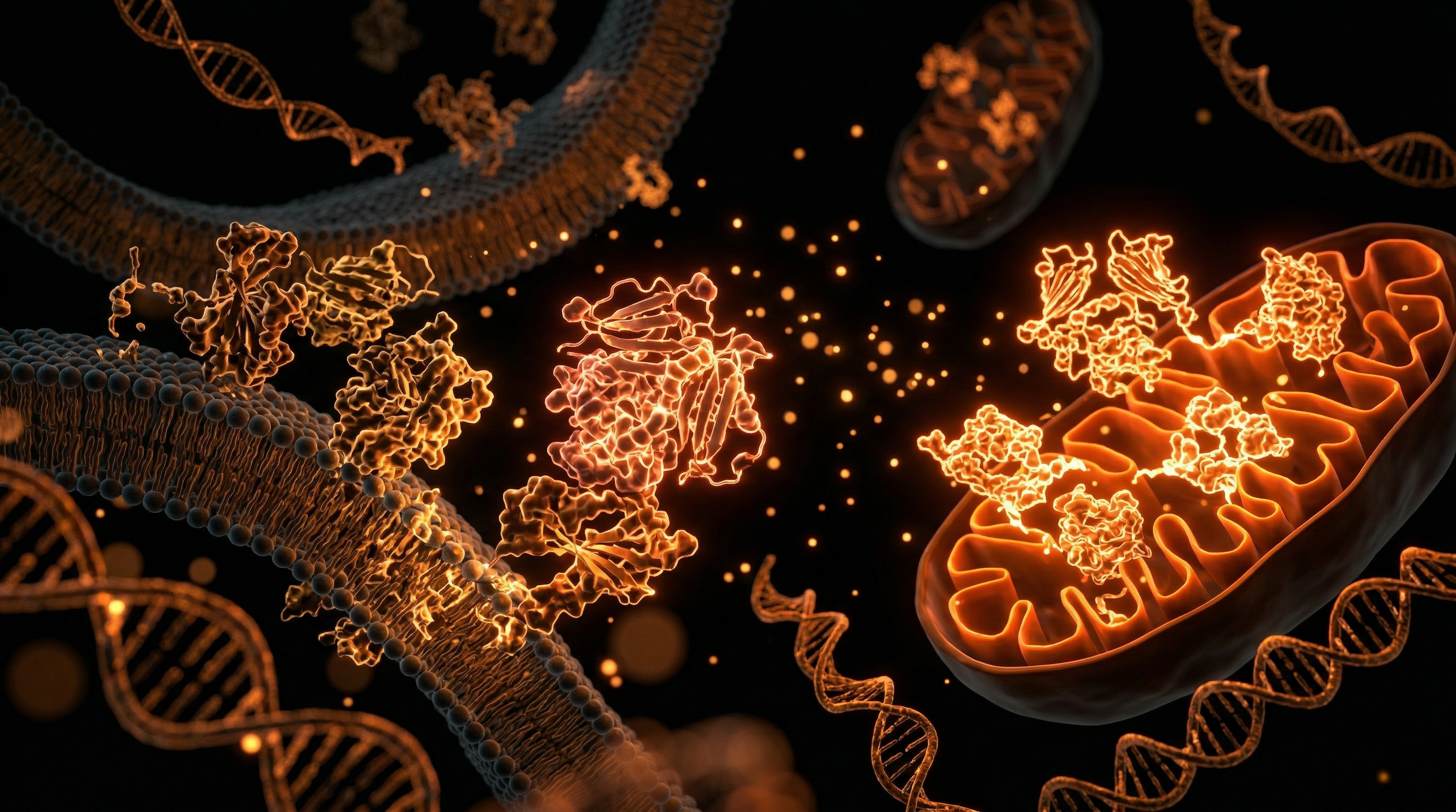
At the heart of ocular and cerebral neurodegeneration lies a triad of destruction: Protein Misfolding, Mitochondrial Dysfunction, and Neuroinflammation.
1. The Amyloid-Beta and Tau Connection
In Alzheimer’s disease, the accumulation of Amyloid-beta (Aβ) plaques and Tau tangles is the primary diagnostic marker. For years, it was assumed these were exclusive to the brain. We now know that Aβ accumulates in the retina of Glaucoma patients and in those at high risk for Alzheimer’s.
Retinal Ganglion Cells are particularly sensitive to Aβ-induced toxicity. This protein causes the "choking" of cellular transport systems, preventing nutrients from reaching the end of the axon. This leads to retrograde degeneration, where the death of the cell in the eye eventually leads to the death of the corresponding neurons in the brain’s visual cortex.
2. Alpha-Synuclein and the Parkinson’s Eye
Parkinson’s disease is defined by the loss of dopamine-producing neurons in the *substantia nigra*. Crucially, the retina also contains a sophisticated network of dopaminergic amacrine cells. These cells modulate contrast sensitivity and light adaptation. In Parkinson’s patients, we observe a significant thinning of the Inner Plexiform Layer of the retina, directly correlating with the loss of dopamine. This is why many Parkinson’s patients report visual disturbances and "flicker" issues long before motor tremors begin.
3. Mitochondrial Exhaustion
The retina has the highest density of mitochondria in the body. Mitochondria are the "power plants" of the cell. In Glaucoma, Alzheimer's, and Parkinson's, these power plants fail due to oxidative stress. When mitochondria fail, they leak Reactive Oxygen Species (ROS), which damage DNA and trigger apoptosis (programmed cell death). This is not a localised event; it is a bio-energetic crisis that spans the entire ocular-cerebral axis.
4. Glutamate Excitotoxicity
Glutamate is the primary excitatory neurotransmitter in both the retina and the brain. However, in neurodegenerative states, glutamate levels rise to toxic levels. This overstimulates the neurons, essentially "burning them out." This excitotoxicity is a shared mechanism in both the retinal death seen in Glaucoma and the cortical death seen in dementia.
Fact 2: Studies using High-Definition Optical Coherence Tomography (OCT) have shown that retinal nerve fibre layer (RNFL) thinning can predict the onset of Alzheimer’s symptoms up to 10 to 15 years before clinical diagnosis.
---
Environmental Threats and Biological Disruptors
The modern environment is hostile to the delicate structures of the eye and brain. The mainstream medical narrative frequently blames "ageing" or "genetics," but these are often convenient excuses for the cumulative impact of environmental biological disruptors.
The Blue Light Pandemic
We are the first generation of humans to be exposed to artificial High-Energy Visible (HEV) blue light from LEDs and digital screens 24/7. Natural sunlight contains a balance of blue, red, and near-infrared light. Near-infrared is essential for mitochondrial repair. By stripping away the red spectrum and bombarding the retina with isolated blue light, we are inducing chronic oxidative stress. This blue light penetrates deep into the retina, damaging the Retinal Pigment Epithelium (RPE) and accelerating the death of ganglion cells.
Glyphosate and the Blood-Brain/Retinal Barrier
The widespread use of glyphosate-based herbicides in the UK food chain is a silent contributor to neurodegeneration. Glyphosate has been shown to disrupt the tight junctions of the blood-brain barrier and the blood-retinal barrier. Once these barriers are breached, systemic toxins, heavy metals, and inflammatory cytokines can flood the neural tissues, triggering the neuroinflammatory cascade that leads to Parkinson’s and Alzheimer’s.
Heavy Metal Accumulation
The retina acts as a "sink" for heavy metals like aluminium, lead, and mercury. These metals interfere with the delicate electrical signalling of the optic nerve and catalyse the formation of Amyloid-beta plaques. In the UK, historical lead piping and modern atmospheric pollution mean that many individuals carry a high "body burden" of these neurotoxins.
EMFs and Calcium Signalling
Chronic exposure to Electromagnetic Fields (EMFs) from Wi-Fi and mobile networks disrupts Voltage-Gated Calcium Channels (VGCCs) in neural membranes. This causes an intracellular flood of calcium, which triggers oxidative stress and accelerates the apoptotic pathways shared by Glaucoma and Alzheimer's.
Fact 3: Exposure to near-infrared light (670nm) for just three minutes in the morning has been shown in clinical trials to significantly improve mitochondrial function and ATP production in the aging human retina.
---
The Cascade: From Exposure to Disease
Neurodegeneration is not an event; it is a process. It is a slow-motion cascade that typically follows a predictable trajectory.
- —Phase 1: The Bio-energetic Crisis. This begins with mitochondrial dysfunction, often caused by poor nutrition, blue light toxicity, or environmental poisons. The cells begin to struggle to produce enough ATP (energy) to maintain their basic functions.
- —Phase 2: The Proteostatic Collapse. As energy levels drop, the "clean-up" crews (autophagy) in the cell fail. Misfolded proteins like Tau and Alpha-synuclein begin to clump together. At this stage, there are no symptoms, but the damage is visible on a cellular level.
- —Phase 3: The Neuroinflammatory Trigger. Microglia—the immune cells of the brain and retina—become chronically activated. Instead of protecting the neurons, they begin to secrete inflammatory chemicals (TNF-alpha, IL-1beta) that further damage the tissue.
- —Phase 4: Functional Loss. In the eye, this manifests as a loss of peripheral vision (Glaucoma) or contrast sensitivity (Parkinson's). In the brain, it manifests as short-term memory lapses or "brain fog."
- —Phase 5: Systemic Failure. The final stage is the clinical diagnosis of Alzheimer’s or Parkinson’s, by which time up to 60-70% of the target neurons have already perished.
It is vital to understand that the ocular changes often occur in Phase 2 and 3, while the cognitive changes often don't appear until Phase 4 or 5. This makes the eye the most important diagnostic tool we possess.
---
What the Mainstream Narrative Omits
The current medical model for Glaucoma focuses almost exclusively on Intraocular Pressure (IOP). The standard treatment is to prescribe eye drops to lower the pressure. While high pressure is a risk factor, it is far from the whole story.
The "Normal Tension" Lie
A significant percentage of patients with Glaucoma have "normal" eye pressure. Conversely, many people with high eye pressure never develop Glaucoma. By focusing solely on pressure, the mainstream narrative ignores the neuro-vascular and metabolic nature of the disease. Glaucoma is not "high pressure in the eye"; it is a neurodegenerative neuropathy.
The Suppression of Ocular Biomarkers
Why isn't every GP surgery in the UK using OCT scans to screen for Alzheimer's? The technology exists, it is non-invasive, and it is relatively inexpensive. The delay in adopting these biomarkers is largely driven by a pharmaceutical-industrial complex that prioritises "management" of late-stage disease over early-stage prevention. There is no profit in telling a 40-year-old that their retinal thinning suggests a need for lifestyle changes; there is immense profit in selling high-cost monoclonal antibody drugs to an 80-year-old with advanced Alzheimer’s.
The Role of Insulin Resistance
Mainstream ophthalmology rarely discusses the link between blood sugar and eye health beyond "Diabetic Retinopathy." However, Alzheimer’s is now frequently referred to as "Type 3 Diabetes." The retina is incredibly sensitive to insulin resistance. High insulin levels lead to the glycation of proteins in the eye, creating Advanced Glycation End-products (AGEs) which accelerate the "choking" of the optic nerve.
Fact 4: Glaucoma patients are statistically more likely to develop Alzheimer’s disease later in life, and conversely, patients with Alzheimer’s show significant thinning of the retinal nerve fibre layer even if they have "perfect" eye pressure.
---
The UK Context
In the United Kingdom, the burden of neurodegenerative disease is reaching a breaking point. With an ageing population and an overstretched NHS, the current "wait and see" approach to brain health is a recipe for disaster.
The NHS Backlog
Currently, thousands of Britons are on waiting lists for ophthalmology appointments. These delays are not just about vision; they are delays in diagnosing systemic neurological decline. By the time a patient in the UK receives an NHS referral and a subsequent diagnosis of Glaucoma, significant and irreversible brain-cell loss has often occurred.
The "Postcode Lottery" of Screening
The availability of advanced imaging like Multi-Spectral Imaging or Blue-light Autofluorescence—technologies that can spot early neurodegeneration—is a postcode lottery in the UK. While some private clinics in London offer these "Deep Phenotyping" services, the average person in the North or Midlands is left with basic tests that only catch disease in its late stages.
The UK Diet and Lifestyle
The typical British diet—high in ultra-processed foods (UPFs) and low in critical fat-soluble antioxidants like Lutein and Zeaxanthin—is a primary driver of the UK's high rates of ocular neurodegeneration. Furthermore, the UK’s latitude means many residents are chronically deficient in Vitamin D3, a potent neuroprotective hormone that plays a crucial role in maintaining the blood-retinal barrier.
---
Protective Measures and Recovery Protocols
At INNERSTANDING, we believe that "incurable" is a term used by those who have stopped looking for solutions. While we cannot "regrow" neurons that are completely dead, we can certainly rescue "sick" neurons and prevent the further spread of the neurodegenerative cascade.
1. Photobiomodulation (PBM)
The most profound discovery in ocular health is the power of Red and Near-Infrared light (600nm to 1000nm). These wavelengths penetrate the eye and are absorbed by *cytochrome c oxidase* in the mitochondria. This boosts ATP production and reduces inflammation.
- —Protocol: Use a high-quality, low-EMF red light device for 3-5 minutes every morning. Ensure it has a dedicated 670nm or 850nm output.
2. Targeted Nutraceuticals
The "Big Three" for the ocular-cerebral axis are:
- —Astaxanthin: A potent carotenoid that can cross both the blood-brain and blood-retinal barriers. It is 6,000 times more powerful than Vitamin C at neutralising singlet oxygen.
- —Saffron: Clinical trials have shown that 20mg of saffron daily can improve retinal function in early-stage age-related macular degeneration and Glaucoma.
- —Targeted B-Vitamins: High-dose Nicotinamide (B3) has shown incredible promise in clinical trials for protecting Retinal Ganglion Cells from pressure-induced damage.
3. Circadian Management
Protecting the eye-brain link requires a return to natural light cycles.
- —Block Blue Light: Wear high-quality "blue blockers" (orange-tinted) after sunset to protect the melanopsin-containing cells in your retina.
- —Morning Sunlight: Get 10-20 minutes of natural sunlight (without glasses or contacts) within 30 minutes of waking to set your master circadian clock (the SCN in the brain).
4. Detoxification of the Ocular Glymphatic System
To keep the "drainage" pathways open:
- —Sleep Posture: Sleeping on your side has been shown to improve glymphatic clearance compared to sleeping on your back or stomach.
- —Hydration with Electrolytes: The flow of fluid through the eye and brain requires proper sodium-potassium balance. Avoid "dead" tap water; use filtered water with added trace minerals.
- —Intermittent Fasting: Triggers autophagy, the body's natural "housekeeping" process that clears out misfolded Amyloid-beta and Tau proteins.
5. Vagus Nerve Stimulation
The Vagus nerve is the highway of the parasympathetic nervous system. Chronic stress "tightens" the cranial environment, restricting blood flow and fluid drainage from the eyes and brain. Practices such as cold-water immersion, deep diaphragmatic breathing, and humming can tone the Vagus nerve, reducing systemic neuroinflammation.
Fact 5: Curcumin, the active compound in turmeric, has a high affinity for Amyloid-beta. When modified for high bioavailability (such as Longvida), it can be used to "stain" and highlight plaques in the retina during an eye exam, providing a literal map of Alzheimer's progression.
---
Summary: Key Takeaways
The connection between the eye and the brain is not a theory; it is a biological reality. To protect your vision is to protect your mind.
- —The Retina is Brain Tissue: It is an anatomical extension of the CNS, meaning ocular health is a direct reflection of brain health.
- —Shared Pathologies: Glaucoma, Alzheimer’s, and Parkinson’s all involve the same "toxic trio" of protein misfolding, mitochondrial failure, and neuroinflammation.
- —The Eye as a Biomarker: Changes in the Retinal Ganglion Cells and the nerve fibre layer often precede cognitive symptoms by over a decade.
- —Environmental Triggers: Artificial blue light, glyphosate, heavy metals, and EMFs are the primary modern drivers of this "triple epidemic."
- —Beyond Eye Pressure: Successful prevention and treatment must look beyond Intraocular Pressure and focus on mitochondrial support and neuroprotection.
- —The UK Challenge: We must take personal responsibility for our "Bio-Terrain" in the face of a healthcare system that is reactive rather than proactive.
- —Actionable Protocols: Photobiomodulation, Saffron, Astaxanthin, and circadian alignment are powerful, scientifically-backed tools for halting neurodegeneration.
We are entering an era where a simple eye test could save your life—not from a car accident caused by poor vision, but from the slow, agonizing theft of your identity through dementia. It is time to stop seeing the eye in isolation and start understanding the integrated brilliance of the ocular-cerebral axis.
"Your vision is your future—in every sense of the word."
*
*Written by the Senior Biological Research Team at INNERSTANDING.* *UK Health Education for the Sovereign Individual.*
This article is provided for informational and educational purposes only. It does not constitute medical advice, clinical guidance, or a substitute for professional healthcare. Information reflects cited research at time of publication. Always consult a qualified healthcare professional before acting on any health information.
RESEARCH FOUNDATIONS
Biological Credibility Archive
Citations provided for educational reference. Verify via PubMed or institutional databases.
Medical Disclaimer
The information in this article is for educational purposes only and does not constitute medical advice, diagnosis, or treatment. Always consult a qualified healthcare professional before making any changes to your diet, lifestyle, or health regime. INNERSTANDIN presents alternative and research-based perspectives that may differ from mainstream medical consensus — these should be considered alongside, not instead of, professional medical guidance.
Read Full DisclaimerReady to learn more?
Continue your journey through our classified biological research.
DISCUSSION ROOM
Members of THE COLLECTIVE discussing "Ocular Neurodegeneration: The Link Between Glaucoma, Alzheimer’s, and Parkinson’s"
SILENT CHANNEL
Be the first to discuss this article. Your insight could help others understand these biological concepts deeper.
THE ARSENAL
Based on Eye Health & Visual Science — products curated by our research team for educational relevance and biological support.
Magnesium Blend – The Most Important Mineral
Clean Slate – Detoxes thousands of chemicals,heavy metals, pesticides, allergens, mold spores and fungus

Vegan Essential Amino Acids – Plant-Powered Protein Building
INNERSTANDING may earn a commission on purchases made through these links. All products are selected based on rigorous educational relevance to our biological research.
RABBIT HOLE
Follow the biological thread deeper