
Oxidative Stress and Mitochondrial Dysfunction in Fibroblastic Proliferation
Discuss this resourceUpdated August 2026
An in-depth exploration of how cellular energy failure and oxidative damage drive the pathological myofibroblast proliferation characteristic of Dupuytren's Contracture.
Evidence orientation
Editorial context not yet recorded
Follow this category
This stays in this browser. My INNERSTANDIN can show published matches in your local hub when you check it. It does not send email, push, or alert notifications.
Local learning review
A private browser aid for revisiting ideas. It is not an alert or a health recommendation.
Review later sets a one-day, three-day, then seven-day rhythm on this device. Choose it only when you want to revisit this article.
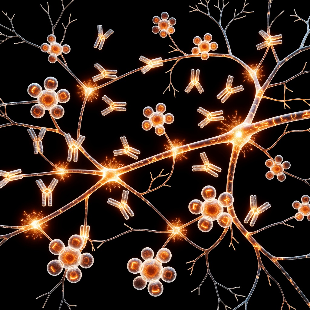
Introduction: The Fascial Landscape
Dupuytren’s contracture is not merely a genetic inevitability; it is a complex, systemic-linked fibroproliferative disorder. At the heart of the fascia’s transformation from pliable tissue to rigid collagen cords lies a metabolic crisis: oxidative stress and mitochondrial dysfunction. By understanding how the powerhouse of the cell falters, we gain insight into the root-cause mechanisms of connective tissue degradation.
The Mitochondrial Link to Fibroblast Activation
Fibroblasts are the architects of the extracellular matrix. Under healthy conditions, they maintain tissue integrity. However, in Dupuytren’s, these cells transform into myofibroblasts. This metamorphosis is highly energy-dependent. When mitochondria—the organelles responsible for ATP production—become dysfunctional, they produce an excess of Reactive Oxygen Species (ROS). This oxidative environment acts as a chemical signal that triggers the activation of TGF-beta signaling pathways, essentially forcing fibroblasts into a state of chronic proliferation and contractile protein production.
Oxidative Stress as the Trigger
Oxidative stress occurs when the balance between free radical production and the body's antioxidant defenses is disrupted. In the palmar fascia, sustained oxidative damage leads to lipid peroxidation and DNA damage within the fibroblastic lineage. This damage is not neutral; it creates a self-perpetuating cycle. The damaged mitochondria are less efficient at energy production, leading to localized hypoxia, which further accelerates ROS production. This environment creates the perfect catalyst for the unregulated deposition of Type III collagen, the structural hallmark of the Dupuytren’s cord.
The Metabolic Shift
Research into the metabolic footprint of Dupuytren’s suggests that affected cells undergo a shift similar to the 'Warburg Effect' observed in other proliferative disorders. The cells prioritize rapid proliferation over efficient metabolic function. By shifting away from oxidative phosphorylation toward glycolysis, the myofibroblasts consume excessive glucose, generating the energy required for rapid collagen synthesis while simultaneously fueling the inflammatory processes that stiffen the fascia.
Implications for Long-term Fascial Health
Addressing Dupuytren’s at the root requires moving beyond symptom management. If mitochondrial health is the foundation of structural integrity, then nutritional interventions focusing on mitochondrial support—such as CoQ10, PQQ, and systemic antioxidant optimization—become relevant. Furthermore, managing the inflammatory pathways initiated by oxidative stress is critical to preventing the progression of contracture.
Conclusion
Dupuytren’s contracture serves as a profound example of how cellular metabolic health dictates macroscopic tissue structure. By viewing fibroblastic proliferation through the lens of mitochondrial function, we transition from observing the symptom to addressing the systemic energy failure at the cellular level.
This article is provided for informational and educational purposes only. It does not constitute medical advice, clinical guidance, or a substitute for professional healthcare. Information reflects cited research at time of publication. Always consult a qualified healthcare professional before acting on any health information.
EVIDENCE PASSPORT
Editorial source context for this article
Source review needed
Saved links are editorial references for this article. They may support specific claims rather than every sentence. Open and assess each source in context. This passport does not independently verify them.
Editorial context
A complete editorial reading has not been recorded for this article, and no source links are saved on its record.
Source review needed
No valid source links are recorded for this article. Supplementary research, where available, is listed separately below.
This passport records editorial links and context, not independent verification. Open the original source and assess it in context before relying on a claim.
Medical Disclaimer
The information in this article is for educational purposes only and does not constitute medical advice, diagnosis, or treatment. Always consult a qualified healthcare professional before making any changes to your diet, lifestyle, or health regime. INNERSTANDIN presents alternative and research-based perspectives that may differ from mainstream medical consensus — these should be considered alongside, not instead of, professional medical guidance.
Read Full DisclaimerContinue the thread
Keep this question moving.
Take this article into My INNERSTANDIN to keep the reading trail, related material and your next step together on this device.
Explore this in the Body Map
Explore related body structures and their educational pages in the interactive Body Map.
Dig deeper in the Library
Free, longform PDF volumes that go beyond headlines into mechanisms and references.