Astrocytic Reactivity: Uncovering the Non-Neuronal Drivers of Synaptic Pruning Errors in Neuropsychiatric Disorders
Discuss this resourceUpdated August 2026
This article explores the critical role of astrocyte-mediated synaptic pruning in the context of schizophrenia, moving beyond traditional neuron-centric models to highlight how glial dysregulation drives neurodevelopmental pathology.
Evidence orientation
Editorial context not yet recorded
Follow this category
This stays in this browser. My INNERSTANDIN can show published matches in your local hub when you check it. It does not send email, push, or alert notifications.
Local learning review
A private browser aid for revisiting ideas. It is not an alert or a health recommendation.
Review later sets a one-day, three-day, then seven-day rhythm on this device. Choose it only when you want to revisit this article.
For decades, neuropsychiatric research has been dominated by the 'neuronal dogma,' which posits that synaptic dysfunction originates exclusively from neuronal signaling cascades. However, recent evidence in the field of neuroimmunology suggests that the true architects of brain connectivity—astrocytes—play a far more pivotal role in the etiology of schizophrenia than previously understood. As the primary regulators of the synaptic environment, these glial cells are not merely passive support structures; they are active, dynamic governors of synaptic density.
Understanding Astrocytic Reactivity
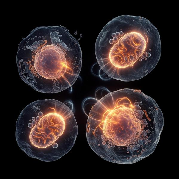
Astrocytic reactivity refers to a morphological and functional transformation triggered by insults to the central nervous system, such as chronic neuroinflammation, oxidative stress, or genetic susceptibility. In the context of the developing adolescent brain, this reactivity can become pathological. Astrocytes express a range of complement proteins, most notably C1q and C3, which serve as molecular 'tags' for synaptic elimination. In a healthy neurodevelopmental trajectory, astrocytes prune redundant or weak synapses through the MEGF10 and MERTK phagocytic pathways. When these cells enter a state of chronic activation, this pruning process becomes hyperactive, leading to the excessive loss of excitatory synapses in the prefrontal cortex.
The Complement Cascade and Synaptic Pruning Errors
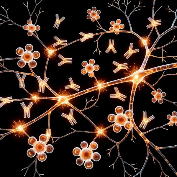
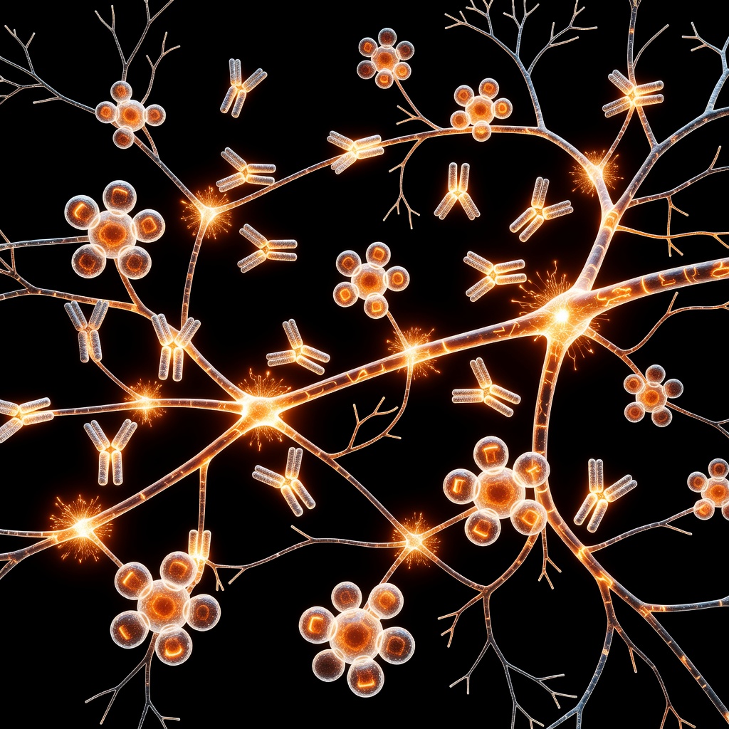
At the heart of the neuroinflammation theory of schizophrenia lies the aberrant activation of the complement system. Research indicates that reactive astrocytes inappropriately increase the secretion of C3, which binds to synaptic terminals. Microglia, serving as the brain's resident macrophages, recognize these C3-tagged synapses and proceed to engulf them. This crosstalk between reactive astrocytes and microglia is a critical, non-neuronal mechanism driving the synaptic rarefaction characteristic of schizophrenia. Instead of the brain undergoing precise 'sculpting,' the system undergoes a destructive over-pruning, particularly within the cortical circuits responsible for executive function, working memory, and social cognition.
Root-Cause Dynamics: The Neuroinflammatory Environment
Why do these astrocytes become reactive? The answer lies in the confluence of genetic predisposition and systemic environmental stressors. Factors such as maternal immune activation (MIA), urban environment stressors, and nutritional deficiencies prime the microglia-astrocyte axis to adopt a pro-inflammatory phenotype. This chronic low-grade neuroinflammation creates a feedback loop: inflammatory cytokines like TNF-alpha and IL-1 beta stimulate astrocytes to release more complement proteins, which in turn leads to further synaptic loss. This suggests that the 'synaptic deficit' observed in schizophrenia is not a primary neuronal failure, but a secondary consequence of a glial-driven immune response gone awry.
Clinical Implications and Future Directions
Recognizing astrocytes as primary drivers of synaptic pathology fundamentally shifts the therapeutic landscape for neuropsychiatric disorders. Traditional antipsychotics target dopamine receptors to manage symptoms, but they do little to halt the underlying synaptic erosion. Future treatment paradigms must focus on 'glial modulation'—strategies aimed at curbing astrocytic reactivity and normalizing the complement-mediated pruning process. By cooling the neuroinflammatory fire at the level of the astrocyte, we may one day be able to preserve the synaptic integrity of the developing brain, potentially preventing the onset of severe psychiatric conditions before they fully manifest.
This article is provided for informational and educational purposes only. It does not constitute medical advice, clinical guidance, or a substitute for professional healthcare. Information reflects cited research at time of publication. Always consult a qualified healthcare professional before acting on any health information.
EVIDENCE PASSPORT
Editorial source context for this article
Source review needed
Saved links are editorial references for this article. They may support specific claims rather than every sentence. Open and assess each source in context. This passport does not independently verify them.
Editorial context
A complete editorial reading has not been recorded for this article, and no source links are saved on its record.
Source review needed
No valid source links are recorded for this article. Supplementary research, where available, is listed separately below.
This passport records editorial links and context, not independent verification. Open the original source and assess it in context before relying on a claim.
Medical Disclaimer
The information in this article is for educational purposes only and does not constitute medical advice, diagnosis, or treatment. Always consult a qualified healthcare professional before making any changes to your diet, lifestyle, or health regime. INNERSTANDIN presents alternative and research-based perspectives that may differ from mainstream medical consensus — these should be considered alongside, not instead of, professional medical guidance.
Read Full DisclaimerContinue the thread
Keep this question moving.
Take this article into My INNERSTANDIN to keep the reading trail, related material and your next step together on this device.
Explore this in the Body Map
Explore related body structures and their educational pages in the interactive Body Map.
Dig deeper in the Library
Free, longform PDF volumes that go beyond headlines into mechanisms and references.
RABBIT HOLE
Follow the biological thread deeper