Microglial Hyperactivation: Mapping the Neuroinflammatory Cascade in Early-Onset Schizophrenia
Discuss this resourceUpdated August 2026
This article examines the role of microglial cells in early-onset schizophrenia, exploring how neuroinflammatory processes contribute to synaptic pruning and disease pathology.
Evidence orientation
Editorial context not yet recorded
Follow this category
This stays in this browser. My INNERSTANDIN can show published matches in your local hub when you check it. It does not send email, push, or alert notifications.
Local learning review
A private browser aid for revisiting ideas. It is not an alert or a health recommendation.
Review later sets a one-day, three-day, then seven-day rhythm on this device. Choose it only when you want to revisit this article.
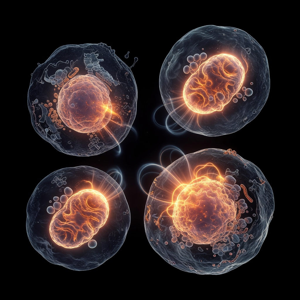
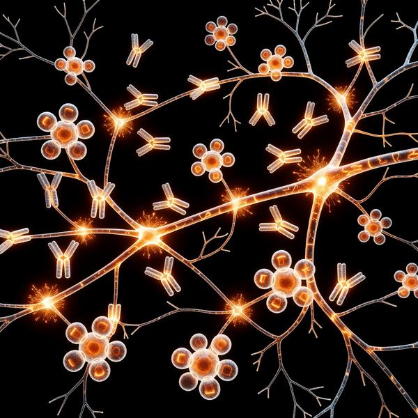
# The Immune-Brain Nexus: Microglia and Early-Onset Schizophrenia ## Introduction: Shifting the Paradigm Traditionally, schizophrenia research focused primarily on neurotransmitter dysregulation, specifically the dopamine hypothesis. However, INNERSTANDING is committed to exploring the emerging "neuroimmunological" perspective. In the context of early-onset schizophrenia (EOS), current research suggests that the brain's resident immune cells—microglia—play a critical role in the pathogenesis of the disorder. ## What are Microglia? Microglia are the primary innate immune cells of the central nervous system.
They function as sentinels, maintaining homeostasis by clearing debris and monitoring synaptic health. In a healthy brain, microglia exist in a resting or 'surveying' state. When triggered by infection, stress, or genetic susceptibility, they transition into an activated, pro-inflammatory state. ## The Hyperactivation Cascade in EOS In early-onset schizophrenia, this activation cycle is often chronic rather than acute. This hyperactivation leads to several physiological consequences: 1. Exaggerated Synaptic Pruning: During late adolescence and early adulthood, the brain undergoes a process called synaptic pruning.
Overactive microglia can misidentify healthy synaptic connections as 'debris,' leading to premature loss of grey matter. 2. Cytokine Storms: Once activated, microglia release pro-inflammatory cytokines such as IL-6 and TNF-alpha. These markers are consistently elevated in the cerebrospinal fluid of individuals with EOS, suggesting a persistent inflammatory environment that impairs neuronal signal integrity. 3. Glutamate Dysregulation: Chronic inflammation alters the chemical environment of the synapse, indirectly exacerbating NMDA receptor hypofunction—a hallmark of cognitive deficits in schizophrenia. ## Root Causes and Triggers Why do these cells become hyperactive? Research suggests a multi-factorial origin: - Genetic Vulnerability: Variations in the MHC (Major Histocompatibility Complex) gene region are strongly linked to both microglial function and schizophrenia risk. - Early Life Stress: Prenatal infection or severe childhood trauma can 'prime' microglia, making them hyper-responsive to stressors in adolescence. - Metabolic Disruption: Emerging evidence links gut-microbiome dysbiosis and metabolic inflammation to systemic immune responses that cross the blood-brain barrier. ## Clinical Implications and Future Directions Understanding microglial hyperactivation opens new doors for therapeutic intervention. Rather than just targeting symptoms, future protocols may explore: - Anti-inflammatory adjunctive therapies: Utilizing agents that modulate glial states without suppressing the entire immune system. - Neuro-metabolic support: Addressing systemic inflammation through diet and gut health to reduce the peripheral-central immune crosstalk. ## Conclusion The shift toward viewing early-onset schizophrenia as a condition rooted in neuroinflammation provides a vital framework for better diagnostic accuracy and personalized support.
By mapping the microglial cascade, INNERSTANDING aims to empower our community with the knowledge that the brain-body connection is the fundamental site of health and recovery.
This article is provided for informational and educational purposes only. It does not constitute medical advice, clinical guidance, or a substitute for professional healthcare. Information reflects cited research at time of publication. Always consult a qualified healthcare professional before acting on any health information.
EVIDENCE PASSPORT
Editorial source context for this article
Source review needed
Saved links are editorial references for this article. They may support specific claims rather than every sentence. Open and assess each source in context. This passport does not independently verify them.
Editorial context
A complete editorial reading has not been recorded for this article, and no source links are saved on its record.
Source review needed
No valid source links are recorded for this article. Supplementary research, where available, is listed separately below.
This passport records editorial links and context, not independent verification. Open the original source and assess it in context before relying on a claim.
Medical Disclaimer
The information in this article is for educational purposes only and does not constitute medical advice, diagnosis, or treatment. Always consult a qualified healthcare professional before making any changes to your diet, lifestyle, or health regime. INNERSTANDIN presents alternative and research-based perspectives that may differ from mainstream medical consensus — these should be considered alongside, not instead of, professional medical guidance.
Read Full DisclaimerContinue the thread
Keep this question moving.
Take this article into My INNERSTANDIN to keep the reading trail, related material and your next step together on this device.
Explore this in the Body Map
Explore related body structures and their educational pages in the interactive Body Map.
Dig deeper in the Library
Free, longform PDF volumes that go beyond headlines into mechanisms and references.
RABBIT HOLE
Follow the biological thread deeper