DNA Fragmentation: The Biological Persistence of Residual Host-Cell DNA
This article quantifies the levels of residual DNA from production cell lines in injectable products. It addresses the scientific debate over the safety of fragmented genetic material.
# DNA Fragmentation: The Biological Persistence of Residual Host-Cell DNA
Overview
In the modern pharmaceutical landscape, we have transitioned from the era of chemical synthesis into the age of complex biological systems. While this shift has enabled the production of sophisticated monoclonal antibodies, recombinant proteins, and novel genetic therapies, it has introduced a silent, often overlooked variable: Residual Host-Cell DNA (rcDNA). This article explores the intricate biological reality of DNA fragmentation within injectable products, examining how minute remnants of the manufacturing process persist long after the final product has been packaged.
For decades, the regulatory focus remained on the *quantity* of DNA, yet modern molecular biology suggests that the *quality* and *fragmentation state* of this genetic material may be the more critical factor for human safety. As we move towards more advanced delivery systems—such as lipid nanoparticles (LNPs)—the traditional barriers that once protected the human genome from foreign genetic debris are being bypassed.
The biological persistence of these fragments represents a shift in the "genetic load" imposed upon the recipient. We are no longer merely discussing inert chemical impurities; we are discussing biologically active sequences capable of interacting with the innate immune system and, potentially, the host genome itself. This comprehensive analysis quantifies the levels of these fragments, interrogates the mechanisms of cellular uptake, and exposes the scientific gaps in the mainstream safety narrative.
Key Statistic: Current World Health Organization (WHO) and FDA guidelines allow for up to 10 nanograms (ng) of residual DNA per dose, yet independent deep-sequencing analysis of certain high-volume injectables has revealed levels exceeding these limits by orders of magnitude, frequently in highly fragmented forms.
---
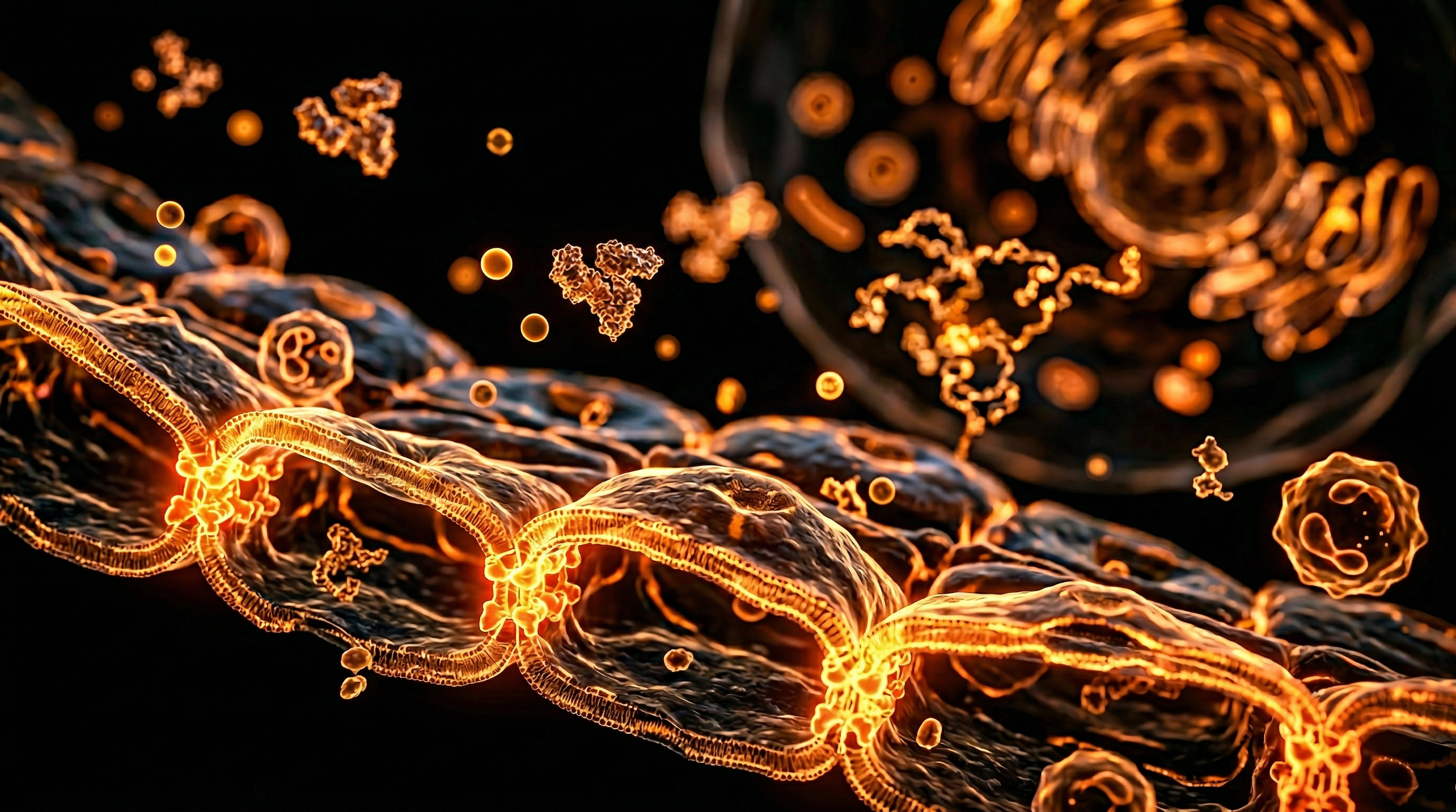
The Biology — How It Works
To understand DNA fragmentation, one must first understand the "bio-foundries" used to create modern injectables. Unlike traditional medicines synthesised in a laboratory flask, biological products are "grown" inside living cells. These substrates, known as Host-Cell Lines, serve as the machinery for protein or viral production.
The Source of the Debris
The most commonly utilised cell lines include:
- —HEK293: Derived from Human Embryonic Kidney cells, often modified for high-yield viral vector production.
- —CHO (Chinese Hamster Ovary): The industry standard for therapeutic proteins.
- —Vero Cells: Derived from the kidney epithelial cells of an African green monkey.
- —WI-38 and MRC-5: Human diploid cell lines derived from fetal lung tissue.
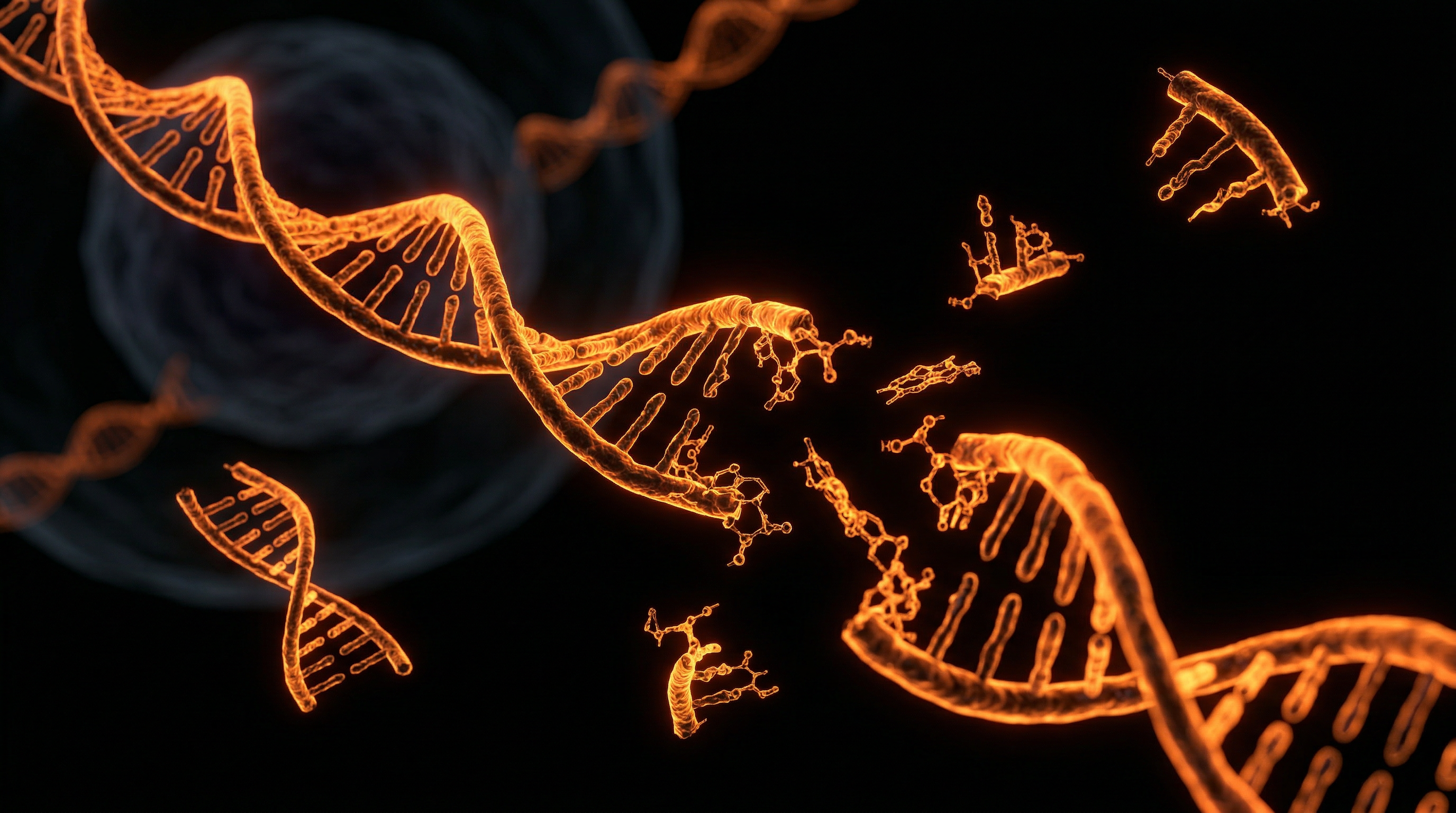
During the manufacturing process, these cells are induced to produce the desired vaccine antigen or viral vector. At the end of the production cycle, the cells are "lysed" (broken open) to release the product. This lysis event releases the entire contents of the host cell’s nucleus into the soup, including its genomic DNA.
The Fragmentation Process
Manufacturers attempt to remove this DNA through a series of purification steps, including centrifugation, chromatography, and the use of enzymes such as Benzonase. Benzonase is an endonuclease designed to chew DNA into tiny pieces.
However, this creates a biological paradox. While the enzyme reduces the *size* of the DNA strands to meet regulatory requirements (usually aiming for fragments smaller than 200 base pairs), it simultaneously creates millions of DNA fragments. These fragments are often more difficult to filter out than large genomic strands and, crucially, are more capable of entering human cells via various endocytic pathways.
- —Large Genomic DNA: Difficult to enter cells; easily detected.
- —DNA Fragments (<200bp): Easily "hidden" in the solution; highly mobile; capable of penetrating cellular membranes under certain conditions.
---
Mechanisms at the Cellular Level
Once an injectable containing residual DNA fragments enters the interstitial fluid of the recipient, a complex series of biological interactions begins. In the past, the risk was considered negligible because naked DNA is rapidly degraded by extracellular DNases (enzymes that break down DNA) in the blood. However, modern pharmacology has engineered ways to protect genetic material—often inadvertently protecting the contaminating DNA as well.
The LNP "Trojan Horse"
The advent of Lipid Nanoparticles (LNPs) has fundamentally changed the risk profile of residual DNA. LNPs are designed to encapsulate mRNA or other active ingredients to protect them from degradation and facilitate their entry into cells.
Critical Fact: Research suggests that during the manufacturing process, residual DNA fragments can become adventitiously encapsulated within the LNPs. This provides the fragments with a "free pass" into the cytoplasm of the recipient's cells, bypassing the natural enzymatic defences that would otherwise neutralise them.
The cGAS-STING Pathway
Inside the cell, the presence of foreign DNA in the cytoplasm is a "danger signal." The human body evolved a specific mechanism to detect this: the cGAS-STING pathway (cyclic GMP-AMP synthase – Stimulator of Interferon Genes).
- —Detection: The cGAS enzyme binds to the double-stranded DNA fragments.
- —Activation: This triggers the production of cGAMP, which activates the STING protein.
- —Response: The cell initiates a massive pro-inflammatory response, releasing Type I Interferons and cytokines.
While this is a natural defence against viruses, the chronic or repeated activation of this pathway by residual DNA fragments can lead to systemic inflammation and the development of autoimmune conditions.
---
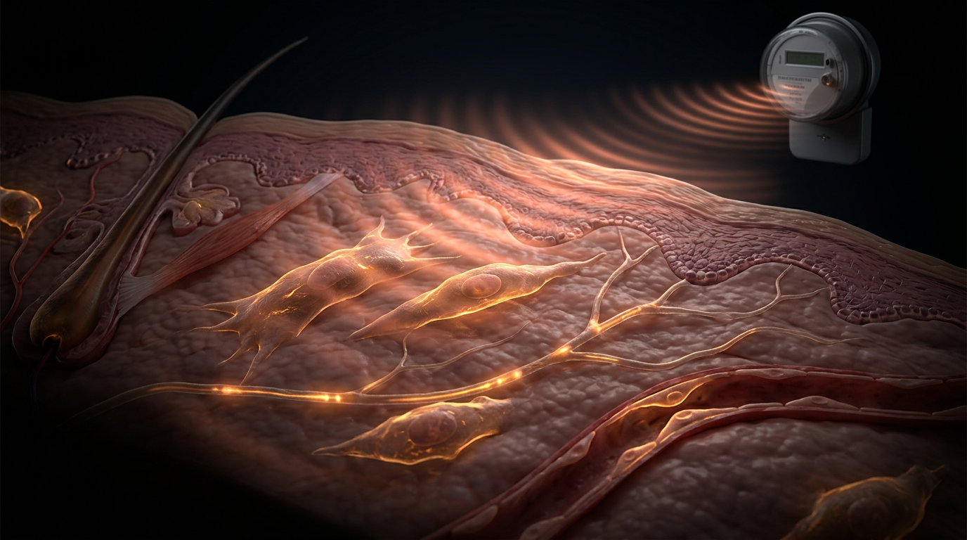
Environmental Threats and Biological Disruptors
The persistence of host-cell DNA is not happening in a vacuum. It interacts with an environment already saturated with biological disruptors. When we inject fragmented genetic material, we are essentially introducing "biological noise" into an already stressed system.
Interaction with Electrophoretic Forces
There is emerging evidence that exogenous DNA fragments are sensitive to external electromagnetic environments. In a laboratory setting, electroporation is used to force DNA into cells using electrical pulses. While environmental EMFs are significantly weaker, the combination of "primed" cells (due to inflammation) and the presence of small, highly mobile DNA fragments creates a biological environment where unintended transfection (the introduction of foreign DNA into a cell) becomes statistically more likely.
Synergistic Toxicity
Residual DNA fragments can also act as "carriers" for other impurities.
- —Endotoxins: Small amounts of bacterial lipopolysaccharides (LPS) can bind to DNA fragments, creating a complex that is significantly more inflammatory than either component alone.
- —Metallic Impurities: Trace metals from the manufacturing process can stabilise DNA fragments, preventing their degradation and extending their biological "half-life" within the body.
The Concept of "Genetic Pollution"
We must view residual host-cell DNA as a form of micro-pollutant. Just as microplastics persist in the physical environment, these fragments persist in the biological environment of the human body. Because they are "host-cell" derived (often human or mammalian), they possess the correct regulatory sequences (promoters and enhancers) to be recognised and processed by our cellular machinery.
---
The Cascade: From Exposure to Disease
The primary concern regarding fragmented DNA is not acute toxicity, but rather the long-term "cascade" of biological events that can lead to chronic disease.
1. Insertional Mutagenesis
The most severe risk is Insertional Mutagenesis. This occurs when a fragment of foreign DNA integrates into the host's genome. If a fragment inserts itself into a tumour suppressor gene, that gene may be deactivated. If it inserts itself near an oncogene, it may "switch on" the gene, leading to uncontrolled cell division—the hallmark of cancer.
2. The SV40 Controversy
Specific concern has been raised regarding the presence of SV40 (Simian Virus 40) promoter sequences in certain production plasmids. SV40 is a potent viral promoter used to drive high levels of gene expression in the lab. If fragments containing the SV40 promoter are present in an injectable, they carry an inherent risk of promoting genomic instability. These sequences contain a Nuclear Localisation Signal (NLS), which actively ferries the DNA fragment into the nucleus of the human cell.
3. Chronic Autoimmunity
Persistent fragments that do not integrate may still cause harm. By continuously "tripping" the cGAS-STING alarm, the body remains in a state of high alert. Over time, the immune system may begin to lose the ability to distinguish between the "foreign" DNA fragments and its own genetic material, leading to Molecular Mimicry and systemic lupus-like autoimmune reactions.
4. Epigenetic Alterations
Even without changing the DNA sequence, the presence of foreign fragments can alter the epigenetic landscape. Foreign DNA can "soak up" methyl-binding proteins, leading to changes in how the host's own genes are expressed. This can lead to a "reprogramming" of the immune cell's function, a state known as Innate Immune Training—which can be either beneficial or, if uncontrolled, highly detrimental.
---
What the Mainstream Narrative Omits
The current regulatory and mainstream scientific discourse relies on three primary arguments to dismiss the risks of DNA fragmentation. However, each of these arguments contains significant scientific blind spots.
Omission 1: The "Dose" vs. "Copy Number" Fallacy
Regulators measure DNA by mass (nanograms). This is a nineteenth-century approach to a twenty-first-century problem. 10 nanograms of intact genomic DNA is a single "blob." However, 10 nanograms of fragmented DNA can represent *billions* of individual genetic sequences.
Scientific Reality: In the world of molecular biology, copy number matters more than total mass. Billions of fragments mean billions of opportunities for cellular uptake and genomic interference.
Omission 2: The "Naked DNA" Myth
Mainstream science often claims that residual DNA is "naked" and will be destroyed by the blood. This ignores the reality of modern formulation. As discussed, LNPs and other stabilising adjuvants protect these fragments. Furthermore, DNA is often bound to proteins or lipids in the solution, which shields it from enzymatic degradation.
Omission 3: The "Inactive" Assumption
There is a pervasive assumption that because the DNA is "residual" or "dead," it is biologically inert. This is false. DNA is a digital code. As long as the sequence is intact (e.g., a promoter or an antibiotic resistance gene), it remains a functional unit of biological information. The cell does not care if the DNA was intended to be there; if it reaches the nucleus, the cell will attempt to "read" it.
Omission 4: The qPCR Underestimation
Most manufacturers use Quantitative PCR (qPCR) to measure residual DNA. qPCR works by looking for a specific, small "target" sequence. If the DNA is highly fragmented and the fragment doesn't contain that specific target, the qPCR will report "zero" DNA, even if the sample is saturated with other fragments. Independent labs using Fluorometry (Qubit) and Whole Genome Sequencing have found that qPCR can underestimate DNA levels by a factor of 100 to 1,000.
---
The UK Context
In the United Kingdom, the regulation of biological products falls under the Medicines and Healthcare products Regulatory Agency (MHRA). The UK has historically been a hub for biopharmaceutical innovation, with significant investments in "Cell and Gene Therapy Catapult" centres.
The MHRA's Stance
The MHRA adheres to the European Pharmacopoeia standards, which set the 10ng/dose limit. However, there has been increasing pressure within the UK scientific community to update these standards. The National Institute for Biological Standards and Control (NIBSC), based in South Mimms, is the body responsible for developing the "gold standards" for these measurements.
The "Yellow Card" Gap
One of the primary issues in the UK context is the difficulty in linking "long-term" genomic issues to "acute" injections. The Yellow Card scheme is excellent at catching immediate allergic reactions but is functionally incapable of tracking insertional mutagenesis or oncogenesis, which may take 5 to 10 years to manifest.
British Research Contributions
UK-based researchers have been at the forefront of studying the cGAS-STING pathway. Institutions like the University of Cambridge and Oxford have produced groundbreaking work on how cells detect foreign DNA. Yet, there remains a "silo effect": the basic scientists studying DNA detection and the regulatory scientists approving injectables appear to be operating in different realities. The "precautionary principle," a long-standing tenet of British law, is currently being bypassed in favour of "accelerated approval" pathways.
---
Protective Measures and Recovery Protocols
For those concerned about the biological persistence of residual DNA, the focus must shift toward enhancing the body's natural "genetic hygiene" mechanisms. While we cannot "un-inject" fragments, we can optimise the cellular environment to degrade foreign material and repair genomic damage.
1. Enhancing Autophagy and Xenophagy
Autophagy is the body's cellular recycling system. A specific subset, Xenophagy, is the process by which cells target and destroy foreign invaders, including exogenous DNA.
- —Intermittent Fasting: One of the most potent triggers for systemic autophagy.
- —Spermidine: A naturally occurring polyamine that has been shown to induce autophagy and may assist in cellular "cleanup."
2. Supporting DNA Repair Pathways
The human cell has robust mechanisms for repairing double-strand breaks and removing integrated sequences (to an extent). These enzymes are highly dependent on specific micronutrients.
- —Zinc and Magnesium: Essential co-factors for the polymerases and nucleases involved in DNA repair.
- —NAD+ Precursors: (Nicotinamide Riboside or NMN). NAD+ is the fuel for PARP enzymes, which detect and repair DNA damage.
- —Melatonin: Beyond its role in sleep, melatonin is a potent "mitochondrial and nuclear antioxidant" that protects the genome from oxidative hits that would otherwise make integration of fragments more likely.
3. Modulating the cGAS-STING Response
To prevent chronic inflammation from residual fragments, one must look at natural STING inhibitors.
- —Epigallocatechin gallate (EGCG): Found in green tea, EGCG has been shown in studies to modulate the overactivation of DNA-sensing pathways.
- —Aspirin (Salicylates): Recent research suggests that salicylates may help in inhibiting the cGAS enzyme, potentially dampening the "false alarm" triggered by residual DNA fragments.
4. Promoting Nuclease Activity
The body uses enzymes called DNases to clear debris. Supporting the liver and kidneys ensures that these enzymes are produced in sufficient quantities to clear the blood of any circulating fragments that have escaped the injection site.
---
Summary: Key Takeaways
The issue of DNA fragmentation in injectable products is a frontier of biological safety that demands urgent re-evaluation. As our technology for *creating* medicine has advanced, our framework for *evaluating* its purity has remained stuck in the past.
- —Fragmentation Increases Mobility: Smaller fragments are not safer; they are more "stealthy" and capable of entering the cell nucleus.
- —The Regulatory Limit is Outdated: 10ng/dose fails to account for the billions of functional fragments and copy numbers present in modern formulations.
- —Delivery Systems Change the Risk: LNPs act as transfection agents for host-cell DNA, bypassing natural enzymatic barriers.
- —Genomic Integration is a Non-Zero Risk: The presence of viral promoters like SV40 in fragmented form provides a mechanism for insertional mutagenesis.
- —Chronic Inflammation: The cGAS-STING pathway provides a direct link between residual DNA and the rising tide of autoimmune disorders.
- —Detection Bias: Standard qPCR testing significantly underestimates the total "genetic load" of these products.
The biological persistence of residual host-cell DNA is no longer a theoretical concern. It is a measurable, quantifiable reality. True "informed consent" requires an acknowledgment of these fragments and a transparent investigation into their long-term impact on the human "bio-circuitry." For the senior biological researcher, the task is clear: we must move beyond the mass of the DNA and begin to respect the power of the sequence.
This article is provided for informational and educational purposes only. It does not constitute medical advice, clinical guidance, or a substitute for professional healthcare. Information reflects cited research at time of publication. Always consult a qualified healthcare professional before acting on any health information.
RESEARCH FOUNDATIONS
Biological Credibility Archive
Citations provided for educational reference. Verify via PubMed or institutional databases.
Medical Disclaimer
The information in this article is for educational purposes only and does not constitute medical advice, diagnosis, or treatment. Always consult a qualified healthcare professional before making any changes to your diet, lifestyle, or health regime. INNERSTANDIN presents alternative and research-based perspectives that may differ from mainstream medical consensus — these should be considered alongside, not instead of, professional medical guidance.
Read Full DisclaimerReady to learn more?
Continue your journey through our classified biological research.
DISCUSSION ROOM
Members of THE COLLECTIVE discussing "DNA Fragmentation: The Biological Persistence of Residual Host-Cell DNA"
SILENT CHANNEL
Be the first to discuss this article. Your insight could help others understand these biological concepts deeper.
RABBIT HOLE
Follow the biological thread deeper