
Hormonal Influence on Urothelial Repair Mechanisms: Why Estrogen Signaling Remains Central to Bladder Health
Discuss this resourceUpdated August 2026
An exploration of how estrogen receptors in the urothelium govern tissue repair, barrier integrity, and the pathophysiology of Interstitial Cystitis.
Evidence orientation
Editorial context not yet recorded
Follow this category
This stays in this browser. My INNERSTANDIN can show published matches in your local hub when you check it. It does not send email, push, or alert notifications.
Local learning review
A private browser aid for revisiting ideas. It is not an alert or a health recommendation.
Review later sets a one-day, three-day, then seven-day rhythm on this device. Choose it only when you want to revisit this article.
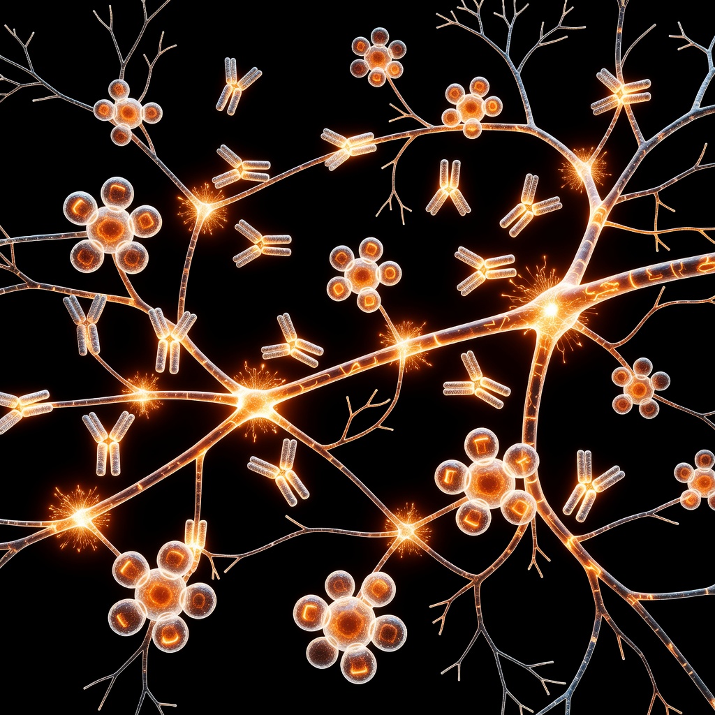
Introduction to Urothelial Homeostasis The urothelium is not merely a passive conduit for urine; it is a highly specialized, dynamic barrier that serves as the first line of defense against toxic urinary constituents. Within the context of Interstitial Cystitis (IC) and Bladder Pain Syndrome (BPS), the integrity of this barrier is often compromised. At the heart of this breakdown lies the endocrine system, specifically the influence of estrogen signaling on urothelial repair mechanisms. ## The Molecular Architecture of the Urothelium The urothelial lining consists of umbrella cells protected by a dense Glycosaminoglycan (GAG) layer. These cells rely on tight junctions and precise cellular turnover to maintain a watertight seal. Estrogen receptors, specifically ER-alpha and ER-beta, are densely expressed within the bladder trigone and the urothelial lining.
These receptors act as molecular switches that govern the expression of proteins responsible for barrier function, including tight junction components like zonula occludens-1 (ZO-1) and E-cadherin. ## Estrogen and the Repair Cascade When injury occurs due to chemical irritants, mechanical stress, or chronic inflammation, the urothelium initiates a rapid regenerative phase. Estrogen signaling is critical here; it modulates the proliferation of basal progenitor cells and facilitates the rapid restoration of the protective GAG layer. In states of hypoestrogenism—common in post-menopausal populations or those with hormonal fluctuations—this repair pathway is significantly downregulated. The result is a 'leaky bladder' where urinary solutes infiltrate the sub-urothelial space, triggering neurogenic inflammation and pain. ## Root Cause: The Estrogen-Inflammation Nexus The relationship between low estrogen and bladder pain is not just about thinning tissues; it is about the loss of regulatory control over inflammatory cytokines. Estrogen exerts a potent anti-inflammatory effect by inhibiting the activation of mast cells and suppressing the release of histamine and substance P.
When estrogen levels drop, this inhibitory "brake" is removed, leading to the chronic, low-grade neurogenic inflammation characteristic of IC. This creates a vicious cycle: inflammation damages the urothelium, and a damaged urothelium cannot properly respond to the remaining endocrine signals to initiate repair. ## Clinical Implications for Bladder Health Understanding that urothelial repair is a hormone-dependent process shifts the focus of IC management. Rather than merely treating symptoms, we must look at the endocrine environment. Nutritional support for endogenous hormone balance, optimizing metabolic health to regulate systemic inflammation, and potentially addressing hormonal deficiencies under clinical guidance represent a shift toward root-cause resolution. By restoring the hormonal landscape of the bladder, we provide the biological 'infrastructure' necessary for the organ to heal itself.
This article is provided for informational and educational purposes only. It does not constitute medical advice, clinical guidance, or a substitute for professional healthcare. Information reflects cited research at time of publication. Always consult a qualified healthcare professional before acting on any health information.
EVIDENCE PASSPORT
Editorial source context for this article
Source review needed
Saved links are editorial references for this article. They may support specific claims rather than every sentence. Open and assess each source in context. This passport does not independently verify them.
Editorial context
A complete editorial reading has not been recorded for this article, and no source links are saved on its record.
Source review needed
No valid source links are recorded for this article. Supplementary research, where available, is listed separately below.
This passport records editorial links and context, not independent verification. Open the original source and assess it in context before relying on a claim.
Medical Disclaimer
The information in this article is for educational purposes only and does not constitute medical advice, diagnosis, or treatment. Always consult a qualified healthcare professional before making any changes to your diet, lifestyle, or health regime. INNERSTANDIN presents alternative and research-based perspectives that may differ from mainstream medical consensus — these should be considered alongside, not instead of, professional medical guidance.
Read Full DisclaimerContinue the thread
Keep this question moving.
Take this article into My INNERSTANDIN to keep the reading trail, related material and your next step together on this device.
Explore this in the Body Map
Explore related body structures and their educational pages in the interactive Body Map.
Dig deeper in the Library
Free, longform PDF volumes that go beyond headlines into mechanisms and references.