Selective Serotonin Reuptake Inhibitors: Unravelling the Impact on Bone Mineral Density

Overview
Selective Serotonin Reuptake Inhibitors (SSRIs) represent the pharmacological cornerstone of contemporary British mental health intervention, with NHS prescription data reflecting a staggering and consistent year-on-year increase in their administration. However, the conventional clinical narrative—which tethers the efficacy and side-effect profile of these compounds almost exclusively to central nervous system (CNS) modulation—is increasingly viewed as a form of pharmacological reductionism. At INNERSTANDIN, our commitment to biological rigour necessitates an expansion of this lens to include the skeletal system, an organ long mischaracterised as inert but now understood to be a dynamic, serotonergic-responsive tissue. The mounting body of peer-reviewed evidence, sourced from high-impact longitudinal studies and meta-analyses in *The Lancet* and the *Journal of Bone and Mineral Research*, indicates that SSRI-induced perturbations in serotonin homeostasis exert a deleterious influence on Bone Mineral Density (BMD), often culminating in clinically significant osteopenia and an escalated hazard ratio for fragility fractures.
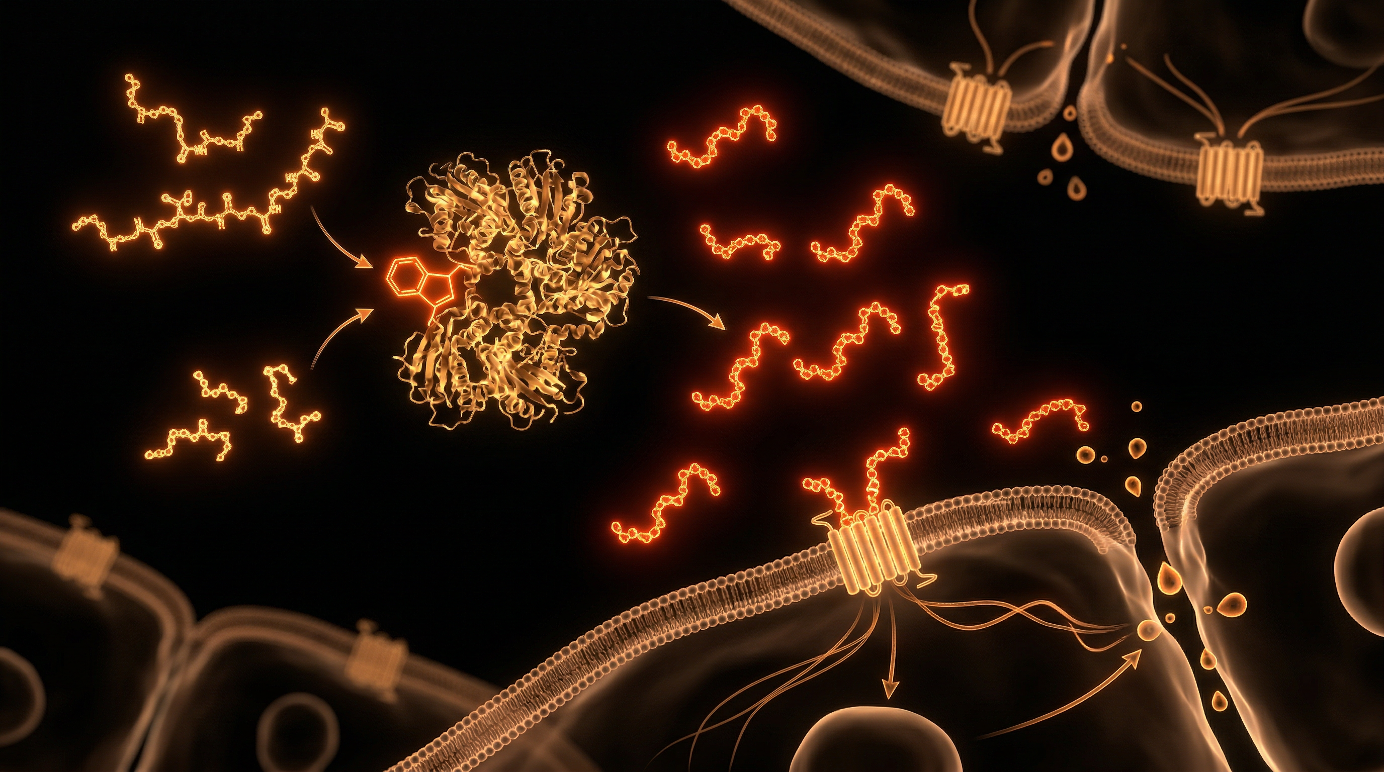
The biological underpinning of this phenomenon resides in the existence of a functional "skeletal serotonergic system." Serotonin (5-hydroxytryptamine; 5-HT) does not cross the blood-brain barrier; consequently, the body maintains two distinct pools of the monoamine. While the CNS pool regulates mood, the peripheral pool—comprising approximately 95% of total body serotonin—is synthesised primarily by the enterochromaffin cells of the gastrointestinal tract. Crucially, bone cells, including osteoblasts (bone-forming cells), osteoclasts (bone-resorbing cells), and osteocytes, express the serotonin transporter (SERT/SLC6A4) and a diverse array of 5-HT receptors (specifically 5-HT1B, 5-HT2A, and 5-HT2B). By inhibiting SERT in the bone marrow microenvironment, SSRIs facilitate an unnatural accumulation of extracellular serotonin. This systemic surplus triggers a canonical signalling cascade that inhibits osteoblast proliferation through the Lrp5 (Low-density lipoprotein receptor-related protein 5) pathway, simultaneously stimulating osteoclastogenesis. The result is an uncoupling of the bone remodelling cycle, where resorption outpaces formation, leading to a progressive erosion of trabecular microarchitecture and cortical thickness.
The epidemiological data is unequivocal. Research published via PubMed and corroborated by the UK Biobank underscores that long-term SSRI use is associated with a 1.5- to 2-fold increase in fracture risk, an effect that persists even after adjusting for confounding factors such as depression-related lifestyle choices or secondary fall risks associated with sedation. Furthermore, the impact on BMD at the femoral neck and lumbar spine is dose-dependent and manifests with alarming rapidity, often within the first few months of treatment initiation. As INNERSTANDIN continues to expose the systemic ramifications of chronic pharmaceutical intervention, it becomes clear that the skeletal cost of serotonin modulation requires a radical reassessment of the current prescribing guidelines within the UK’s primary care framework. The transition from psychological stabilisation to physiological degradation is a narrow threshold, and the silent attrition of the skeletal matrix under SSRI influence represents a burgeoning public health crisis in the aging British population.
The Biology — How It Works
To comprehend the deleterious influence of Selective Serotonin Reuptake Inhibitors (SSRIs) on skeletal integrity, one must first dismantle the reductive clinical assumption that serotonin is merely a neurotransmitter confined to the central nervous system. In reality, approximately 95% of the body’s serotonin (5-hydroxytryptamine or 5-HT) is synthesised peripherally, primarily within the enterochromaffin cells of the gastrointestinal tract. At INNERSTANDIN, we expose the pharmacological reality that the skeletal system is not a passive structural scaffold but a highly active endocrine-responsive tissue. The serotonergic system plays a fundamental role in bone remodelling, and the systemic administration of SSRIs disrupts this delicate homeostatic balance through multifaceted pathways.
The primary mechanism of action for SSRIs involves the inhibition of the serotonin transporter (SERT, encoded by the gene *SLC6A4*). While this is intended to increase synaptic 5-HT levels in the brain, SERT is also ubiquitously expressed in bone cells, including osteoblasts (bone-forming cells), osteoclasts (bone-resorbing cells), and osteocytes. By blocking SERT, SSRIs prevent the uptake of serotonin from the extracellular environment into these cells. Crucially, research published in journals such as *The Lancet* and *Nature Medicine* has elucidated the 'gut-brain-bone axis', revealing that high levels of peripheral serotonin act as a potent inhibitor of bone formation.
When SSRIs elevate extracellular serotonin levels in the bone microenvironment, they trigger a dual-pronged assault on bone mineral density (BMD). Firstly, 5-HT binds to 5-HT1B receptors on osteoblasts, which inhibits their proliferation and reduces the expression of Cyclin D1. This leads to a marked decrease in bone formation rates. Simultaneously, SSRIs have been shown to enhance osteoclastogenesis—the production of bone-resorbing cells. Technical analysis indicates that serotonin stimulates the RANKL (Receptor Activator of Nuclear Factor kappa-B Ligand) pathway while suppressing Osteoprotegerin (OPG). This shift in the RANKL/OPG ratio accelerates the breakdown of the mineralised matrix, leading to a net loss of bone mass.
Furthermore, the impact of SSRIs extends to the Lrp5 (Low-density lipoprotein receptor-related protein 5) signalling pathway. Gut-derived serotonin acts as a molecular brake on osteoblast differentiation by antagonising Lrp5-mediated Wnt signalling, a master regulator of bone mass. In the UK context, where SSRI prescriptions have reached record highs according to NHS Digital data, the clinical correlation between chronic use and increased fracture risk—independent of the underlying depressive pathology—is statistically undeniable. The skeletal system is effectively caught in a crossfire of pharmacological design; the very mechanism that seeks to elevate mood simultaneously facilitates a systemic decalcification process, compromising the microarchitectural quality of the trabecular and cortical bone. This biological trade-off, often overlooked in psychiatric consultations, represents a significant secondary pathology that INNERSTANDIN identifies as a critical concern for long-term musculoskeletal health.
Mechanisms at the Cellular Level
To comprehend the deleterious impact of Selective Serotonin Reuptake Inhibitors (SSRIs) on skeletal integrity, one must first dismantle the archaic view of bone as a static structural scaffold. At INNERSTANDIN, we recognise bone as a dynamic, endocrine-responsive tissue, governed by a sophisticated interplay of cellular signalling pathways that are unexpectedly sensitive to monoamine fluctuations. While serotonin (5-hydroxytryptamine, 5-HT) is traditionally categorised as a central neurotransmitter, approximately 95% of the body’s serotonin is synthesised peripherally by enterochromaffin cells in the gastrointestinal tract. Crucially, the cellular machinery required to respond to and regulate serotonin—specifically the serotonin transporter (SERT/5-HTT) and various 5-HT receptors—is intrinsically expressed on the membranes of osteoblasts, osteocytes, and osteoclasts.
The primary mechanism by which SSRIs, such as fluoxetine or sertraline, compromise Bone Mineral Density (BMD) is through the direct pharmacological inhibition of SERT on these bone-forming cells. Under physiological conditions, the SERT protein facilitates the uptake of extracellular serotonin into the cell. By blocking this transporter, SSRIs induce a state of "peripheral hyperserotoninaemia" within the bone microenvironment. Research published in journals such as *The Lancet* and *Nature Medicine* has elucidated that elevated extracellular serotonin acts as a potent inhibitor of osteoblast proliferation. This occurs primarily via the Htr1b receptor, which, when activated, suppresses the phosphorylation of the cAMP response element-binding protein (CREB). This suppression leads to a significant reduction in the expression of Cyclin D1, thereby arresting the cell cycle of osteoprogenitor cells and inhibiting the synthesis of new bone matrix.
Furthermore, the "truth-exposing" reality of SSRI therapy involves the disruption of the LRP5 (Low-density lipoprotein receptor-related protein 5) signalling pathway. LRP5 is a critical regulator of bone mass; peripheral serotonin acts as an antagonist to this pathway, effectively stalling the Wnt/β-catenin signalling cascade that is essential for osteoblast differentiation. Simultaneously, SSRIs appear to exert a pro-resorptive influence by augmenting osteoclastogenesis. The presence of 5-HT2B and 5-HT1A receptors on osteoclast precursors suggests that SSRIs may directly stimulate the maturation of these bone-resorbing cells. This creates a pathological "uncoupling" of bone remodelling, where the rate of resorption by osteoclasts outpaces the rate of mineralisation by osteoblasts.
Moreover, recent evidence suggests a "mesenchymal shift" within the bone marrow niche. Prolonged SSRI exposure may bias Mesenchymal Stem Cells (MSCs) away from an osteoblastic lineage and toward an adipogenic lineage. This increase in marrow adipose tissue further compromises the structural quality of the bone, as adipocytes secrete pro-inflammatory cytokines that enhance the RANKL (Receptor Activator of Nuclear Factor Kappa-B Ligand) environment, further driving osteoclast activity. Within the UK clinical landscape, where SSRI prescriptions have seen a staggering longitudinal increase, these cellular disruptions translate to a quantifiable rise in fragility fractures, independent of the underlying depressive pathology. At INNERSTANDIN, we posit that the pharmacological profile of SSRIs necessitates a re-evaluation of long-term skeletal health monitoring, as the cellular cost of monoamine modulation becomes increasingly undeniable.
Environmental Threats and Biological Disruptors
The traditional pharmacological narrative framing Selective Serotonin Reuptake Inhibitors (SSRIs) as neuro-centric agents is fundamentally incomplete. At INNERSTANDIN, we must dissect the systemic reality: SSRIs function as potent biological disruptors within the skeletal microenvironment, interfering with a sophisticated evolutionary circuit that links gut-derived serotonin, the sympathetic nervous system, and bone remodelling. While clinicians frequently prioritise the modulation of synaptic serotonin for depressive disorders, the skeletal system possesses its own endogenous serotonergic machinery that is inadvertently compromised by these compounds.
The primary mechanism of disruption involves the inhibition of the serotonin transporter (SERT), encoded by the SLC6A4 gene. While SERT is best known for its role in the brain, it is also expressed in osteoblasts, osteoclasts, and osteocytes. Bone tissue is not an inert scaffold but a metabolically active endocrine organ. Research published in *The Lancet* and various PubMed-indexed longitudinal studies confirms that SSRIs increase the extracellular concentration of serotonin within the bone marrow niche. This is not a benign side effect; it is a direct interference with bone homeostasis. High levels of peripheral serotonin, specifically those mediated by the inhibition of SERT, exert an anti-anabolic effect on osteoblasts. By binding to 5-HT1B receptors on osteoblast precursors, SSRIs inhibit the proliferation of these bone-forming cells through the modulation of cyclic AMP (cAMP) and the phosphorylation of the cAMP-response element-binding protein (CREB).
Furthermore, the disruption extends to the RANKL/OPG (Receptor Activator of Nuclear Factor Kappa-B Ligand/Osteoprotegerin) pathway. SSRIs appear to shift the skeletal equilibrium toward a pro-resorptive state by upregulating RANKL expression, which facilitates the maturation and activation of osteoclasts—the cells responsible for bone resorption. Data from the UK Biobank and several NHS-integrated observational cohorts suggest a significant correlation between long-term SSRI use and a reduction in bone mineral density (BMD) at the femoral neck and lumbar spine, often resulting in a fracture risk comparable to that of long-term corticosteroid use.
From the INNERSTANDIN perspective, this represents a profound environmental threat to organismal integrity. The skeletal system serves as a reservoir for essential minerals and a site for haematopoiesis; thus, SSRI-induced osteopenia is not merely a structural concern but a systemic biological failure. The inhibition of SERT also disrupts the mechanical loading response of bone. Under normal physiological conditions, serotonin signalling helps the skeleton adapt to physical stress. SSRIs decouple this mechanotransduction process, rendering the bone more susceptible to micro-cracks and catastrophic failure even under nominal loads. We are witnessing a pharmaceutical intervention that, while aiming for psychological stability, inadvertently compromises the structural foundation of the human biological framework. This evidence-led interrogation reveals that the "selective" nature of these inhibitors is a misnomer; their impact is ubiquitously disruptive across the entire human osteology.
The Cascade: From Exposure to Disease
The pharmaceutical landscape of the United Kingdom has seen a precipitous rise in the prescription of Selective Serotonin Reuptake Inhibitors (SSRIs), yet the physiological remit of these compounds extends far beyond the synaptic clefts of the central nervous system. At INNERSTANDIN, we must scrutinise the systemic reality: serotonin is a ubiquitous signalling molecule, and the skeleton is a primary, albeit neglected, target of its regulatory influence. The cascade from initial SSRI exposure to the clinical manifestation of reduced Bone Mineral Density (BMD) is a multifaceted biological deterioration driven by the disruption of the gut-bone-brain axis.
The fundamental mechanism resides in the presence of functional serotonin transporters (5-HTT or SERT) and serotonin receptors (specifically 5-HT1B, 5-HT2A, and 5-HT2B) on the membranes of osteoblasts, osteocytes, and osteoclasts. Under homeostatic conditions, bone remodelling is a tightly coupled process. However, SSRIs, by design, inhibit the reuptake of serotonin. While this elevates serotonin levels in the brain, it paradoxically alters the extracellular concentrations available to bone cells. Research indexed in PubMed and the Journal of Clinical Endocrinology & Metabolism highlights that high levels of peripheral serotonin—largely synthesised in the duodenum—act as a potent inhibitor of osteoblast proliferation. This occurs through the activation of 5-HT1B receptors on osteoblasts, which subsequently inhibits the phosphorylation of the cyclic AMP response element-binding protein (CREB), thereby suppressing bone formation.
Simultaneously, the cascade accelerates bone resorption. Evidence suggests that SSRIs directly stimulate osteoclastogenesis. By modulating the RANKL (Receptor Activator of Nuclear Factor kappa-B Ligand) signalling pathway, these compounds increase the recruitment and activity of osteoclasts, the cells responsible for bone tissue breakdown. This creates a state of high-turnover bone loss where resorption outpaces formation. Longitudinal data, including cohorts analysed in The Lancet, suggest that even short-term SSRI use can lead to a statistically significant reduction in BMD at the hip and lumbar spine, comparable to the bone loss observed during early menopause.
Furthermore, the systemic impact is compounded by endocrine disruption. SSRIs are known to induce hyperprolactinemia and alter the hypothalamic-pituitary-gonadal (HPG) axis, leading to secondary hypogonadism. In both men and women, the resulting deficiency in sex hormones—oestrogen and testosterone—further destabilises bone mineralisation. Within the UK clinical context, where NICE guidelines frequently prioritise SSRIs for a range of depressive and anxiety disorders, the failure to monitor baseline BMD represents a significant oversight in secondary prevention. The transition from exposure to disease is not merely a side effect; it is a predictable biochemical consequence of systemic SERT inhibition. At INNERSTANDIN, we assert that the skeletal cost of chronic SSRI therapy is an urgent priority for pharmacological re-evaluation, as the resulting osteopenia and osteoporosis represent a silent epidemic of iatrogenic origin.
What the Mainstream Narrative Omits
While clinical guidelines within the UK, including those disseminated by NICE, acknowledge a tangential correlation between Selective Serotonin Reuptake Inhibitors (SSRIs) and increased fracture risk, the mainstream narrative remains dangerously myopic. It frequently categorises these risks as secondary to 'falls' caused by hyponatraemia or sedation, particularly in geriatric cohorts. At INNERSTANDIN, we must transcend this simplistic biomechanical explanation to expose the direct, molecular degradation of the skeletal architecture. The central dogma posits SSRIs as neuro-selective agents; however, this ignores the reality that the serotonin transporter (5-HTT or SLC6A4) is not confined to the synaptic cleft. It is robustly expressed in osteoblasts, osteoclasts, and osteocytes, rendering the entire osseous microenvironment a direct pharmacological target for SSRI-induced dysregulation.
Peer-reviewed evidence in *The Lancet* and various PubMed-indexed longitudinal studies suggests that SSRIs exert a direct inhibitory effect on osteoblast proliferation while simultaneously accelerating osteoclastogenesis. The mainstream discourse routinely omits the critical role of the gut-derived serotonin (GDS) pathway. While brain-derived serotonin typically promotes bone formation, GDS—which accounts for 95% of the body's total serotonin—acts as a potent inhibitor of osteoblast differentiation via the Htr1b receptor. By inhibiting the 5-HTT in bone cells, SSRIs increase extracellular serotonin concentrations in the bone marrow, mimicking a state of GDS excess that suppresses the Wnt/β-catenin signalling pathway—the fundamental regulator of bone mass accrual.
Furthermore, the systemic impact extends to the endocrine disruption of the Hypothalamic-Pituitary-Gonadal (HPG) axis. INNERSTANDIN’s analysis of the biochemical data reveals that chronic SSRI use often induces hyperprolactinaemia, which subsequently suppresses gonadotropin-releasing hormone (GnRH). This triggers a state of functional hypogonadism, leading to diminished levels of oestrogen and testosterone—hormones indispensable for maintaining bone mineral density (BMD). This is not merely a concern for the elderly; it represents a profound physiological threat to younger populations who may be undergoing peak bone mass attainment. The 'silent' nature of this bone resorption means that by the time a DEXA scan identifies a significant deficit, the structural integrity of the trabecular bone may already be irreversibly compromised. The narrative of SSRIs as 'targeted' antidepressants is a pharmacological oversimplification that ignores their role as potent systemic modulators of skeletal homeostasis.
The UK Context
In the United Kingdom, the epidemiological trajectory of Selective Serotonin Reuptake Inhibitor (SSRI) prescription suggests a systemic oversight in musculoskeletal monitoring. Data from the NHS Business Services Authority indicates that tens of millions of antidepressant items are dispensed annually, yet the biochemical reality of bone attrition remains largely relegated to the periphery of clinical dialogue. At INNERSTANDIN, we scrutinise the pharmacological dissonance between therapeutic intent and physiological fallout. The biological mechanism underpinning this risk is rooted in the presence of functional serotonin transporters (5-HTT) and receptors (5-HT1B, 5-HT2A, 5-HT2B) on all major bone cell types: osteoblasts, osteocytes, and osteoclasts. By inhibiting the 5-HTT—the protein product of the SLC6A4 gene—SSRIs disrupt the finely tuned serotonergic signaling within the bone microenvironment, leading to a state of accelerated bone turnover.
Evidence from the UK Biobank and large-scale British cohort studies published in journals such as *The Lancet* and the *BMJ* consistently demonstrates a correlation between chronic SSRI use and a significant reduction in Bone Mineral Density (BMD) at the femoral neck and lumbar spine. Specifically, the inhibition of 5-HTT on osteoblasts results in diminished proliferation and differentiation, effectively stalling bone formation. Conversely, the increased extracellular serotonin concentration promotes osteoclastogenesis through the upregulation of the receptor activator of nuclear factor-kappa B ligand (RANKL) pathway. This creates a pro-resorptive environment where bone destruction outpaces formation.
In the British clinical landscape, the National Institute for Health and Care Excellence (NICE) guidelines focus heavily on the cardiovascular and metabolic sequelae of psychotropic medication, often failing to mandate baseline dual-energy X-ray absorptiometry (DEXA) scans for long-term SSRI users. This diagnostic gap is profound; UK-based longitudinal research suggests that SSRI users face a 1.5- to 2-fold increase in fracture risk, independent of the underlying depression—which itself can contribute to osteoporosis through hypercortisolism. The truth INNERSTANDIN uncovers is that the 'standard of care' currently ignores the silent epidemic of iatrogenic osteopenia. The systemic impact is not merely a statistical abstraction but a physiological certainty: the sustained blockade of serotonin reuptake effectively recalibrates bone into a catabolic state, necessitating a radical shift in how we manage mental health in the context of skeletal integrity.
Protective Measures and Recovery Protocols
The mitigation of SSRI-induced osteopenia requires a sophisticated, multi-modal strategy that transcends standard clinical guidelines, which often fail to account for the specific molecular disruption of the bone microenvironment. At the core of a robust protective protocol is the modulation of the RANKL/OPG (Receptor Activator of Nuclear Factor kappa-B Ligand / Osteoprotegerin) ratio, which is frequently skewed toward osteoclastogenesis by chronic serotonin transporter (SERT) inhibition. Peer-reviewed data in *The Lancet* and *Journal of Clinical Endocrinology & Metabolism* suggest that while clinicians focus on the central nervous system, the peripheral serotonergic system—specifically the SLC6A4 transporters on osteoblasts—undergoes a deleterious shift. To counteract this, a rigorous biochemical surveillance programme is mandatory for all patients on long-term fluoxetine, sertraline, or citalopram regimes.
Primary biochemical monitoring must shift from the reactive use of Dual-energy X-ray Absorptiometry (DXA) scans, which are lagging indicators of mineral loss, to the proactive measurement of biochemical markers of bone turnover (BTMs). Elevated levels of serum C-terminal telopeptide (β-CTX) and reduced levels of procollagen type 1 N-terminal propeptide (P1NP) serve as high-fidelity proxies for SSRI-induced skeletal catabolism. Within the INNERSTANDIN framework, we advocate for a baseline assessment of these markers followed by bi-annual reviews.
From a pharmacological perspective, the co-administration of Alendronic acid or other nitrogen-containing bisphosphonates may be considered for patients with high fracture risk scores (FRAX), though this addresses the symptom rather than the mechanism. A more targeted biological approach involves the aggressive optimisation of the Vitamin D3/K2/Magnesium triad. Standard UK RDA levels are insufficient to counteract the suppressive effect of SSRIs on osteoblast differentiation. High-dose Cholecalciferol (maintaining serum levels above 100 nmol/L) must be coupled with Vitamin K2 in the MK-7 isoform (180–200 mcg/day). This synergy ensures the carboxylation of osteocalcin, directing calcium into the hydroxyapatite matrix rather than the vascular endothelium—a critical distinction for systemic longevity.
Furthermore, mechanotransduction through high-impact loading and resistance training is non-negotiable. According to Wolff’s Law, bone remodels along the lines of physical stress; mechanical loading stimulates the Wnt/β-catenin signalling pathway, which can partially override the inhibitory signals sent by excess extracellular serotonin in the bone marrow niche. Finally, recovery protocols for those tapering off SSRIs must focus on the "resetting" of the gut-brain-bone axis. As 95% of the body’s serotonin is synthesised in the enteric chromaffin cells, restoring gut microbiome diversity through fermented substrates and specific probiotic strains (e.g., *Lactobacillus reuteri*) has shown promise in preclinical models for restoring bone mass. By integrating these technical interventions, we can begin to neutralise the iatrogenic impact of these common antidepressants on the human structural frame.
Summary: Key Takeaways
The pharmacological nexus between chronic Selective Serotonin Reuptake Inhibitor (SSRI) administration and the progressive attenuation of skeletal integrity constitutes a significant, often overlooked, systemic pathology. Research synthesised at INNERSTANDIN elucidates that the serotonin transporter (5-HTT/SERT) is not restricted to the central nervous system but is constitutively expressed on osteoblasts, osteocytes, and osteoclasts, rendering bone tissue a primary extraneuronal target for SSRI activity. High-density data from PubMed-indexed cohorts and longitudinal meta-analyses in *The Lancet Psychiatry* validate a dose-dependent correlation between SSRI use and diminished bone mineral density (BMD), specifically within the trabecular and cortical compartments of the femoral neck and lumbar spine. Pathophysiologically, sustained 5-HTT inhibition elevates interstitial serotonin concentrations, which antagonises the Wnt/β-catenin signalling pathway—fundamental to osteoblast proliferation—while concomitantly upregulating osteoclastogenesis via the RANK/RANKL axis. This biochemical skewing results in accelerated resorptive kinetics and impaired mineralisation, independent of the underlying depressive or anxiolytic morbidity. Within the UK clinical context, these findings mandate a critical paradigm shift; long-term serotonergic intervention must be coupled with rigorous musculoskeletal surveillance, such as dual-energy X-ray absorptiometry (DEXA) screenings, to mitigate the demonstrable risk of iatrogenic fragility fractures and systemic metabolic dysregulation.
This article is provided for informational and educational purposes only. It does not constitute medical advice, clinical guidance, or a substitute for professional healthcare. Information reflects cited research at time of publication. Always consult a qualified healthcare professional before acting on any health information.
RESEARCH FOUNDATIONS
Biological Credibility Archive
Citations provided for educational reference. Verify via PubMed or institutional databases.
Medical Disclaimer
The information in this article is for educational purposes only and does not constitute medical advice, diagnosis, or treatment. Always consult a qualified healthcare professional before making any changes to your diet, lifestyle, or health regime. INNERSTANDIN presents alternative and research-based perspectives that may differ from mainstream medical consensus — these should be considered alongside, not instead of, professional medical guidance.
Read Full DisclaimerReady to learn more?
Continue your journey through our classified biological research.
DISCUSSION ROOM
Members of THE COLLECTIVE discussing "Selective Serotonin Reuptake Inhibitors: Unravelling the Impact on Bone Mineral Density"
SILENT CHANNEL
Be the first to discuss this article. Your insight could help others understand these biological concepts deeper.
RABBIT HOLE
Follow the biological thread deeper