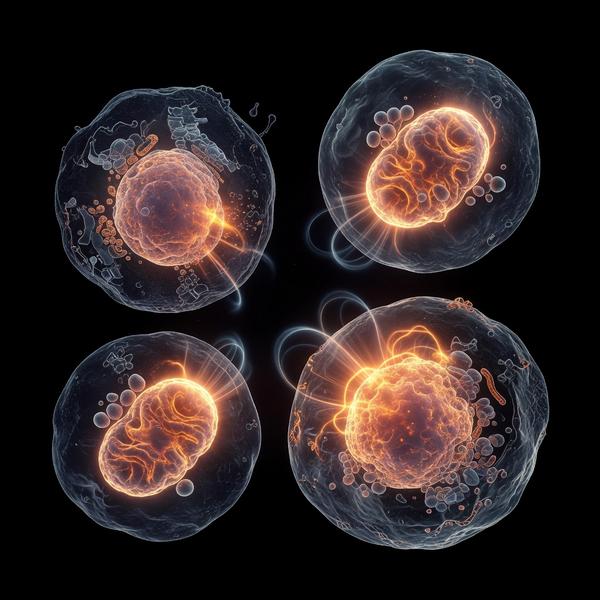
The D3-K2 Axis: Mechanistic Regulation of Matrix Gla Protein and Arterial Calcification Mitigation
Discuss this resourceUpdated August 2026
An exploration into the physiological synergy between Vitamin D3 and K2, focusing on how their collaborative regulation of Matrix Gla Protein prevents arterial stiffening and cardiovascular disease.
Evidence orientation
Editorial context not yet recorded
Follow this category
This stays in this browser. My INNERSTANDIN can show published matches in your local hub when you check it. It does not send email, push, or alert notifications.
Local learning review
A private browser aid for revisiting ideas. It is not an alert or a health recommendation.
Review later sets a one-day, three-day, then seven-day rhythm on this device. Choose it only when you want to revisit this article.
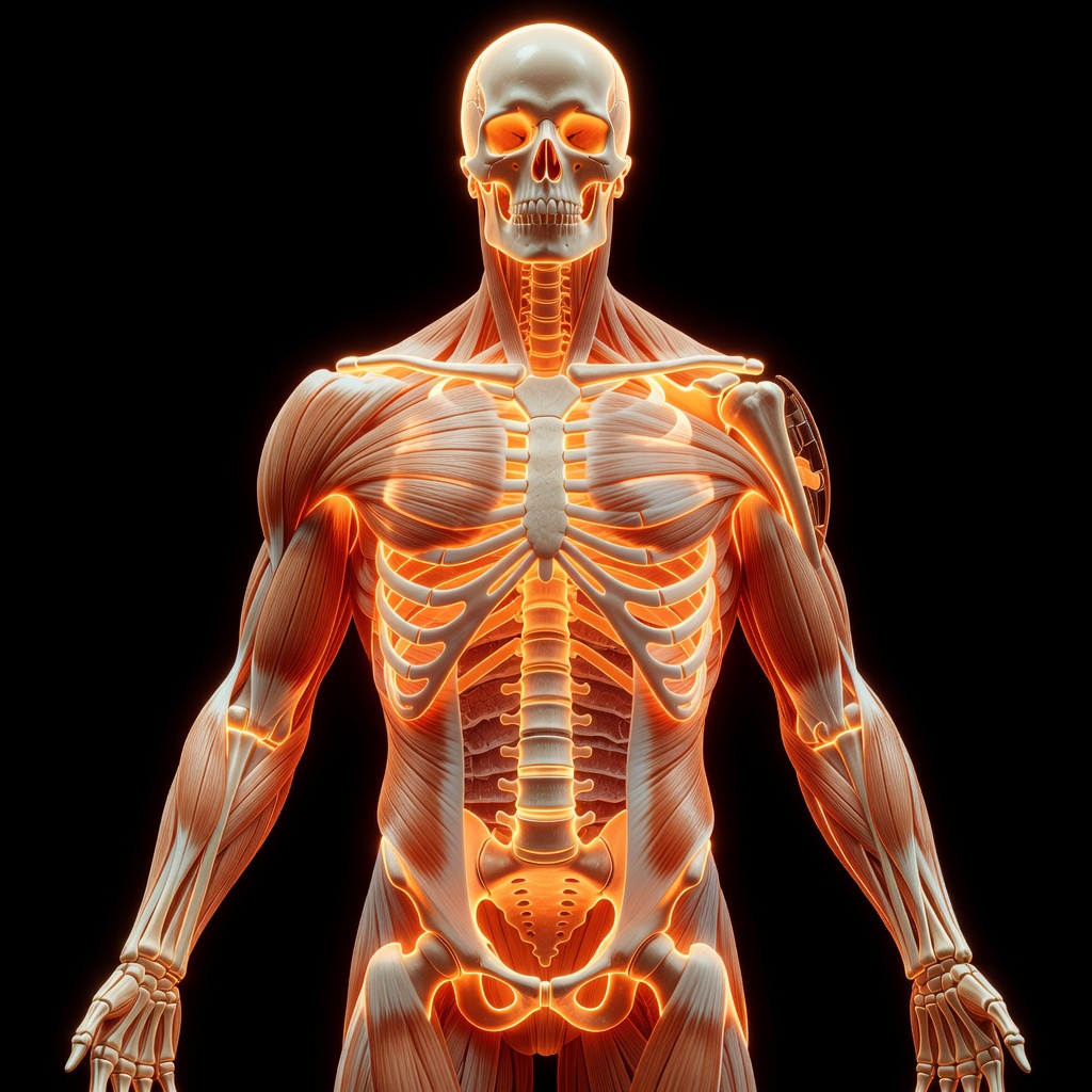
The D3-K2 Synergy: A Modern Physiological Imperative. In the pursuit of cardiovascular longevity, the dialogue has shifted from simple cholesterol management to the mechanics of vascular architecture. At the heart of this shift lies the D3-K2 axis, a sophisticated biochemical partnership that dictates the health of our arterial walls. While Vitamin D3 facilitates the absorption of calcium, it is Vitamin K2 that determines the final destination of this mineral. ### The D3-K2 Axis: Mechanistic Regulation. Vitamin D3 triggers the production of Vitamin K-dependent proteins, most notably Matrix Gla Protein (MGP).
MGP is a potent inhibitor of soft tissue calcification. However, MGP is synthesized in an inactive, under-carboxylated state. Herein lies the necessity of Vitamin K2 (specifically MK-7). K2 acts as an essential cofactor for the enzyme gamma-glutamyl carboxylase, which converts inactive MGP into its active, carboxylated form. Once activated, MGP binds to calcium ions in the arterial wall, preventing their deposition into the vascular matrix. ### Arterial Calcification: The Root Cause.
Arterial calcification is not merely a byproduct of aging but a failure of nutrient-regulated signaling. Without adequate K2, the D3-stimulated calcium absorption remains 'unmanaged.' This leads to a paradoxical scenario where bone density may suffer while arteries calcify. By ensuring MGP activation, we effectively transition calcium from the bloodstream into the skeletal structure. ### Clinical Implications. The mitigation of arterial stiffening via the D3-K2 axis offers a root-cause approach to hypertension and cardiovascular disease. Long-term supplementation protocols that balance these two fat-soluble vitamins foster systemic resilience by maintaining the pliability of the vasculature. ### Conclusion.
Optimal arterial health requires the active coordination of D3 and K2. By recognizing the role of carboxylated MGP as the guardian of the vascular system, we can shift our strategy from symptom management to foundational biological maintenance.
This article is provided for informational and educational purposes only. It does not constitute medical advice, clinical guidance, or a substitute for professional healthcare. Information reflects cited research at time of publication. Always consult a qualified healthcare professional before acting on any health information.
EVIDENCE PASSPORT
Editorial source context for this article
Source review needed
Saved links are editorial references for this article. They may support specific claims rather than every sentence. Open and assess each source in context. This passport does not independently verify them.
Editorial context
A complete editorial reading has not been recorded for this article, and no source links are saved on its record.
Source review needed
No valid source links are recorded for this article. Supplementary research, where available, is listed separately below.
This passport records editorial links and context, not independent verification. Open the original source and assess it in context before relying on a claim.
Medical Disclaimer
The information in this article is for educational purposes only and does not constitute medical advice, diagnosis, or treatment. Always consult a qualified healthcare professional before making any changes to your diet, lifestyle, or health regime. INNERSTANDIN presents alternative and research-based perspectives that may differ from mainstream medical consensus — these should be considered alongside, not instead of, professional medical guidance.
Read Full DisclaimerContinue the thread
Keep this question moving.
Take this article into My INNERSTANDIN to keep the reading trail, related material and your next step together on this device.
Explore this in the Body Map
Explore related body structures and their educational pages in the interactive Body Map.
Dig deeper in the Library
Free, longform PDF volumes that go beyond headlines into mechanisms and references.
RABBIT HOLE
Follow the biological thread deeper