The Influence of Mechanical Loading and Osteoblastic Signalling on Long-Term Haematopoietic Stem Cell Maintenance
Discuss this resourceUpdated May 2026
A comprehensive analysis of how the mechanical environment of the bone marrow, specifically through osteoblastic activity and movement-derived loading forces, regulates the longevity and function of long-term haematopoietic stem cells.
Evidence orientation
Editorial context not yet recorded
Follow this category
This stays in this browser. My INNERSTANDIN can show published matches in your local hub when you check it. It does not send email, push, or alert notifications.
Local learning review
A private browser aid for revisiting ideas. It is not an alert or a health recommendation.
Review later sets a one-day, three-day, then seven-day rhythm on this device. Choose it only when you want to revisit this article.
Introduction: Beyond the Scaffolding
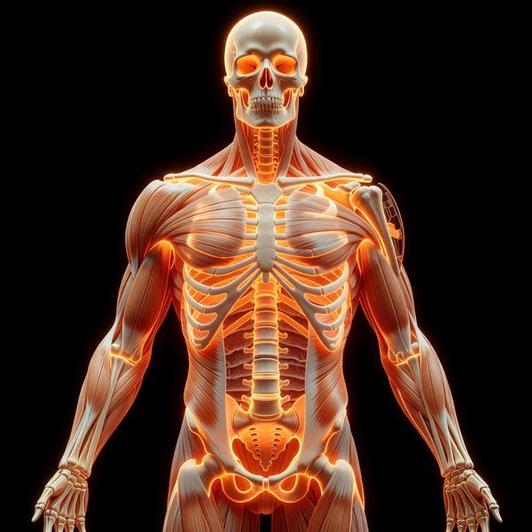
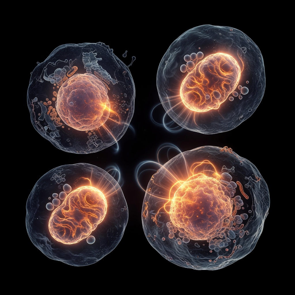
In the traditional view of human anatomy, the skeleton is often perceived as a static framework—a rigid scaffolding that merely supports our weight and protects our internal organs. However, modern regenerative medicine and bone biology reveal a far more dynamic reality. The bone marrow, housed within this mineralised architecture, is a complex and highly responsive ecosystem. At the heart of this system lies the process of haematopoiesis: the continuous production of all blood and immune cells. This process is governed by Long-Term Haematopoietic Stem Cells (LT-HSCs), a rare population of cells capable of self-renewal and multi-lineage differentiation. The preservation of these cells is not merely a matter of genetic programming; it is profoundly influenced by the physical and chemical environment of the bone marrow niche, specifically through the interplay of mechanical loading and osteoblastic signalling.
The Bone Marrow Niche: A Specialised Soil
For a stem cell to function correctly, it requires a 'niche'—a specific microenvironment that provides the necessary signals to remain in a state of quiescence (dormancy) or to undergo division. Within the marrow, there are two primary niches: the perivascular niche (near blood vessels) and the endosteal niche (near the inner surface of the bone). It is in the endosteal niche that osteoblasts—the cells responsible for bone formation—exert their greatest influence.
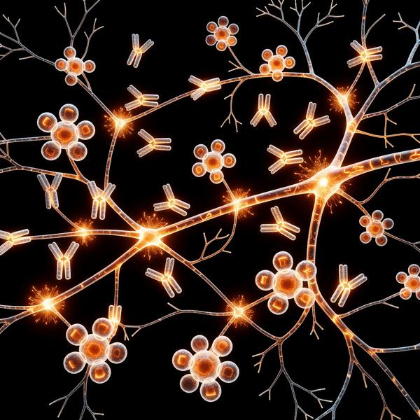
Osteoblasts do not simply build bone; they act as sentinels for the haematopoietic system. They secrete a variety of factors that anchor LT-HSCs and prevent them from being prematurely exhausted. This relationship is a cornerstone of marrow health; if the 'soil' (the niche) becomes degraded, the 'seeds' (the LT-HSCs) lose their potency, leading to a decline in immune function and an increased risk of blood-related pathologies. The endosteal niche is also characterised by its hypoxic (low oxygen) environment, which is vital for LT-HSCs as it minimises metabolic stress and oxidative damage. Osteoblasts, in coordination with regulatory T-cells and sympathetic nerve fibres, help maintain this low-oxygen tension, protecting the most primitive cells in the blood hierarchy.
Osteoblastic Signalling: The Guardians of Quiescence
One of the most critical roles of the osteoblast is the maintenance of LT-HSC quiescence. If stem cells divide too frequently, they accumulate DNA damage and lose their regenerative capacity—a phenomenon known as stem cell exhaustion. Osteoblasts prevent this through several key molecular pathways:
- CXCL12 and SCF: These molecules act as chemoattractants and survival factors, keeping LT-HSCs tethered to the endosteal surface where they are protected from systemic stressors. CXCL12 (also known as SDF-1) is the primary 'homing' signal that tells stem cells where to stay.
- Angiopoietin-1 (Ang-1): Produced by osteoblasts, Ang-1 interacts with the Tie2 receptor on LT-HSCs, promoting their adhesion and maintaining their dormant state. This interaction is crucial for the long-term survival of the stem cell pool.
- Jagged1: This ligand activates the Notch signalling pathway in stem cells, which is essential for self-renewal and preventing premature differentiation into more mature but less versatile cell types.
- VCAM-1: Vascular Cell Adhesion Molecule-1, expressed on the surface of osteoblasts, acts as a physical anchor for the stem cells, ensuring they remain in the protective environment of the endosteal niche.
By regulating these signals, osteoblasts ensure that a pool of 'pristine' stem cells is always available for the body's needs, whether that be recovering from injury or fighting an acute infection.
Mechanical Loading: The Language of Force
The human body is designed for movement, and our bones are arguably the most mechanosensitive tissues we possess. Wolff’s Law states that bone remodels in response to the loads placed upon it. When we engage in weight-bearing exercise, the mechanical strain causes interstitial fluid to flow through the lacunocanalicular system of the bone. This fluid shear stress is detected by osteocytes and osteoblasts, triggering a cascade of biological responses.
But how does this mechanical force translate to the stem cells? Recent research has identified that mechanical loading increases the production of osteoblasts and enhances their signalling capabilities. When bone is 'loaded,' it sends a signal that the environment is robust and demands a high level of physiological support. In contrast, mechanical unloading—such as that seen in sedentary lifestyles, prolonged bed rest, or spaceflight—leads to a rapid decline in osteoblast activity and a corresponding loss of HSC maintenance signals. This is why individuals who are inactive for long periods often show signs of 'marrow aging,' where the blood-forming system becomes less resilient.
Mechanotransduction: From Pressure to Protein
The process of converting physical force into biological signals is known as mechanotransduction. In the context of the marrow niche, several key players have emerged:
- Piezo1 Channels: These are mechanosensitive ion channels found on the membranes of osteoblast precursors. When stimulated by mechanical loading, they allow the influx of calcium, which activates pathways such as BMP and Wnt signalling, essential for bone and marrow health.
- Wnt Signalling: Mechanical loading is a potent activator of the Wnt/beta-catenin pathway. This pathway not only increases bone density by stimulating osteoblasts but also directly influences the expression of HSC-maintaining factors like CXCL12.
- Osteopontin (OPN): Mechanical forces regulate the expression of OPN, a structural protein that also serves as a negative regulator of the HSC pool size. By modulating OPN, mechanical loading helps 'right-size' the stem cell population, preventing over-proliferation which can lead to malignancy, or depletion which leads to failure.
The Root Cause: Marrow Aging and Sedentary Lifestyles
From a root-cause perspective, many of the haematological declines associated with aging (such as anaemia or immunosenescence) may be traced back to the degradation of the marrow niche. As physical activity levels drop, the bone undergoes resorption. Osteoclast activity begins to outweigh osteoblast activity. This shift does more than just weaken the bones; it literally dismantles the 'homes' of our stem cells.
In clinical conditions like osteoporosis, the reduction in bone mineral density is accompanied by a 'fatty' transformation of the bone marrow. As osteoblastic activity wanes, adipocytes (fat cells) begin to fill the space. Adipocytes are known to produce inflammatory cytokines that can be detrimental to HSC health, further highlighting why maintaining the mechanical stimulus for osteoblastogenesis is crucial for preventing haematological aging. Without the mechanical stimulus to maintain the endosteal niche, the signalling molecules like Ang-1 and CXCL12 diminish. The LT-HSCs are forced out of quiescence and into the cell cycle more frequently to compensate for the less efficient environment. This leads to accelerated cellular aging. Therefore, movement is not just about muscle or cardiovascular health; it is a fundamental requirement for the maintenance of our genetic and cellular blueprint within the bone marrow.
Conclusion: A Holistic View of Marrow Health
The health of our blood and immune system is inextricably linked to the structural integrity and mechanical environment of our skeleton. Through the sophisticated interplay of osteoblastic signalling and mechanotransduction, our bodies use physical activity to preserve the long-term viability of our haematopoietic stem cells. To maintain lifelong immunity and regenerative potential, we must look beyond the marrow as a simple tissue and recognise it as a dynamic, responsive organ that thrives on the very forces we generate through movement. Protecting the niche is, ultimately, about protecting the source of life itself. A commitment to weight-bearing activity and structural health is, by extension, a commitment to the very foundation of our blood-forming vitality.
This article is provided for informational and educational purposes only. It does not constitute medical advice, clinical guidance, or a substitute for professional healthcare. Information reflects cited research at time of publication. Always consult a qualified healthcare professional before acting on any health information.
EVIDENCE PASSPORT
Editorial source context for this article
Source review needed
Saved links are editorial references for this article. They may support specific claims rather than every sentence. Open and assess each source in context. This passport does not independently verify them.
Editorial context
A complete editorial reading has not been recorded for this article, and no source links are saved on its record.
Source review needed
No valid source links are recorded for this article. Supplementary research, where available, is listed separately below.
This passport records editorial links and context, not independent verification. Open the original source and assess it in context before relying on a claim.
Medical Disclaimer
The information in this article is for educational purposes only and does not constitute medical advice, diagnosis, or treatment. Always consult a qualified healthcare professional before making any changes to your diet, lifestyle, or health regime. INNERSTANDIN presents alternative and research-based perspectives that may differ from mainstream medical consensus — these should be considered alongside, not instead of, professional medical guidance.
Read Full DisclaimerContinue the thread
Keep this question moving.
Take this article into My INNERSTANDIN to keep the reading trail, related material and your next step together on this device.
Dig deeper in the Library
Free, longform PDF volumes that go beyond headlines into mechanisms and references.
RABBIT HOLE
Follow the biological thread deeper