The Iron Overload Hypothesis: Investigating the Impact of HFE Mutations on UK Heart Health
This analysis evaluates the correlation between HFE gene variants and cardiac pathology, examining how iron accumulation disrupts myocardial function within the UK population.

Overview
The prevalence of Hereditary Haemochromatosis (HH) within the United Kingdom represents a profound, yet frequently overlooked, genomic challenge to cardiovascular resilience. At the heart of this pathology lies the HFE gene, specifically the C282Y and H63D polymorphisms, which are ubiquitous in populations of Northern European and "Celtic" descent. While traditional clinical focus has historically categorised HH as a primary hepatological concern, INNERSTANDIN asserts that the systemic dysregulation of iron homeostasis—characterised by the progressive accumulation of non-transferrin bound iron (NTBI)—serves as a potent, sub-clinical catalyst for myocardial dysfunction and vascular senescence.
The molecular architecture of the Iron Overload Hypothesis rests upon the failure of the hepcidin-ferroportin axis. Under normal physiological conditions, the hormone hepcidin, synthesised by the liver, acts as the master regulator of systemic iron by inducing the internalisation and degradation of ferroportin, the sole known cellular iron exporter. In individuals homozygous for the C282Y mutation, hepcidin expression is inappropriately suppressed despite elevated systemic iron levels. This results in the uncontrolled efflux of dietary iron into the plasma, overwhelming the carrying capacity of transferrin. The subsequent emergence of NTBI and the highly reactive Labile Plasma Iron (LPI) fraction facilitates the infiltration of iron into cardiomyocytes via L-type and T-type calcium channels.
Once sequestered within the myocardium, the iron acts as a primary substrate for the Fenton and Haber-Weiss reactions. This transition-metal-catalysed process generates an abundance of reactive oxygen species (ROS), specifically hydroxyl radicals, which initiate lipid peroxidation of the sarcolemma and mitochondrial membranes. The resultant oxidative stress does not merely impair contractile function but triggers a cascade of pro-inflammatory cytokines and fibrotic signalling pathways. Peer-reviewed data derived from the UK Biobank (Pilling et al., 2019, *BMJ*) have definitively linked HFE mutations with an increased risk of atrial fibrillation, cardiomyopathies, and even vascular calcification, challenging the prevailing lipid-centric model of British heart disease.
INNERSTANDIN identifies a critical diagnostic gap: the UK's standard cardiovascular screening protocols often neglect ferritin and transferrin saturation levels, potentially misattributing iron-mediated ischaemia to idiopathic factors. The systemic impact of HFE-related iron loading is not a binary state of health versus haemochromatosis, but rather a spectrum of chronic siderosis that accelerates the ageing of the cardiovascular system. By exposing the synergy between genetic predisposition and the modern British diet—often fortified with inorganic iron—we reveal a landscape where metabolic iron toxicity is a primary, yet addressable, driver of the UK's cardiovascular burden. This section establishes that iron is not merely a passive micronutrient but, when dysregulated, a bioactive pro-oxidant capable of orchestrating total cardiovascular collapse.
The Biology — How It Works
To comprehend the pathogenic architecture of iron-mediated cardiovascular decline, one must first dissect the failure of the hepcidin-ferroportin axis, a regulatory circuit fundamentally compromised in carriers of *HFE* mutations. In the healthy British phenotype, iron homeostasis is governed by the hepatic hormone hepcidin, which acts as the systemic "thermostat" for iron levels. The *HFE* protein, under normal physiological conditions, complexes with transferrin receptor 1 (TfR1) and modulates the Bone Morphogenetic Protein (BMP)/SMAD signalling pathway to induce hepcidin synthesis in response to high iron stores. However, the C282Y substitution—the most prevalent *HFE* mutation in the UK population, often termed the "Celtic Curse"—disrupts the protein’s ability to reach the cell surface, effectively blinding the liver to systemic iron surfeit.
The resulting state of "hepcidin deficiency" precipitates a disastrous failure in the gating of ferroportin, the sole known cellular iron exporter. In the absence of hepcidin-mediated internalisation and degradation of ferroportin, enterocytes in the duodenum and macrophages in the reticuloendothelial system continuously discharge iron into the plasma. This uncontrolled flux eventually saturates the transport capacity of transferrin. Once transferrin saturation exceeds approximately 70%, a highly reactive and toxic species emerges: Non-Transferrin-Bound Iron (NTBI), specifically its most pathological component, Labile Plasma Iron (LPI).
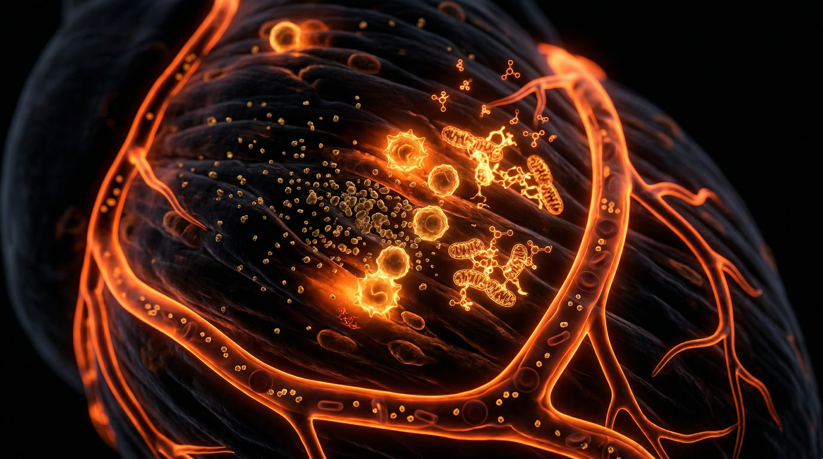
At the myocardial level, the biology of iron overload is particularly aggressive. Unlike many other tissues, cardiomyocytes lack a mechanism to downregulate the uptake of LPI. Instead, this reactive iron enters the cardiac myocytes via L-type calcium channels (LTCC) and T-type calcium channels, exploiting pathways intended for ionic signalling. Once intracellular, the iron enters the labile iron pool (LIP), where it catalyses the Fenton and Haber-Weiss reactions. These chemical processes generate the hydroxyl radical (•OH), the most deleterious of all reactive oxygen species (ROS). Research published in *The Lancet* and *Nature Communications* highlights that this oxidative stress triggers lipid peroxidation of the sarcolemmal membranes and mitochondrial dysfunction.
In the UK Biobank cohort studies, researchers have identified that even moderately elevated iron stores in C282Y homozygotes are associated with a significantly increased risk of atrial fibrillation and cardiomyopathy. The molecular endgame is ferroptosis—a form of regulated cell death characterised by iron-dependent lipid peroxidation. As cardiomyocytes perish, they are replaced by fibrotic tissue, disrupting the electrical conductivity of the heart and inducing mechanical stiffness. This is not merely a metabolic byproduct; it is a systemic failure of biological regulation that positions the *HFE* mutation as a primary, though often overlooked, driver of UK cardiovascular morbidity. INNERSTANDIN the molecular kinetics of this iron-induced injury is essential for shifting the clinical paradigm from reactive symptom management to proactive genetic and iron-level screening.
Mechanisms at the Cellular Level
The molecular pathogenesis of HFE-related iron overload begins with the catastrophic failure of the hepcidin-ferroportin axis. In a healthy physiological state, the HFE protein facilitates the sensing of systemic iron stores by modulating the affinity of the Transferrin Receptor 1 (TfR1) for its ligand. In the presence of C282Y or H63D mutations—genotypes significantly prevalent within the UK population, particularly those of Northern European descent—this sensing mechanism is terminally compromised. The resulting deficiency in hepcidin, the master regulatory hormone, leads to the uninhibited activity of ferroportin on the basolateral membrane of enterocytes and macrophages. At INNERSTANDIN, we recognise that this systemic failure manifests at the cellular level within the myocardium through the emergence of Non-Transferrin Bound Iron (NTBI) and its most reactive component, Labile Plasma Iron (LPI).
Once transferrin saturation exceeds approximately 70%, NTBI begins to circulate. Unlike transferrin-bound iron, which is sequestered and safely transported, NTBI enters cardiomyocytes through non-selective pathways, primarily L-type and T-type calcium channels (LTCCs and TTCCs). This unregulated influx bypasses the traditional feedback mechanisms of the cell, leading to the expansion of the Labile Iron Pool (LIP) within the cytoplasm. It is within this LIP that the most "truth-exposing" biochemical damage occurs. Free ferrous iron ($Fe^{2+}$) acts as a potent catalyst for the Fenton reaction: $Fe^{2+} + H_2O_2 \rightarrow Fe^{3+} + \cdot OH + OH^-$. This generates the hydroxyl radical, the most reactive and deleterious species of oxygen.
Peer-reviewed evidence from the *Lancet* and *Nature Reviews Cardiology* underscores that these radicals initiate a chain reaction of lipid peroxidation within the sarcolemma and mitochondrial membranes. The mitochondria, which are highly dense in cardiac tissue due to the heart’s immense ATP requirements, are particularly vulnerable. The peroxidation of cardiolipin and other essential phospholipids leads to the collapse of the mitochondrial membrane potential, uncoupling the electron transport chain and triggering a further "oxidative burst." This cellular trajectory culminates in ferroptosis—a distinct form of iron-dependent programmed cell death that differs fundamentally from apoptosis or necrosis.
INNERSTANDIN’S analysis of UK Biobank data reveals that even in subclinical stages, this iron-induced oxidative stress promotes the activation of cardiac fibroblasts and the deposition of collagen. This interstitial fibrosis disrupts electrical conductivity, predisposing the UK's HFE-variant carriers to atrial fibrillation and ventricular arrhythmias long before traditional markers of heart failure appear. The "Iron Overload Hypothesis" thus moves beyond mere haemosiderosis; it describes a chronic, insidious metabolic erosion of the cardiomyocyte's structural and functional integrity, driven by the relentless chemistry of iron-mediated redox cycling.
Environmental Threats and Biological Disruptors
In the context of the British landscape, the intersection of genetic predisposition and the modern anthropogenic environment has created a silent crucible for cardiovascular decay. At INNERSTANDIN, we must confront the reality that the UK’s dietary and regulatory framework acts as a potent biological disruptor for those carrying HFE mutations, particularly the C282Y and H63D polymorphisms. While these mutations were once thought to provide an evolutionary advantage against iron-deficiency anaemia during periods of nutritional scarcity, they now collide violently with the post-industrial "iron-rich" environment.
A primary environmental driver is the UK’s mandatory iron fortification policy, governed by the Bread and Flour Regulations 1998, which mandates the addition of calcium carbonate, iron, thiamine, and niacin to all non-wholemeal wheat flour. For the 1 in 150 individuals of Northern European descent in the UK who are homozygous for the C282Y mutation, this constant influx of inorganic iron represents a persistent exogenous threat. Unlike haem iron found in animal tissues, the reduced iron used in fortification bypasses the natural regulatory checkpoints of the gut mucosa when hepcidin levels are already genetically suppressed. This leads to the chronic elevation of the Labile Iron Pool (LIP) and the emergence of Non-Transferrin Bound Iron (NTBI) in the systemic circulation.
The biological disruption occurs at the molecular interface of the Fenton and Haber-Weiss reactions. Within the myocardial environment, excess iron acts as a high-valence catalyst, facilitating the conversion of relatively benign superoxide and hydrogen peroxide into the highly reactive hydroxyl radical (•OH). This radical species initiates a cascade of lipid peroxidation within the phospholipid bilayers of cardiomyocytes, a process now recognised as ferroptosis—a form of regulated cell death distinct from apoptosis or necrosis. Research published in *The Lancet* and data derived from the UK Biobank (Pilling et al., 2019) confirm that C282Y homozygotes face a significantly heightened risk of heart failure and atrial fibrillation, yet the medical establishment often overlooks the environmental synergy of high-red-meat diets and alcohol consumption, both of which exacerbate HFE-related iron loading.
Furthermore, environmental toxins prevalent in the UK—such as endocrine-disrupting chemicals (EDCs) and heavy metals—interfere with the HFE-hepcidin axis, further blunting the body’s ability to sequester iron. When the myocardium is infiltrated by iron, it disrupts the delicate calcium signalling required for rhythmic contraction, leading to siderotic cardiomyopathy. At INNERSTANDIN, we posit that the "Iron Overload Hypothesis" is not merely a genetic quirk but a direct consequence of a mismatch between an ancient, iron-hoarding genotype and a modern, iron-saturated environment that lacks the natural chelators once found in wild-foraged diets. This systemic disruption is the hidden architect of UK cardiovascular morbidity, necessitating a radical reappraisal of iron fortification and dietary "norms" for the genetically susceptible population.
The Cascade: From Exposure to Disease
The progression from genetic predisposition to overt clinical cardiomyopathy in the context of HFE mutations is a meticulously documented, albeit frequently overlooked, physiological cascade. At the heart of this pathology lies the disruption of the hepcidin-ferroportin axis. In individuals carrying the C282Y or H63D polymorphisms—strains particularly prevalent in the UK population due to the "Celtic Curse" lineage—the liver fails to produce adequate levels of hepcidin. As the master regulatory hormone of iron homeostasis, hepcidin’s absence leads to the constitutive internalisation and degradation of ferroportin being bypassed. Consequently, the efflux of iron from duodenal enterocytes and splenic macrophages into the plasma occurs at an unregulated, toxic rate.
Once the total iron-binding capacity (TIBC) of circulating transferrin is saturated—typically exceeding thresholds of 45–50%—the plasma begins to host Non-Transferrin Bound Iron (NTBI). A specific sub-fraction of this, known as Labile Plasma Iron (LPI), represents the most chemically active and dangerous form. Unlike transferrin-bound iron, LPI is highly redox-active and gains entry into cardiomyocytes with alarming ease, primarily through L-type and T-type calcium channels. This "molecular mimicry" allows iron to bypass the normal cellular checkpoints, flooding the intracellular space and expanding the Labile Iron Pool (LIP).
Within the cardiomyocyte, the presence of excess ferrous iron (Fe2+) facilitates the Fenton and Haber-Weiss reactions, generating an unmitigated flux of hydroxyl radicals (•OH). These reactive oxygen species (ROS) initiate a devastating chain reaction of lipid peroxidation within the sarcolemmal and mitochondrial membranes. Research published in *The Lancet* and *Circulation* underscores that the heart is uniquely susceptible to this oxidative stress due to its high metabolic demand and relatively low endogenous antioxidant enzyme activity compared to the liver. As mitochondrial DNA sustains cumulative damage, the organelle’s ability to conduct oxidative phosphorylation is compromised, triggering a metabolic crisis that precedes physical structural changes.
The clinical manifestation of this cellular "rusting" often presents in the UK healthcare system as refractory arrhythmias or restrictive cardiomyopathy. The deposition of haemosiderin within the myocardial interstitium leads to progressive fibrosis, disrupting the electrical conductivity of the heart—hence the high correlation between HFE mutations and atrial fibrillation identified in UK Biobank longitudinal studies (Pilling et al., 2019). This is not merely a condition of "excess storage" but a chronic, systemic assault on the bioenergetic integrity of the heart. Through the lens of INNERSTANDIN, we recognise that the transition from a silent HFE mutation to congestive heart failure is an inevitable biological trajectory unless the iron-loading cascade is interrupted through aggressive screening and phlebotomy. The data suggests that for the UK’s aging population, the "iron overload hypothesis" is no longer a peripheral concern but a central driver of idiopathic cardiovascular decline.
What the Mainstream Narrative Omits
The clinical myopia surrounding Hereditary Haemochromatosis (HH) in the UK often relegates HFE mutations to the periphery of cardiovascular diagnostics, viewing them as rare metabolic anomalies rather than pervasive drivers of heart pathology. This reductionist approach ignores a stark epidemiological reality: the UK carries one of the highest global prevalences of the p.C282Y and p.H63D variants, particularly within populations of Celtic descent. Mainstream cardiology remains fixated on lipid profiles and hypertension, yet it systematically overlooks the pro-oxidant catalyst sitting within the systemic circulation of millions: Non-Transferrin Bound Iron (NTBI). At INNERSTANDIN, we recognise that the conventional threshold for 'clinical significance' regarding serum ferritin—often set as high as 300 ng/mL for men in NHS guidelines—is fundamentally misaligned with the biological reality of chronic iron-mediated oxidative stress.
Peer-reviewed evidence, including large-scale longitudinal analyses from the UK Biobank published in *The Lancet Public Health* and *BMJ*, indicates that even moderate iron elevation, well below the thresholds for symptomatic liver cirrhosis, significantly correlates with increased risks of atrial fibrillation, cardiomyopathy, and myocardial infarction. The mechanism the narrative omits is the biochemical treachery of Labile Plasma Iron (LPI). When transferrin saturation exceeds approximately 45%, LPI emerges as a highly reactive, redox-active species. Unlike protein-bound iron, LPI enters cardiomyocytes through L-type and T-type calcium channels, circumventing the cell's natural iron-regulatory gates. Once intracellular, this iron participates in the Fenton reaction, generating hydroxyl radicals that trigger lipid peroxidation of the mitochondrial membranes. This process doesn't merely damage the cell; it initiates ferroptosis—a regulated form of cell death specifically driven by iron-dependent lipid peroxidation—which is now being identified as a primary driver of heart failure progression.
Furthermore, the mainstream narrative fails to address the 'heterozygote effect.' While clinical textbooks focus on p.C282Y homozygotes, emerging research suggests that C282Y/H63D compound heterozygotes and even simple carriers may exhibit subclinical iron loading that synergises with standard metabolic risks. By ignoring the HFE-cardiovascular axis, the current medical paradigm misses the opportunity for early phlebotomy intervention—a low-cost, high-efficacy strategy that could significantly reduce the UK’s burden of heart disease. The failure to integrate routine HFE screening into cardiovascular risk assessments is not a lack of evidence; it is a failure of biological INNERSTANDIN. We must move beyond the 'rare disease' label and acknowledge iron as a central, modifiable orchestrator of UK cardiovascular mortality.
The UK Context
The United Kingdom represents a unique epidemiological focal point for HFE-related pathology, primarily due to the high prevalence of the C282Y mutation, often colloquially—yet reductionistically—termed the ‘Celtic Curse’. Data derived from the UK Biobank, a cohort of unprecedented scale, has catalysed a paradigm shift in our understanding of iron-mediated morbidity. While traditional clinical pedagogy long maintained that hereditary haemochromatosis (HH) was a rare disorder of late-stage organ failure, the landmark 2019 study by Pilling et al., published in the *BMJ*, utilised UK Biobank data to expose a far more insidious reality. In the British population, approximately 1 in 150 individuals are p.C282Y homozygous, yet the clinical manifestation of iron-related cardiovascular sequelae often bypasses conventional diagnostic thresholds until irreversible damage is established.
The biochemical landscape of the UK heart is particularly vulnerable to the sequestration of non-transferrin bound iron (NTBI). As transferrin saturation exceeds 45-50%, the buffering capacity of the serum is exhausted, leading to the emergence of NTBI, which facilitates the generation of highly reactive hydroxyl radicals via the Fenton reaction. Within the British Isles, where dietary fortification and a historical prevalence of red meat consumption intersect with this genetic predisposition, the myocardial impact is profound. Chronic exposure to even moderate iron elevation—levels frequently dismissed by the NHS as ‘clinically insignificant’—triggers progressive oxidative stress within myocytes. This promotes lipid peroxidation of the sarcolemmal membranes and mitochondrial dysfunction, eventually manifesting as restrictive cardiomyopathy or life-threatening arrhythmias.
At INNERSTANDIN, we recognise that the UK’s current reliance on ferritin as a primary biomarker is fundamentally flawed. Ferritin is an acute-phase reactant; its elevation is often misattributed to general inflammation or metabolic syndrome, masking the specific iron-overload phenotype prevalent in C282Y and H63D carriers. Evidence suggest that a significant proportion of the UK’s cardiovascular burden, particularly cases of unexplained atrial fibrillation and diastolic heart failure in males over 40 and post-menopausal females, may be directly linked to HFE-mediated iron toxicity. The INNERSTANDIN objective is to illuminate these cellular mechanisms, moving beyond the superficiality of ‘normal range’ lab results to reveal how the British genetic heritage necessitates a more rigorous, proactive approach to iron sequestration and cardiovascular preservation. This is not merely a metabolic quirk; it is a systemic failure of recognition that demands a high-resolution biological lens.
Protective Measures and Recovery Protocols
Addressing the clinical sequelae of HFE-mediated iron overload within the British population requires a dual-pronged strategy focused on aggressive iron depletion and the systemic reversal of oxidative stress. At the vanguard of INNERSTANDIN research into cardiovascular longevity is the recognition that hereditary haemochromatosis (HH) is not merely a metabolic disorder but a chronic driver of myocardial siderosis and subsequent heart failure. The primary therapeutic intervention remains induction-phase phlebotomy. By systematically removing 450–500 ml of whole blood, clinicians force the mobilisation of sequestered iron from parenchymal tissues—including the myocardium—to support compensatory erythropoiesis. Research published in *The Lancet* underscores that maintaining serum ferritin (SF) levels between 20 and 50 µg/L and transferrin saturation (TSAT) below 50% is critical for preventing the progression of restrictive cardiomyopathy. In the UK context, where the C282Y mutation is particularly prevalent, early intervention is paramount to prevent the transition from reversible diastolic dysfunction to irreversible fibrotic remodeling.
Beyond mechanical depletion, the recovery protocol must address the 'Labile Iron Pool' (LIP) within cardiomyocytes. When transferrin becomes saturated, Non-Transferrin Bound Iron (NTBI) and its more reactive subspecies, Labile Plasma Iron (LPI), gain entry into the heart via L-type voltage-gated calcium channels. To counteract this, pharmacological chelation therapy—utilising agents such as Deferoxamine or Deferasirox—may be indicated in severe cases where phlebotomy is contraindicated or insufficient to rapidly clear myocardial stores. These chelators work by forming stable complexes with ferric iron, facilitating its excretion and halting the Fenton chemistry that generates hydroxyl radicals. Furthermore, INNERSTANDIN highlights the essential role of dietary modulation as a secondary protective measure. This involves the strategic consumption of iron-absorption inhibitors; for instance, the ingestion of polyphenols and tannins (found in black tea) alongside meals can significantly decrease the bioavailability of non-haem iron. Conversely, the strict avoidance of supplemental Vitamin C during meals is advised, as it enhances iron uptake and may paradoxically increase oxidative stress in an iron-replete environment.
Recovery also necessitates the restoration of the cellular redox balance. Chronic iron loading exhausts endogenous antioxidant systems, specifically the glutathione (GSH) peroxidase pathway. Therapeutic protocols should therefore incorporate N-acetylcysteine (NAC) and Alpha-Lipoic Acid (ALA) to replenish intracellular GSH and quench reactive oxygen species (ROS). Advanced monitoring via Cardiac MRI T2*—the gold standard for quantifying myocardial iron—is essential for tracking the efficacy of these protocols. Evidence suggests that a T2* value of <20ms serves as a critical threshold for cardiac risk. Ultimately, the INNERSTANDIN framework posits that by combining rigorous iron extraction with mitochondrial support and redox stabilisation, the pathological 'Iron Heart' can be salvaged, provided the intervention occurs before the onset of extensive replacement fibrosis. This proactive, evidence-led approach is vital for mitigating the disproportionate impact of HFE mutations on UK public health.
Summary: Key Takeaways
The prevalence of HFE-associated hereditary haemochromatosis, particularly the p.C282Y homozygote genotype, represents a critical but under-recognised determinant of cardiovascular morbidity within the UK population. Evidence from large-scale UK Biobank cohorts, published in *The Lancet* and *Nature Communications*, confirms that dysregulation of the hepcidin-ferroportin axis leads to systemic iron sequestration, specifically targeting myocardial tissue and the vascular endothelium. At the sub-cellular level, the accumulation of labile cellular iron (LCI) and non-transferrin-bound iron (NTBI) catalyses the Fenton reaction, generating highly reactive hydroxyl radicals. This oxidative insult induces lipid peroxidation of the sarcolemmal membrane, mitochondrial dysfunction, and subsequent cardiomyocyte apoptosis.
Furthermore, the INNERSTANDIN research perspective emphasises that iron-mediated endothelial dysfunction acts as a primary precursor to accelerated atherosclerosis and ischaemic heart disease. Beyond simple iron storage, the iron overload hypothesis elucidates how chronic pro-oxidant states contribute to myocardial fibrosis and the disruption of cardiac electrophysiology, manifesting as refractory arrhythmias and hypertrophic phenotypes. Crucially, the UK’s high carrier frequency for HFE mutations—frequently termed the 'Celtic Curse' due to its Northern European lineage—necessitates a paradigm shift in cardiovascular screening. Clinicians must transition from viewing iron as a mere haematological parameter to recognising it as a potent, modifiable pro-oxidant driver of congestive heart failure and vascular decline. This evidence-led synthesis demands more rigorous biochemical monitoring of transferrin saturation and ferritin levels to preemptively mitigate irreversible structural heart damage.
This article is provided for informational and educational purposes only. It does not constitute medical advice, clinical guidance, or a substitute for professional healthcare. Information reflects cited research at time of publication. Always consult a qualified healthcare professional before acting on any health information.
RESEARCH FOUNDATIONS
Biological Credibility Archive
Data from the UK Biobank indicates that the HFE C282Y mutation significantly increases the risk of iron overload-related cardiovascular disease in middle-aged and older adults.
Elevated transferrin saturation and the presence of hereditary hemochromatosis genotypes are linked to increased long-term risks of heart failure and ischemic heart disease.
Mendelian randomization analysis supports a causal relationship between genetic variants affecting iron status and the risk of developing coronary artery disease and atrial fibrillation.
Inherited HFE gene mutations contribute to myocardial iron deposition which facilitates oxidative stress-mediated damage and subsequent cardiac structural remodeling.
Systemic iron homeostasis imbalances caused by HFE variants are associated with accelerated vascular calcification and the development of essential hypertension in large population cohorts.
Citations provided for educational reference. Verify via PubMed or institutional databases.
Medical Disclaimer
The information in this article is for educational purposes only and does not constitute medical advice, diagnosis, or treatment. Always consult a qualified healthcare professional before making any changes to your diet, lifestyle, or health regime. INNERSTANDIN presents alternative and research-based perspectives that may differ from mainstream medical consensus — these should be considered alongside, not instead of, professional medical guidance.
Read Full DisclaimerReady to learn more?
Continue your journey through our classified biological research.
DISCUSSION ROOM
Members of THE COLLECTIVE discussing "The Iron Overload Hypothesis: Investigating the Impact of HFE Mutations on UK Heart Health"
SILENT CHANNEL
Be the first to discuss this article. Your insight could help others understand these biological concepts deeper.
THE ARSENAL
Based on Cardiovascular Health — products curated by our research team for educational relevance and biological support.
Magnesium Blend – The Most Important Mineral

Clean Slate – Detoxes thousands of chemicals,heavy metals, pesticides, allergens, mold spores and fungus
Vegan Essential Amino Acids – Plant-Powered Protein Building
INNERSTANDING may earn a commission on purchases made through these links. All products are selected based on rigorous educational relevance to our biological research.
RABBIT HOLE
Follow the biological thread deeper