
Autophagy: Understanding the Biological Mechanism of Cellular Self-Cleaning
Updated August 2026
Learn how autophagy allows the body to recycle damaged components and maintain cellular homeostasis. This guide explains the science behind cellular renewal and its implications for preventing neurodegeneration.
Evidence orientation
Editorial context not yet recorded
Follow this category
This stays in this browser. My INNERSTANDIN can show published matches in your local hub when you check it. It does not send email, push, or alert notifications.
Local learning review
A private browser aid for revisiting ideas. It is not an alert or a health recommendation.
Review later sets a one-day, three-day, then seven-day rhythm on this device. Choose it only when you want to revisit this article.
Overview
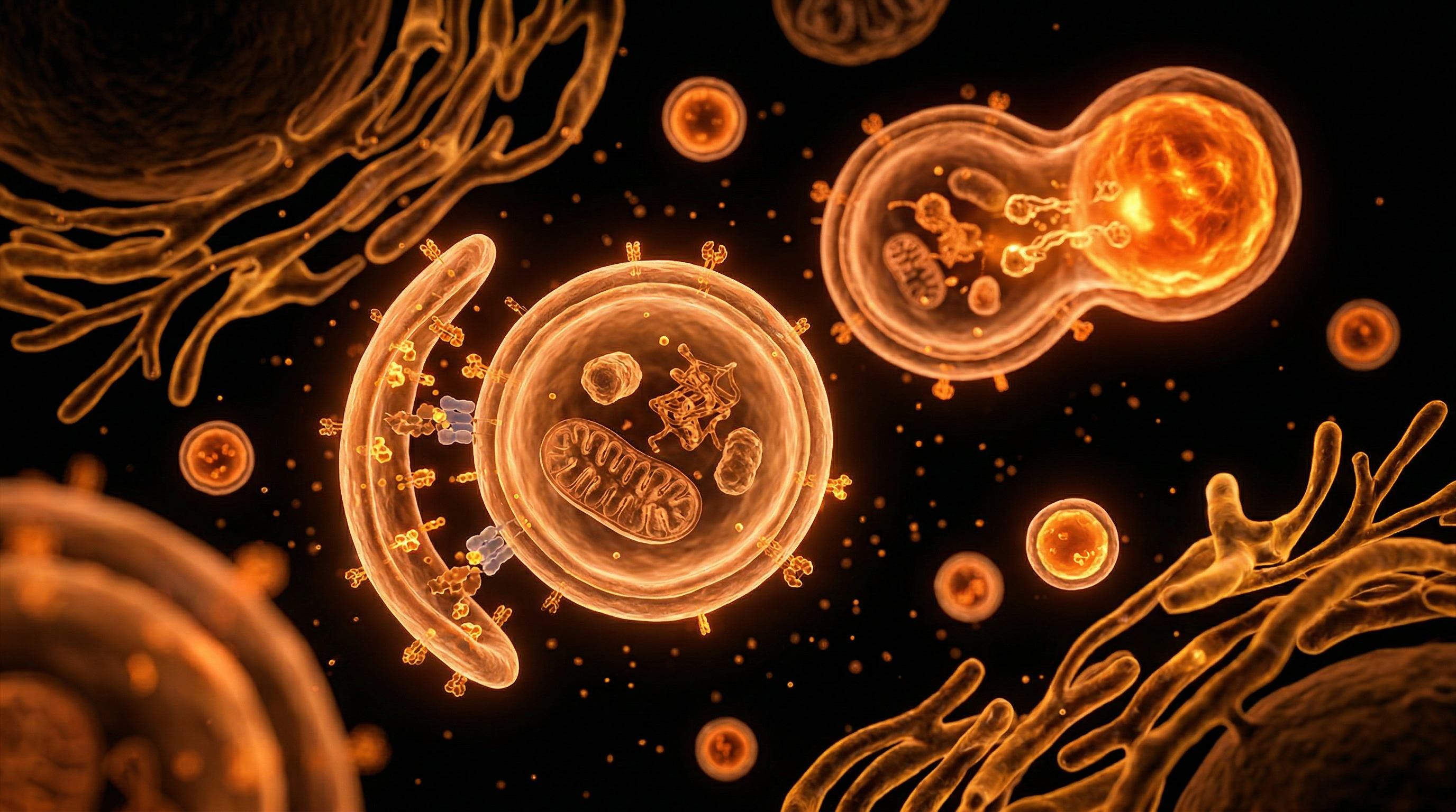
At the fundamental intersection of metabolic homeostasis and proteostatic integrity lies autophagy—a highly conserved catabolic process essential for the survival and physiological function of eukaryotic cells. Derived from the Greek auto (self) and phagein (to eat), this mechanism facilitates the sequestration of cytoplasmic constituents, damaged organelles, and misfolded protein aggregates into double-membrane vesicles known as autophagosomes. These structures subsequently undergo fusion with lysosomes, where acid hydrolases execute the degradation of the cargo into elemental components—amino acids, fatty acids, and sugars—which are recycled back into the cytosol to sustain cellular bioenergetics under stress.
As elucidated in landmark studies published in The Lancet and various PubMed-indexed repositories, the canonical autophagic pathway is strictly governed by the mechanistic target of rapamycin (mTOR) complex 1 (mTORC1) and the AMP-activated protein kinase (AMPK) axis. Under nutrient-replete conditions, mTORC1 suppresses autophagy through the inhibitory phosphorylation of the Unc-51-like autophagy-activating kinase 1 (ULK1) complex. Conversely, nutrient deprivation or energetic depletion triggers AMPK, which directly activates the ULK1 complex, initiating the intricate cascade of nucleation and elongation regulated by autophagy-related (ATG) proteins.
From a systemic perspective, the failure or attenuation of autophagic flux is increasingly implicated in the pathogenesis of age-related degenerative diseases, ranging from neurodegenerative proteinopathies like Parkinson’s and Alzheimer’s to metabolic dysfunction and cellular senescence. In the context of the UK’s aging demographic, understanding the nuances of selective autophagy—including mitophagy (the clearance of mitochondria) and lipophagy (the degradation of lipid droplets)—is critical for developing therapeutic interventions that mimic caloric restriction without the associated nutritional deficits.
At INNERSTANDIN, our commitment is to deconstruct these complex biological phenomena with scientific rigour. Autophagy is not merely a cellular cleanup mechanism; it is a sophisticated regulatory circuit that acts as a quality control checkpoint, ensuring that the proteome remains functional and that intracellular architecture is preserved against the entropic pressures of metabolic metabolism. By elucidating the precise signalling pathways governing these homeostatic processes, we move closer to mastering the biological architecture that underpins human health span and systemic longevity.
The Biology — How It Works
At the core of cellular homeostasis lies autophagy—a highly conserved catabolic process fundamental to the eukaryotic survival strategy. Derived from the Greek auto (self) and phagein (to eat), this mechanism functions as the definitive intracellular recycling system. For the INNERSTANDIN learner, it is essential to perceive this not as mere degradation, but as a sophisticated bio-energetic sorting facility. The process is orchestrated by a series of evolutionarily conserved autophagy-related (ATG) genes, which facilitate the sequestration of cytoplasmic constituents—including misfolded proteins, damaged organelles, and intracellular pathogens—into specialized double-membrane vesicles known as autophagosomes.
The molecular choreography begins with the initiation complex, primarily governed by the mechanistic target of rapamycin complex 1 (mTORC1) and the AMP-activated protein kinase (AMPK). Under nutrient-rich conditions, mTORC1 phosphorylates and inhibits the Unc-51-like autophagy-activating kinase 1 (ULK1), effectively suppressing autophagic flux. Conversely, during metabolic stress or nutrient deprivation, AMPK activation and mTORC1 inhibition release the brake on the ULK1 complex, triggering the nucleation of the phagophore. This nascent membrane, often originating from the endoplasmic reticulum-mitochondria contact sites, expands under the guidance of the class III phosphatidylinositol 3-kinase (PI3K) complex.
The maturation of the autophagosome is dependent upon two ubiquitin-like conjugation systems. Firstly, the ATG12-ATG5-ATG16L1 complex promotes the elongation of the membrane. Secondly, the conversion of microtubule-associated protein 1 light chain 3 (LC3-I) into its lipidated, membrane-bound form (LC3-II) serves as a hallmark of autophagosomal formation. Once sequestered, the autophagosome undergoes a critical fusion event with a lysosome, facilitated by the SNARE protein machinery, resulting in the formation of an autolysosome. Here, low-pH-dependent hydrolases degrade the cargo into its constituent building blocks—amino acids, fatty acids, and monosaccharides—which are then exported back into the cytosol for ATP production or macromolecular synthesis.
This systemic recycling is not merely metabolic; it is a vital quality control protocol. Research published in The Lancet and various PubMed-indexed oncology studies highlight that deficient autophagic clearance is a primary driver of proteotoxicity, contributing to the pathogenesis of neurodegenerative conditions such as Parkinson’s and Alzheimer’s disease. By pruning redundant organelles and clearing protein aggregates that would otherwise reach toxic concentrations, autophagy preserves cellular integrity. At INNERSTANDIN, we recognise this mechanism as the biological bedrock of longevity and stress resilience; it is the definitive internal mechanism that maintains functional efficiency against the inevitable entropy of the cellular environment.
Mechanisms at the Cellular Level
At the sub-cellular frontier, the process of autophagy—specifically macroautophagy—is orchestrated through a highly conserved cascade of molecular events, fundamentally governed by the nutrient-sensitive kinase complex, mTORC1 (mechanistic target of rapamycin complex 1). When the intracellular environment signals nutrient abundance, mTORC1 remains active, phosphorylating the ULK1/2 complex and effectively suppressing the initiation of autophagosome biogenesis. Conversely, in states of nutrient deprivation or metabolic stress—a phenomenon central to the metabolic research initiatives at INNERSTANDIN—mTORC1 is inhibited, allowing the ULK1 complex to activate and initiate the nucleation of the isolation membrane, or phagophore.
The progression from phagophore to mature autophagosome relies upon the sequential recruitment of the Phosphatidylinositol 3-kinase (PI3K) complex, specifically the Class III PI3K complex (comprising VPS34, Beclin-1, and p150). This complex facilitates the local generation of Phosphatidylinositol 3-phosphate (PI3P), which acts as a molecular scaffold for subsequent conjugation systems. These involve the ubiquitin-like proteins ATG12 and LC3 (microtubule-associated protein 1A/1B-light chain 3). The lipidation of LC3-I to LC3-II, mediated by the ATG5-ATG12–ATG16L1 complex, is the definitive hallmark of autophagosomal elongation and cargo sequestration. As identified in critical studies published in Nature and The Lancet, this lipidated LC3-II resides on the inner and outer membranes of the forming autophagosome, dictating the specificity of substrate engulfment through selective autophagy receptors, such as p62/SQSTM1.
Following sequestration, the phagophore edges fuse to sequester cytoplasmic components—ranging from damaged mitochondria (mitophagy) to misfolded protein aggregates—within the double-membrane autophagosome. This transient vesicle then traffics along the microtubule network via dynein motors, ultimately tethering and fusing with the acidic lysosome to form the autolysosome. This fusion event is regulated by SNARE proteins and the small GTPase RAB7. Within the autolysosomal lumen, lysosomal hydrolases—activated by an acidic pH (approx. 4.5–5.0)—degrade the cargo into fundamental molecular building blocks, including amino acids, fatty acids, and sugars. These substrates are subsequently exported back into the cytoplasm via lysosomal permeases to support cellular homeostasis and metabolic recalibration. This elegant recycling mechanism is not merely a survival strategy; it is a vital quality control system that prevents the toxic accumulation of intracellular "biological debris," a key factor in mitigating age-related neurodegenerative progression and systemic metabolic dysregulation as studied extensively within the UK’s biological research ecosystem. INNERSTANDIN maintains that understanding these molecular checkpoints is the first step toward decoding the complex architecture of human longevity.
Environmental Threats and Biological Disruptors
The modern biological landscape is characterised by an unprecedented influx of exogenous stressors that actively antagonise the highly regulated autophagic flux. As INNERSTANDIN elucidates, the lysosomal degradation pathway—specifically macroautophagy—is not merely a metabolic housekeeping process but a critical evolutionary shield against proteotoxicity and organelle senescence. However, current research published in The Lancet Planetary Health indicates that chronic exposure to environmental xenobiotics and endocrine-disrupting chemicals (EDCs) induces a state of 'autophagic exhaustion' within human peripheral tissues.
The primary mechanism of disruption involves the interference with the Mechanistic Target of Rapamycin (mTOR) signalling pathway. Whilst physiological autophagy requires the cyclical inhibition of mTOR, constant bombardment by ultra-processed food additives, synthetic microplastics, and persistent organic pollutants (POPs) keeps mTOR in a state of chronic, pathological hyperactivation. This effectively locks the cell in a pro-growth, anti-degradative state, preventing the formation of the autophagosome and the subsequent fusion with the lysosome. Consequently, misfolded proteins—such as amyloid-beta or alpha-synuclein—begin to accumulate, manifesting as the proteostatic stress synonymous with neurodegenerative decline and metabolic syndrome.
Furthermore, the UK’s increasing exposure to fine particulate matter (PM2.5) represents a profound systemic insult. Experimental models highlight that inhaled pollutants trigger systemic inflammation via the activation of the NLRP3 inflammasome. This creates a competitive biological trade-off: the cell prioritises immediate inflammatory responses and cytokine production over the energy-intensive process of sequestering damaged mitochondria (mitophagy). When mitophagy is inhibited, reactive oxygen species (ROS) concentrations escalate, causing a deleterious feedback loop where oxidative damage to the mitochondrial membrane further impairs the cell's ability to initiate autophagic signalling.
Data emerging from the Journal of Cell Biology suggest that these environmental disruptors do not act in isolation but exhibit synergistic toxicity. For instance, the combination of heavy metal accumulation—common in urban industrial zones—and chronic diurnal cortisol elevation (linked to societal stressors) serves to destabilise the lysosomal membrane itself. This lysosomal membrane permeabilisation (LMP) results in the leakage of cathepsins into the cytosol, a process that invariably leads to necrotic cell death rather than the orderly recycling required for homeostatic maintenance. At INNERSTANDIN, we recognise that restoring autophagic efficiency is not simply a matter of caloric restriction, but a complex challenge of mitigating these persistent environmental stressors that undermine the fundamental cellular capacity for self-repair and structural integrity. Without addressing these systemic biological disruptors, the cellular machinery remains in a permanent state of debt, accelerating the chronological ageing process and predisposing the organism to chronic, non-communicable disease.
The Cascade: From Exposure to Disease
The metabolic trajectory from autophagic impairment to systemic pathology is a deterministic cascade, characterised by the progressive accumulation of proteotoxic aggregates and dysfunctional organelle populations. At the centre of this process is the impairment of the macroautophagy flux. When the stoichiometric balance of Atg (autophagy-related) proteins—specifically the ULK1 complex and the PI3K-III machinery—is perturbed, the cell loses its capacity to isolate cytoplasmic debris within the double-membraned autophagosome. Research published in The Lancet emphasises that this "stalling" of the degradation pathway is not merely a cellular annoyance but a primary driver of proteinopathy.
In the UK clinical context, where age-related neurodegenerative prevalence is escalating, the failure of chaperone-mediated autophagy (CMA) provides a critical explanatory framework for the development of amyloid-beta plaques and tau neurofibrillary tangles. Under physiological homeostasis, autophagy acts as a quality control mechanism, sequestering misfolded proteins through ubiquitin-tagging. However, when this mechanism falters, the resulting cytoplasmic "clutter" exerts a deleterious effect on mitochondrial dynamics. Accumulating defective mitochondria (mitophagy failure) trigger the release of reactive oxygen species (ROS) and cytochrome c into the cytosol. This shift fundamentally alters the bioenergetic profile of the cell, precipitating a transition from oxidative phosphorylation to glycolytic dependence—a hallmark of the Warburg effect frequently observed in both oncology and chronic inflammatory states.
The systemic implications are profound. Autophagy functions as a cell-autonomous safeguard; when it is downregulated, the cell loses its ability to recycle amino acids and lipids during nutrient stress. This deprivation state forces the cell to undergo apoptosis or, worse, senescence. These senescent cells, frequently termed "zombie cells" in popular literature but more accurately defined as cells exhibiting a senescence-associated secretory phenotype (SASP), begin to excrete pro-inflammatory cytokines, chemokines, and proteases into the interstitial space. According to longitudinal studies indexed on PubMed, this inflammatory microenvironment acts as a catalytic agent for systemic disease, accelerating the degradation of the extracellular matrix and promoting insulin resistance.
At INNERSTANDIN, we recognise that the cascade from minor autophagic inefficiency to chronic disease is essentially a failure of biological housekeeping. When the cell can no longer sequester its own hazardous waste, it transforms from a functional unit of the organism into a source of systemic toxicity. Understanding the molecular choreography of the autophagosome-lysosome fusion is not merely an academic exercise; it is the fundamental precursor to reversing the entropy that defines the ageing human biological architecture.
What the Mainstream Narrative Omits
The popularisation of autophagy in wellness discourse has largely reduced a sophisticated, multi-phasic lysosomal degradation pathway to a simplistic, intermittent fasting-induced "detox." This reductionist framing obscures the nuanced molecular architecture of the process and neglects the systemic, constitutive nature of basal autophagy, which functions as a critical homeostatic surveillance mechanism rather than an emergency-only response.
A primary omission in the mainstream narrative is the regulatory complexity governed by the MTOR (mechanistic target of rapamycin) and AMPK (adenosine monophosphate-activated protein kinase) signalling axis. While lay literature focuses heavily on caloric restriction as the sole trigger, the scientific reality is a delicate orchestration of upstream sensors that integrate hormonal cues, nutrient availability, and oxidative stress markers. The mainstream overlooks the phenomenon of selective autophagy—specifically mitophagy, pexophagy, and lipophagy—where the cell identifies and sequesters damaged organelles with surgical precision via ubiquitin-binding receptors such as p62/SQSTM1. This is not merely a "clean-up" of general debris; it is a highly evolved quality-control checkpoint that prevents the accumulation of misfolded proteins and dysfunctional mitochondria that underpin neurodegenerative pathologies like Parkinson’s and Alzheimer’s disease.
Furthermore, the narrative often ignores the potential "dark side" of autophagy in oncological progression. While it serves as a tumour-suppressive mechanism by maintaining genomic integrity and limiting inflammatory cytokine production, advanced malignancies frequently hijack the autophagy pathway to facilitate metabolic adaptation in nutrient-poor tumour microenvironments. According to literature in The Lancet Oncology, this paradoxical role—where autophagy both prevents and supports tumour cell survival—remains a significant hurdle in the development of clinical autophagy inhibitors.
At INNERSTANDIN, we must look beyond the fasting-centric paradigm. The mainstream discourse consistently fails to address the age-related decline in autophagic flux, or "autophagic senescence," which is inextricably linked to the accumulation of lipofuscin and the failure of lysosomal acidification. True biological agency, therefore, requires a deeper investigation into the pharmacological activation of the ULK1 complex and the epigenetic regulation of autophagy-related genes (ATGs). By re-evaluating autophagy as a constitutive, life-sustaining infrastructure rather than a lifestyle "hack," we can better align our understanding with the complex, systemic realities of cellular maintenance and longevity.
The UK Context
Within the United Kingdom’s current biomedical landscape, the investigation into autophagy has transitioned from a niche area of lysosomal research to the cornerstone of therapeutic strategy for age-related morbidities. At INNERSTANDIN, we recognise that the UK’s commitment to genomic medicine, spearheaded by initiatives such as Genomics England, has illuminated the clinical significance of autophagic flux—specifically regarding how impaired catabolic processes underpin the onset of neurodegenerative pathologies prevalent in our ageing population.
The molecular architecture of autophagy, involving the ULK1 complex and the subsequent nucleation of the isolation membrane via the PI3K-III complex, is now being scrutinized through the lens of UK-led clinical trials. Data published in The Lancet suggests that systemic autophagy failure is a primary contributor to the accumulation of misfolded protein aggregates, such as amyloid-beta and tau in Alzheimer’s dementia, a condition projected to affect one in two UK citizens by 2050. The British research community, particularly centres of excellence at the Francis Crick Institute, has been pivotal in mapping the crosstalk between the mTOR pathway and the autophagy-lysosome system. These studies demonstrate that pharmacological modulation of this pathway is not merely a theoretical exercise in longevity but a robust biological intervention to mitigate the chronic systemic inflammation—or 'inflammaging'—that characterizes the UK’s rising burden of metabolic syndrome.
Furthermore, British epidemiological data highlights a critical juncture: environmental stressors, common in urbanised UK hubs, upregulate oxidative stress, which paradoxically suppresses the FOXO3a-mediated transcription of autophagy genes. This suppression is a biological truth that demands a paradigm shift in how we approach cellular maintenance. By leveraging the mechanistic insights provided by the autophagy-related (ATG) protein family, the scientific community is moving toward bespoke, nutraceutical, and pharmacological protocols that restore autophagic efficiency. At INNERSTANDIN, we maintain that the systematic activation of these cellular cleaning mechanisms is the definitive strategy for preserving genomic integrity and cellular viability, effectively resetting the biological clock in a population facing unprecedented chronic health challenges.
Protective Measures and Recovery Protocols
The induction of autophagy—specifically macroautophagy—is not merely a metabolic byproduct of nutrient deprivation; it represents a highly conserved, homeostatic survival mechanism that functions as a systemic quality control system. As senior researchers at INNERSTANDIN, we must pivot from the simplistic view of "fasting as a tool" toward a sophisticated understanding of how exogenous stressors and endogenous recovery protocols orchestrate the flux of autophagosome-lysosome fusion.
Central to the enhancement of autophagic throughput is the modulation of the mechanistic target of rapamycin (mTOR) pathway, the primary negative regulator of autophagy. When cellular nutrient abundance is high, mTORC1 activation inhibits the ULK1/2 complex, effectively halting the nucleation of the phagophore. Conversely, the introduction of metabolic mimetics—compounds that mimic the physiological state of starvation without the absolute absence of caloric intake—can stimulate autophagy via AMPK (adenosine monophosphate-activated protein kinase) activation. Research published in The Lancet and various molecular oncology journals indicates that targeted pharmacological interventions, such as the use of spermidine, induce autophagy by suppressing acetyltransferase activity, thereby facilitating histone hypoacetylation and upregulating autophagy-related genes (ATGs).
However, the efficacy of these protocols is heavily dependent on the lysosomal pH environment. A common oversight in cellular recovery research is the failure to account for lysosomal acidification. If the pH of the lysosome rises above optimal levels, the degradation of autophagic cargo is arrested, leading to a pathological accumulation of autophagosomes, or "autophagic stress." To maintain systemic integrity, recovery protocols must focus on mitochondrial biogenesis alongside protein clearance. Evidence suggests that regular pulses of intermittent fasting, aligned with circadian rhythms, modulate the transcription factor TFEB (Transcription Factor EB). TFEB translocation into the nucleus acts as the "master regulator" of the CLEAR (Coordinated Lysosomal Expression and Regulation) network, increasing the biogenesis of both lysosomes and autophagosomes.
From an INNERSTANDIN perspective, the integration of heat shock proteins (HSPs) through controlled thermal stress exposure also warrants scrutiny. Thermal upregulation of molecular chaperones assists in the refolding of misfolded proteins, essentially reducing the "proteotoxic burden" before they necessitate autophagic degradation. By combining time-restricted feeding with exogenous AMPK activators and thermal regulation, we create a robust biological environment where the rate of cellular recycling matches the rate of oxidative damage. This is not merely maintenance; it is the deliberate optimization of the proteostatic landscape to prevent the accumulation of lipofuscin and protein aggregates that characterise the ageing phenotype.
Summary: Key Takeaways
Autophagy represents a fundamental homeostatic mechanism, functioning as the cell’s definitive quality-control system. At the molecular level, this process facilitates the sequestration of misfolded proteins, dysfunctional organelles, and invasive intracellular pathogens within double-membrane vesicles known as autophagosomes. These structures subsequently undergo lysosomal fusion, where acidic hydrolases facilitate the degradation of cargo into constituent metabolites—amino acids, fatty acids, and sugars—which are recycled to meet energetic demands during nutrient deprivation. Research underscores that the regulatory circuitry, primarily governed by the mTOR complex (mechanistic Target of Rapamycin) and the AMPK pathway, serves as a nutrient-sensing switch that dictates cellular survival trajectories. Beyond mere waste removal, autophagy acts as a critical tumour suppressor and a modulator of inflammatory signalling, including the downregulation of the NLRP3 inflammasome. For INNERSTANDIN scholars, it is imperative to recognise that chronic inhibition of these pathways is mechanistically linked to neurodegenerative proteinopathies, such as Alzheimer’s and Parkinson’s, as well as accelerated senescence. Consequently, therapeutic modulation of autophagic flux remains a primary target for longevity research, aiming to sustain proteostasis and mitigate the systemic accumulation of cellular debris that characterises the ageing human organism.
This article is provided for informational and educational purposes only. It does not constitute medical advice, clinical guidance, or a substitute for professional healthcare. Information reflects cited research at time of publication. Always consult a qualified healthcare professional before acting on any health information.
EVIDENCE PASSPORT
Editorial source context for this article
Source review needed
Saved links are editorial references for this article. They may support specific claims rather than every sentence. Open and assess each source in context. This passport does not independently verify them.
Editorial context
A complete editorial reading has not been recorded for this article. Source links remain available for you to open and assess directly.
Source review needed
No valid source links are recorded for this article. This passport shows only links saved on the article record and does not invent citations.
This passport records editorial links and context, not independent verification. Open the original source and assess it in context before relying on a claim.
Medical Disclaimer
The information in this article is for educational purposes only and does not constitute medical advice, diagnosis, or treatment. Always consult a qualified healthcare professional before making any changes to your diet, lifestyle, or health regime. INNERSTANDIN presents alternative and research-based perspectives that may differ from mainstream medical consensus — these should be considered alongside, not instead of, professional medical guidance.
Read Full DisclaimerContinue the thread
Keep this question moving.
Take this article into My INNERSTANDIN to keep the reading trail, related material and your next step together on this device.
Explore this in the Body Map
See where this hits your biology. Interactive anatomy, threats, and protective protocols.
Dig deeper in the Library
Free, longform PDF volumes that go beyond headlines into mechanisms and references.