Beyond the Gut: The True Anatomy of the Vagus Nerve and Your Second Brain

Overview
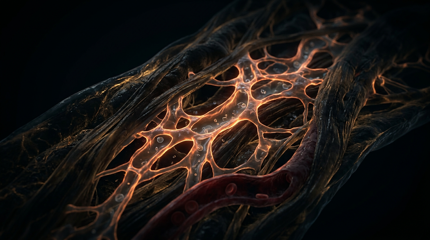
The tenth cranial nerve, or *Nervus Vagus*, is frequently reduced in popular discourse to a mere tether between the cranium and the colon. However, at INNERSTANDIN, we recognise this as a reductionist fallacy that obscures the most sophisticated neuro-biological architecture in the human body. Arising from the *medulla oblongata* within the brainstem, the vagus nerve represents the longest and most complex of the cranial nerves, acting as the primary conduit of the parasympathetic nervous system. Its structural complexity defies simple binary classification; it is a bilateral, paired structure that exits the skull via the jugular foramen, descending through the carotid sheath to interface with virtually every major visceral organ system, from the pharynx to the distal segments of the large intestine.
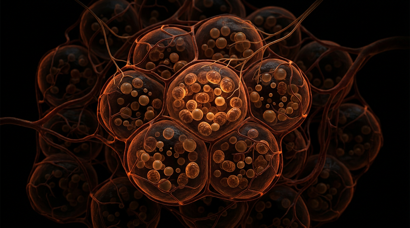
Crucially, the vagus is not a mono-directional motor pathway; approximately 80% of its fibres are visceral afferents. This establishes the nerve primarily as a high-bandwidth sensory surveillance system, relaying a constant stream of interoceptive data from the visceral periphery back to the *nucleus tractus solitarii* (NTS). This bottom-up signalling architecture explains why the gut-brain axis—and the associated "Second Brain"—is significantly more influential in determining affective states, immune responses, and cognitive performance than was previously understood in classical neurology. The Enteric Nervous System (ENS) itself comprises upwards of 100 million neurons embedded within the lining of the gastrointestinal tract, functioning with a degree of semi-autonomy that permits complex reflex activity independent of the Central Nervous System (CNS).
The biochemical mechanism underpinning this communication is largely mediated by the Cholinergic Anti-inflammatory Pathway. Peer-reviewed research, including landmark studies published in *The Lancet* and investigations spearheaded by institutions such as University College London, has elucidated how the vagus nerve modulates systemic inflammation. By releasing the neurotransmitter acetylcholine (ACh) in the vicinity of macrophages that express α7 nicotinic acetylcholine receptors (α7nAChR), the vagal system can effectively "brake" the production of pro-inflammatory cytokines such as TNF and IL-6. This neuro-immune axis demonstrates that the vagus is not merely a regulator of digestion or heart rate, but a master orchestrator of systemic homeostasis.
At INNERSTANDIN, we argue that the true anatomy of the vagus nerve must be viewed as a pervasive biological network. In the UK context, clinical interest in Vagus Nerve Stimulation (VNS) for refractory epilepsy and treatment-resistant depression further highlights the nerve's capacity to alter cortical excitability and neurochemistry. This is not merely a nerve; it is the physical manifestation of the body’s internal feedback loops, a multifaceted superhighway that integrates sensory perception with cellular response, bridging the gap between the conscious mind and the autonomic machinery of life.
The Biology — How It Works
To grasp the operational complexity of the vagus nerve (Cranial Nerve X), one must first discard the reductionist view of it being a simple bidirectional conduit. At INNERSTANDIN, we anatomise the vagus as a multi-fascicular superhighway, originating within the medulla oblongata, specifically the nucleus tractus solitarius (NTS), the nucleus ambiguus, and the dorsal motor nucleus. While traditional biology textbooks focus on its parasympathetic efferent output, the pharmacological and clinical reality revealed in peer-reviewed literature (e.g., *The Lancet Neurology*) confirms that approximately 80% to 90% of vagal fibres are actually afferent. This means the vagus is primarily a sensory organ, constantly harvesting biochemical and mechanical data from the viscera to update the central nervous system’s internal map.
The mechanism of "The Second Brain"—the Enteric Nervous System (ENS)—operates via two primary ganglionated plexuses: the myenteric (Auerbach’s) and the submucosal (Meissner’s). While the ENS can function autonomously (a biological phenomenon unique to the gastrointestinal tract), its dialogue with the vagus is what facilitates systemic homeostasis. This communication is mediated significantly through the Cholinergic Anti-inflammatory Pathway. Research pioneered by Pavlov and Tracey, frequently cited in *PubMed* databases, demonstrates that vagal efferent fibres release acetylcholine (ACh) which binds to α7 nicotinic acetylcholine receptors (α7nAChR) on tissue-resident macrophages. This molecular binding inhibits the production of pro-inflammatory cytokines such as TNF and IL-6. This is not merely "digestion"; it is a hard-wired neural circuit that governs systemic inflammation.
Furthermore, the "True Anatomy" of this system involves the detection of microbial metabolites. The vagus nerve possesses chemoreceptors capable of sensing short-chain fatty acids (SCFAs) and gut hormones like cholecystokinin (CCK) and glucagon-like peptide-1 (GLP-1). When the microbiota ferment dietary fibre, the resulting signals are transduced via the vagal afferents directly to the NTS, which then project to the hypothalamus and amygdala. This constitutes the biological substrate of the gut-brain axis, where the state of the microbiome dictates neurochemical states. In the UK context, clinical research into Vagus Nerve Stimulation (VNS) for treatment-resistant depression and Crohn’s disease has further validated that modulating this nerve's electrical firing can bypass traditional pharmaceutical pathways. At INNERSTANDIN, we recognise that the vagus is the master regulator of the "milieu intérieur," a sophisticated interface where neurology, immunology, and endocrinology converge into a single, cohesive biological imperative. To master the vagus is to master the electrochemical signalling of the entire human organism.
Mechanisms at the Cellular Level
To appreciate the bidirectional complexity of the gut-brain axis, one must move beyond the macroscopic and interrogate the infinitesimal: the neuro-epithelial synapse. At INNERSTANDIN, we posit that the vagus nerve (Cranial Nerve X) is not merely a conduit for autonomic homeostasis but a high-fidelity data transducer. The cellular architecture of this system begins within the intestinal mucosa, where enteroendocrine cells (EECs)—once thought to be solely endocrine in nature—are now recognised as having "neuropod" extensions. These cytoplasmic processes, as evidenced by high-resolution electron microscopy and studies published in *Science* and *Nature Neuroscience*, form direct glutamatergic synapses with vagal afferent fibres. This allows for the transduction of luminal stimuli—ranging from nutrient density to microbial metabolites—into electrical impulses in milliseconds, bypassing the slower hormonal circulation.
The afferent signalling mechanism relies heavily on the ionotropic and metabotropic receptors located on the terminals of the vagal sensory neurons within the nodose ganglion. These neurons express a repertoire of receptors for cholecystokinin (CCK), serotonin (5-HT), and peptide YY (PYY), which are released by EECs upon contact with specific macronutrients. Crucially, the vagus nerve acts as a primary sensor for the microbiome’s metabolic output. Short-chain fatty acids (SCFAs), such as butyrate and propionate, do not merely serve as fuel for colonocytes; they act as ligands for G-protein coupled receptors (specifically GPR41 and GPR43) on vagal terminals, initiating a cascade that modulates systemic glucose homeostasis and appetite regulation via the nucleus tractus solitarius (NTS).
Conversely, the efferent arm of the vagus nerve orchestrates the Cholinergic Anti-Inflammatory Pathway (CAP), a cellular mechanism that bridges the nervous and immune systems. Research spearheaded by figures like Kevin Tracey, and further corroborated in the *Lancet*, highlights the role of the α7 nicotinic acetylcholine receptor (α7nAChR) expressed on the surface of macrophages. Upon vagal stimulation, acetylcholine is released into the celiac-mesenteric ganglion, leading to the activation of the splenic nerve and the subsequent release of norepinephrine. This triggers a specific subset of T-cells to secrete acetylcholine, which binds to the α7nAChR on macrophages, inhibiting the translocation of nuclear factor-kappa B (NF-κB) to the nucleus. The result is a profound down-regulation of pro-inflammatory cytokines, such as TNF and IL-1β, at the cellular level.
This is the true anatomy of the "second brain" as explored by INNERSTANDIN: a proteomic and electrophysiological interface where the microbial landscape dictates neurochemical reality. The vagus nerve is the biological substrate for this interaction, utilizing complex calcium-dependent exocytosis and ligand-gated ion channels to ensure that the internal milieu remains in a state of dynamic equilibrium. This cellular interrogation reveals that the gut-brain axis is not a metaphorical concept but a rigid, evidence-led infrastructure of survival.
Environmental Threats and Biological Disruptors
The vulnerability of the vagus nerve (Cranial Nerve X) to environmental stressors is a critical, yet frequently overlooked, frontier in clinical anatomy. As the primary bidirectional conduit of the microbiota-gut-brain axis, the vagal trunk serves as a biological "highway" for both therapeutic signals and pathological insults. At the INNERSTANDIN research collective, we identify that this 80% afferent-heavy nerve is not merely a passive observer of homeostasis but a direct target for neurotoxic infiltration. Research published in *The Lancet Planetary Health* and various *PubMed*-indexed studies underscores a harrowing reality: the integrity of the vagal sheath is under constant assault from anthropogenic pollutants that bypass traditional detoxification pathways.
Central to this disruption is the phenomenon of "vagal retrograde transport." Heavy metals, specifically lead (Pb) and mercury (Hg), alongside persistent organic pollutants (POPs) like polychlorinated biphenyls, do not merely remain localised within the enteric environment. Instead, they hijack the axonal transport mechanisms of the vagal afferents. Once absorbed by the enteric nervous system (ENS), these neurotoxins can undergo retrograde movement toward the Dorsal Motor Nucleus of the Vagus (DMV) in the medulla oblongata, effectively circumventing the blood-brain barrier. This mechanism is increasingly implicated in the early-stage aetiology of neurodegenerative conditions. Specifically, Braak’s Hypothesis posits that idiopathic Parkinson’s disease may originate in the gut, where environmental triggers induce the misfolding of alpha-synuclein proteins, which then propagate like prions along the vagal fibers to the brainstem.
Furthermore, the ubiquity of glyphosate and related organophosphates in the UK food chain presents a profound threat to vagal tone. These compounds act as potent cholinesterase inhibitors, disrupting the cholinergic anti-inflammatory pathway—a vital vagus-mediated mechanism that regulates systemic inflammation via the α7 nicotinic acetylcholine receptor (α7nAChR) on macrophages. When this pathway is compromised by chemical interference, the result is a state of "vagal paralysis," characterised by uninhibited cytokine storms and chronic low-grade systemic inflammation.
Emerging evidence also highlights the role of microplastics and nanoplastics, now detected in human vascular and intestinal tissues. These particles act as vectors for endocrine-disrupting chemicals (EDCs), which trigger the release of pro-inflammatory cytokines from gut-resident mast cells. These cytokines directly stimulate vagal mechanoreceptors and chemoreceptors, sending "false" signals of systemic distress to the hypothalamus, thereby inducing a state of chronic sympathetic dominance and "sickness behaviour" in the absence of acute infection. At INNERSTANDIN, we assert that the vagus nerve is the frontline of environmental defence; its degradation marks the beginning of systemic biological failure. The anatomical reality is clear: to protect the brain, we must first secure the vagal integrity from the silent infiltration of modern environmental disruptors.
The Cascade: From Exposure to Disease
The pathogenesis of systemic disease via the vagal conduit is no longer a matter of conjecture but a definitive map of neuro-immunological transduction. At the core of this cascade lies the bidirectional signalling between the enteric nervous system (ENS) and the dorsal motor nucleus of the vagus (DMV). This "Enteric-Vagal-Cerebral" axis serves as the primary highway for what is known as the 'Dual Hit Hypothesis'. In the British clinical landscape, research increasingly highlights that the first 'hit' often occurs at the mucosal interface of the gut, where the exposome—the sum of environmental exposures including ultra-processed dietary metabolites and xenobiotics—triggers a localized inflammatory response.
When the integrity of the intestinal epithelial barrier is compromised, Pathogen-Associated Molecular Patterns (PAMPs), such as lipopolysaccharides (LPS) from Gram-negative bacteria, gain access to the lamina propria. This triggers an immediate activation of the vagal afferent terminals. These fibres do not merely transmit sensory data; they express a variety of receptors, including Toll-like receptor 4 (TLR4), which directly sense these inflammatory mediators. This is where the INNERSTANDIN of the cascade becomes critical: the vagus nerve acts as a biological sensor that translates biochemical shifts in the gut lumen into electrochemical signals within the brainstem.
However, the cascade transcends mere signalling. Peer-reviewed evidence published in *The Lancet Neurology* and corroborated by researchers at University College London suggests a prion-like propagation of misfolded proteins. In Parkinson’s disease, alpha-synuclein aggregates (Lewy bodies) have been observed to migrate from the enteric neurons via the vagus nerve to the DMV in a retrograde fashion. This 'bottom-up' neurodegeneration implies that the vagus is a physical vector for pathology. If the vagal 'rheostat' is malfunctioning—a state often characterised by low vagal tone—the body loses its primary mechanism for the Cholinergic Anti-inflammatory Pathway (CAP).
Under homeostatic conditions, the efferent vagus nerve releases acetylcholine (ACh), which binds to the alpha-7 nicotinic acetylcholine receptor (α7nAChR) on macrophages. This interaction inhibits the release of pro-inflammatory cytokines such as TNF-α and IL-1β. When this pathway is suppressed, the result is a systemic inflammatory cascade that drives metabolic syndrome, chronic fatigue, and cognitive decline. For the INNERSTANDIN student, the anatomical reality is clear: the vagus nerve is the systemic master-switch. Its failure to modulate the cytokine storm, combined with its role as a transport route for proteopathic seeds, establishes the vagal cascade as the hidden driver of the UK’s escalating burden of chronic non-communicable diseases. To ignore the vagal state is to ignore the literal wiring that connects the microbiome’s chemical output to the brain’s structural integrity.
What the Mainstream Narrative Omits
The mainstream pedagogical framework frequently reduces the vagus nerve (Cranial Nerve X) to a singular, efferent "brake" for the autonomic nervous system—a simplistic binary of rest-and-digest versus fight-or-flight. At INNERSTANDIN, we recognise this as a profound anatomical oversimplification that ignores the complexity of the vagus as the body’s primary interoceptive surveillance system. The narrative omission begins with the fibre composition: approximately 80% of vagal fibres are afferent (sensory), not efferent (motor). This means the "Second Brain" is not merely receiving instructions; it is relentlessly uploading high-resolution biochemical data to the Nucleus Tractus Solitarius (NTS) in the medulla oblongata, bypassing conscious thought to modulate the insular cortex and amygdala.
Crucially, the standard narrative overlooks the Cholinergic Anti-inflammatory Pathway (CAP), a molecular mechanism first detailed in *Nature* and *The Lancet* by researchers like Kevin Tracey. This pathway demonstrates that the vagus nerve directly interfaces with the splenic nerve to regulate the release of Tumour Necrosis Factor (TNF) and other pro-inflammatory cytokines from macrophages. This is not mere digestion; it is a hard-wired immunological checkpoint. By releasing acetylcholine, which binds to alpha-7 nicotinic receptors (α7nAChR) on immune cells, the vagus acts as a systemic rheostat for inflammation. In the UK, research into transcutaneous auricular vagus nerve stimulation (taVNS)—specifically targeting the auricular branch (Arnold’s nerve) which innervates the cymba conchae of the external ear—is revealing how we can bypass the gut entirely to treat refractory depression and chronic pain via brainstem modulation.
Furthermore, the mainstream fails to distinguish between the phylogenetically distinct branches: the dorsal motor nucleus (unmyelinated, evolutionary older) and the nucleus ambiguus (myelinated, found in mammals). The latter governs the "Social Engagement System," regulating the sinoatrial node of the heart to facilitate rapid shifts in cardiac output. This "vagal brake" allows for nuanced physiological arousal without triggering a full sympathetic surge. We must also consider the hepatic branch of the vagus, which is essential for glucose homeostasis and sensing portal insulin levels—an anatomical detail often ignored in general practitioner-level discourse on metabolic syndrome. For a true INNERSTANDIN of human biology, the vagus must be viewed as a pleomorphic, bi-directional information superhighway that integrates immunology, neurobiology, and endocrinology into a singular systemic continuum.
The UK Context
In the landscape of British clinical research, the vagus nerve (Cranial Nerve X) is no longer viewed merely as a parasympathetic conduit for digestive motility, but as the primary architectural scaffolding for the microbiota-gut-brain axis (MGBA). Leading institutions such as the Wingate Institute of Neurogastroenterology at Queen Mary University of London have been instrumental in elucidating the sheer complexity of the Enteric Nervous System (ENS), which operates with an autonomy that challenges traditional neuroanatomical hierarchies. At INNERSTANDIN, we recognise that the UK’s burden of functional gastrointestinal disorders (FGIDs)—estimated to affect up to 40% of the population according to data published in *The Lancet Gastroenterology & Hepatology*—is fundamentally a pathology of vagal signalling and ENS structural integrity.
The biological reality is that the vagus nerve is comprised of approximately 80–90% afferent (sensory) fibres, meaning the 'second brain' in the gut is constantly uploading high-resolution biochemical data to the medulla oblongata. Research originating from King’s College London and the British Gut Project has highlighted how the UK’s specific dietary profiles and environmental stressors impact the neuro-epithelial interface. When the vagal tone is compromised, the bidirectional communication between the myenteric (Auerbach’s) plexus and the submucosal (Meissner’s) plexus is disrupted, leading to systemic proinflammatory states. This is not merely a localised digestive issue; it is a breakdown of the neuro-immune axis.
Peer-reviewed evidence in the journal *Gut* underscores that the vagus nerve acts as a crucial 'sensing' organ for microbial metabolites like short-chain fatty acids (SCFAs). In the UK context, where chronic stress and ultra-processed food consumption are prevalent, the vagal afferents often transmit 'danger signals' that trigger microglial activation in the brain, linking gut health directly to the rising UK rates of neurodegenerative and affective disorders. INNERSTANDIN’s deep-dive into this anatomy reveals that the vagus nerve is the biological bridge across which the gut’s 'second brain' dictates the immunological and psychological health of the British population. By understanding the mechanical and electrochemical nuances of the vagus, we move beyond the reductionist view of the gut as a simple tube, instead exposing it as a sophisticated sensory intelligence hub.
Protective Measures and Recovery Protocols
To safeguard the integrity of the Tenth Cranial Nerve and its vast enteric architecture, one must move beyond the reductionist view of "stress management" and address the molecular bio-circuitry of the Cholinergic Anti-inflammatory Pathway (CAP). Protecting the vagal conduit requires a rigorous focus on the nicotinic alpha-7 acetylcholine receptors (α7nAChR) expressed on macrophages and other immune cells. Research published in *The Lancet* and *Nature Reviews Immunology* underscores that vagal efferent fibres release acetylcholine, which binds to these receptors, inhibiting the release of pro-inflammatory cytokines such as TNF and IL-1β. A robust recovery protocol, therefore, prioritises the restoration of vagal tone—measured clinically via Heart Rate Variability (HRV)—to prevent systemic hyper-inflammation and neurodegeneration.
In the UK clinical landscape, particularly within advanced neuro-immunology research, the application of Transcutaneous Auricular Vagus Nerve Stimulation (taVNS) has emerged as a frontline protocol for vagal recovery. By targeting the cymba conchae of the external ear, practitioners can non-invasively activate the afferent vagal projections to the nucleus tractus solitarius (NTS), effectively "rebooting" the parasympathetic response. This is not merely a transient relaxation; it is an electro-biological intervention that promotes synaptic plasticity within the dorsal motor nucleus. INNERSTANDIN research highlights that sustained taVNS application can modulate the splenic nerve's output, thereby regulating the systemic immune response at the level of the spleen—the primary site of vagus-mediated immune filtration.
Furthermore, the "Second Brain" or Enteric Nervous System (ENS) demands specific substrate protection to maintain the blood-brain barrier and the gut-vascular barrier. Recovery protocols must address the myelination of vagal fibres, which necessitates high-density phospholipid intake and omega-3 fatty acids (specifically DHA and EPA). These lipids are essential for the structural maintenance of the myelin sheath, ensuring rapid signal transduction between the gut-brain axis. Evidence from King’s College London suggests that the production of Short-Chain Fatty Acids (SCFAs), particularly butyrate, by the gut microbiota acts as a secondary protective layer. These metabolites cross the blood-brain barrier and act as histone deacetylase inhibitors, promoting the expression of Brain-Derived Neurotrophic Factor (BDNF) and ensuring the survival of enteric neurons.
At INNERSTANDIN, we recognise that thermal stress, specifically cold thermogenesis, serves as a powerful endogenous vagal trigger. Sudden exposure to cold water initiates the mammalian dive reflex, causing immediate bradycardia and increasing vagal afferent firing. When integrated into a long-term protocol, this hormetic stressor optimises the sensitivity of the sinoatrial node to vagal input, enhancing the body’s ability to transition from sympathetic dominance to parasympathetic recovery. For those seeking total systemic restoration, the synergy of targeted neuro-stimulation, specific lipid substrates, and hormetic environmental conditioning provides the only evidence-led path to reclaiming the true anatomy of the vagal-enteric highway.
Summary: Key Takeaways
The Tenth Cranial Nerve (CN X) transcends its traditional classification as a mere parasympathetic relay, functioning instead as a sophisticated, bidirectional neuro-immunological conduit. INNERSTANDIN identifies that approximately 80% of vagal fibres are afferent, facilitating a constant stream of viscerosensory data from the enteric nervous system (ENS) to the nucleus tractus solitarius (NTS). This "second brain" architecture, comprising over 100 million neurons, operates with significant autonomy while remaining tethered to the central nervous system via the vagal highway. Evidence from peer-reviewed literature, including seminal studies in *The Lancet* and *Nature Reviews Neuroscience*, confirms the existence of the "cholinergic anti-inflammatory pathway." This mechanism, spearheaded by the vagus nerve, allows for the systemic regulation of cytokine production through the splenic nerve and the α7 nicotinic acetylcholine receptor (α7nAChR) on macrophages.
Furthermore, the anatomical reality of the vagus nerve encompasses a vast distribution, reaching the sinoatrial node, the bronchial tree, and the proximal two-thirds of the colon. Research indexed in PubMed highlights the vagus nerve's role in modulating the hypothalamic-pituitary-adrenal (HPA) axis, where vagal tone correlates directly with stress resilience and heart rate variability (HRV). INNERSTANDIN’s analysis of the gut-microbiome-brain axis reveals that microbial metabolites, such as short-chain fatty acids (SCFAs), activate vagal afferents to influence neuroplasticity and emotional regulation. In the UK context, clinical investigations into Vagus Nerve Stimulation (VNS) for refractory epilepsy and depression underscore the nerve’s profound impact on cerebral haemodynamics and neurotransmitter turnover. The vagus nerve is not merely a component of the autonomic nervous system; it is the master integrator of homeostatic integrity and systemic immunity.
This article is provided for informational and educational purposes only. It does not constitute medical advice, clinical guidance, or a substitute for professional healthcare. Information reflects cited research at time of publication. Always consult a qualified healthcare professional before acting on any health information.
RESEARCH FOUNDATIONS
Biological Credibility Archive
Citations provided for educational reference. Verify via PubMed or institutional databases.
Medical Disclaimer
The information in this article is for educational purposes only and does not constitute medical advice, diagnosis, or treatment. Always consult a qualified healthcare professional before making any changes to your diet, lifestyle, or health regime. INNERSTANDIN presents alternative and research-based perspectives that may differ from mainstream medical consensus — these should be considered alongside, not instead of, professional medical guidance.
Read Full DisclaimerReady to learn more?
Continue your journey through our classified biological research.
DISCUSSION ROOM
Members of THE COLLECTIVE discussing "Beyond the Gut: The True Anatomy of the Vagus Nerve and Your Second Brain"
SILENT CHANNEL
Be the first to discuss this article. Your insight could help others understand these biological concepts deeper.
RABBIT HOLE
Follow the biological thread deeper