Cartilage Repair: Why Joints Struggle to Regenerate Naturally
Cartilage lacks a direct blood supply, making natural regeneration difficult after injury. We look at how stem cell-seeded hydrogels are providing new hope for UK arthritis patients.
# Cartilage Repair: Why Joints Struggle to Regenerate Naturally
Overview
The human body is an extraordinary feat of biological engineering, capable of knitting together fractured bone, regenerating the liver, and constantly replacing the epithelial lining of the gut. Yet, within our joints lies a silent, shimmering tissue that defies this regenerative norm: articular cartilage. This pearly-white, low-friction substance, known technically as hyaline cartilage, is the primary interface of our mobility. It allows the knee, hip, and shoulder to glide under immense loads without pain.
However, once this tissue is compromised—whether through acute trauma or the slow attrition of time—it possesses almost zero capacity for self-repair. For decades, the medical establishment has viewed cartilage degradation as an inevitable consequence of "wear and tear," a mechanical breakdown akin to a tyre losing its tread. But this perspective is dangerously reductive.
At INNERSTANDING, we view the failure of cartilage regeneration not merely as a mechanical oversight of evolution, but as a complex biological impasse. We are currently facing a "joint health crisis" in the United Kingdom, with over 10 million people suffering from arthritis and related conditions. The mainstream solution remains the "hardware model": wait for the joint to fail and replace it with plastic and metal.
This article serves as a deep-dive investigation into why cartilage is biologically "locked" in a non-regenerative state and how the emerging field of stem cell-seeded hydrogels and bio-scaffolding is finally beginning to pick the lock. We will expose the environmental disruptors that accelerate this decay and challenge the narrative that joint replacement is the only eventual outcome for the ageing population.
---
The Biology — How It Works
MSM Sulphur – Nature’s Forgotten Mineral
MSM Sulphur provides a high-purity, bioavailable source of a mineral that has largely vanished from modern diets due to industrial farming. It supports essential biological processes and structural health, restoring a vital nutrient your body needs to function optimally.
Vetting Notes
Pending
To understand why cartilage fails to heal, we must first appreciate its unique, almost "alien" biological structure. Unlike most tissues in the human body, articular cartilage is avascular, aneural, and alymphatic.
The Lack of Vascularity: The Primary Barrier
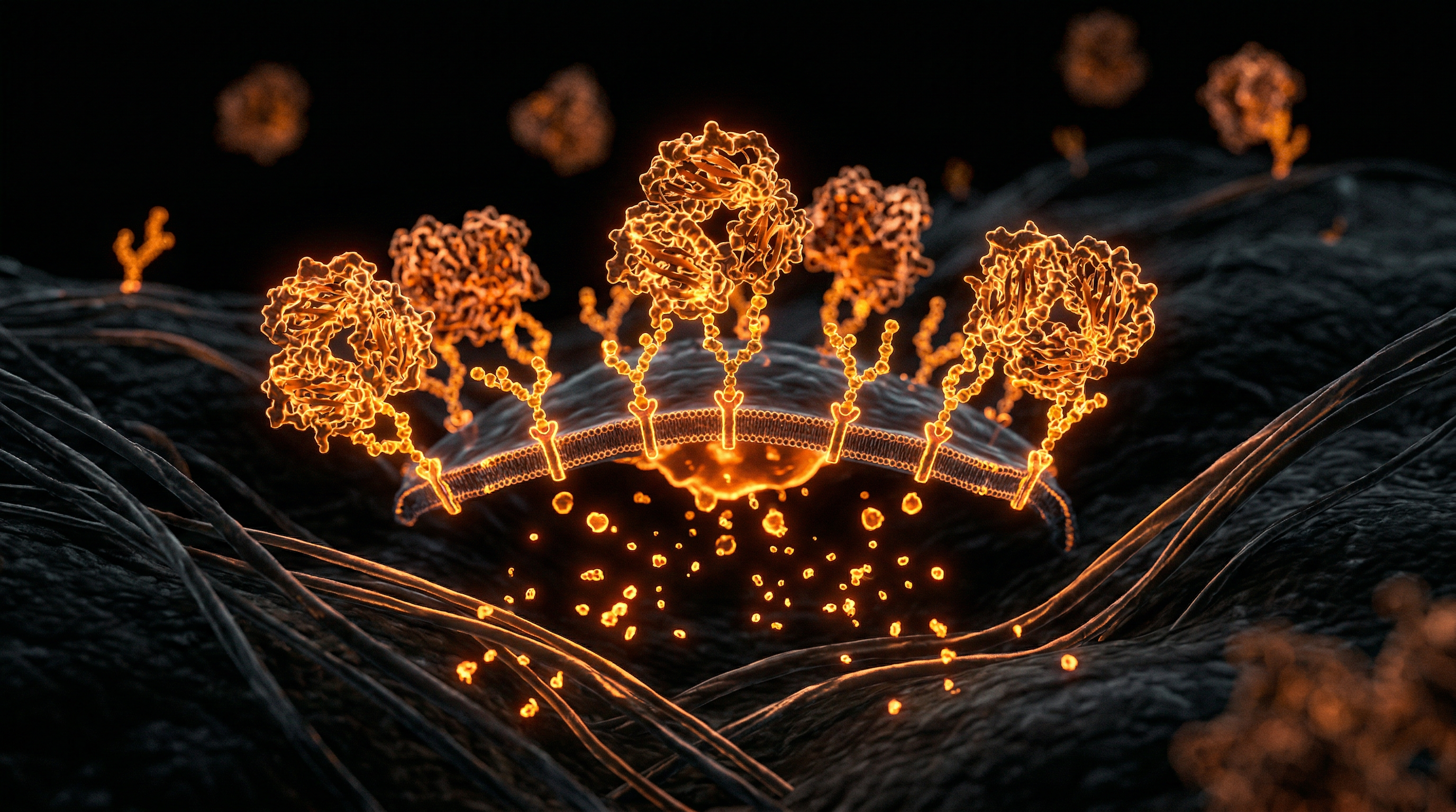
In almost every other part of the body, healing is driven by the blood. When you cut your skin, blood brings oxygen, nutrients, and, crucially, mesenchymal stem cells (MSCs) and inflammatory mediators to the site to initiate the repair cascade. Cartilage has no blood vessels. It sits in a state of permanent "physiological hypoxia" (low oxygen).
Because there is no direct blood supply, there is no "emergency response team" to arrive when a lesion occurs. The tissue relies entirely on diffusion from the surrounding synovial fluid—the lubricating oil of the joint. This diffusion is a slow, inefficient process, governed by the mechanical loading of the joint (the "pump" action of walking), which pushes nutrients in and waste products out.
The Extracellular Matrix (ECM)
Cartilage is not primarily made of cells; it is made of a dense, highly organised Extracellular Matrix (ECM). The cells that do live there, called chondrocytes, are sparse, representing only about 1% to 5% of the total tissue volume.
Callout Fact: Chondrocytes are the only cell type found in healthy articular cartilage. They are entombed within small cavities called lacunae, isolated from their neighbours, making intercellular communication—a requirement for coordinated healing—almost impossible.
The ECM is composed of:
- —Type II Collagen: Provides structural framework and tensile strength.
- —Proteoglycans (mainly Aggrecan): Large molecules that trap water, providing the "cushion" or compressive strength.
- —Water: Making up 70–80% of the weight, held in place by the negative charge of the proteoglycans.
This structure creates a high osmotic pressure, allowing the joint to withstand forces several times the body's weight. However, this very density acts as a physical barrier. Even if the body tries to send repair cells to a site of injury, they cannot easily penetrate this dense, pressurized meshwork of collagen and sugar-chains.
---
Mechanisms at the Cellular Level
At the heart of the regeneration failure is the metabolic quiescence of the chondrocyte. In a growing child, chondrocytes are active, building the skeleton. In an adult, they switch to a "maintenance mode." They produce just enough matrix to replace what is naturally lost to friction, but they lack the "proliferative capacity" to fill a gap or a hole.
The Problem of Differentiation
When a deep injury occurs—one that reaches the subchondral bone—the body *does* try to heal it. Blood from the bone enters the gap, bringing stem cells. However, instead of creating smooth, durable hyaline cartilage, these cells almost always differentiate into fibrocartilage.
- —Fibrocartilage is rich in Type I Collagen (the kind found in skin and scars) rather than the Type II Collagen required for joints.
- —It is mechanically inferior, stiffer, and lacks the water-binding capacity of the original tissue.
- —Within months or years, this "patch" typically fails, leading to further degradation.
Mechanotransduction: The Double-Edged Sword
Chondrocytes are governed by mechanotransduction—the process by which cells convert mechanical loads into biochemical signals.
- —Healthy Loading: Moderate walking or cycling signals the chondrocyte to produce more ECM.
- —Static Loading/Obesity: Constant, heavy pressure signals the chondrocyte to release catabolic enzymes (enzymes that break down tissue).
- —Shear Stress: Sudden, twisting injuries tear the collagen framework, and once the framework is broken, the chondrocytes undergo apoptosis (programmed cell death).
Important Statistic: Once a chondrocyte dies, it is not replaced. We are born with a finite "bank account" of these cells, and we spend them throughout our lives with no way to deposit more—until now.
---
Environmental Threats and Biological Disruptors
While the lack of blood supply is the "built-in" reason for poor healing, modern environmental factors are accelerating the rate of cartilage death to unprecedented levels. We are seeing "old age" joint diseases in people in their 30s and 40s.
Microplastics and the Synovium
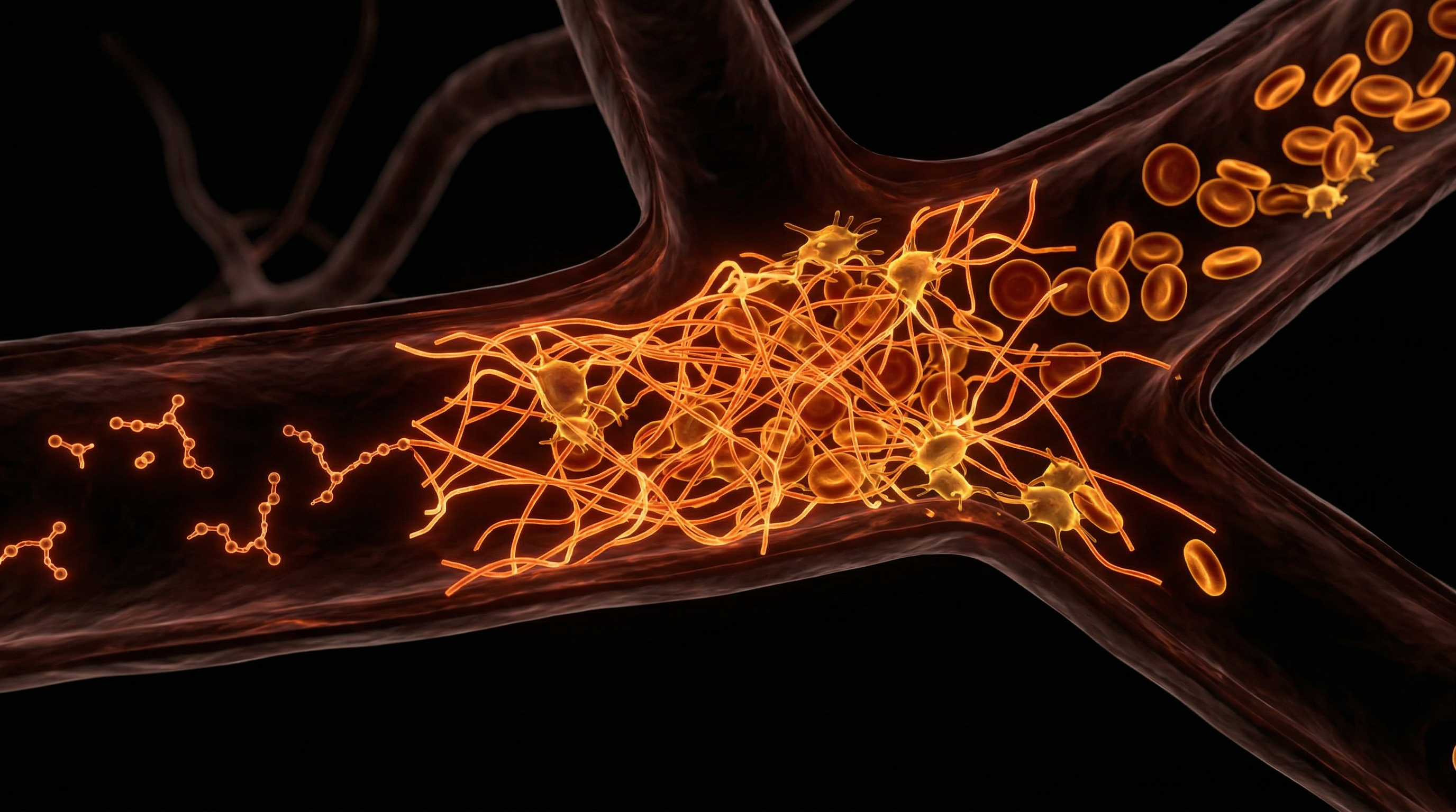
Recent pilot studies have identified microplastics and nanoplastics within human synovial fluid. These particles trigger a chronic, low-grade inflammatory response. When the immune system detects these foreign particles in the joint space, it releases cytokines like IL-1β and TNF-α. These "messenger molecules" tell the chondrocytes to stop making new matrix and start producing MMPs (Matrix Metalloproteinases)—enzymes that literally digest the joint from the inside out.
The "Sugar-Joint" Connection: Advanced Glycation End-products (AGEs)
Mainstream rheumatology often ignores the role of systemic metabolic health in cartilage. High blood sugar levels lead to the formation of Advanced Glycation End-products (AGEs). These molecules create "cross-links" in the collagen of the cartilage, making it brittle and prone to cracking like old rubber. A diet high in ultra-processed foods (UPFs) is not just making the UK population obese; it is chemically altering the structural integrity of their joints.
Endocrine Disruptors
Chemicals such as BPA (Bisphenol A) and certain PFAS (forever chemicals) have been shown to interfere with the signalling of growth factors like TGF-β (Transforming Growth Factor beta). TGF-β is the primary signal that tells a stem cell to become a chondrocyte. When this signal is "jammed" by environmental toxins, the body’s internal repair kit is effectively disabled.
---
The Cascade: From Exposure to Disease
The path from a healthy joint to a "bone-on-bone" nightmare is a predictable, yet devastating, cascade. It is rarely a single event, but a failure of biological "check and balances."
- —Phase 1: The Micro-tear. A small disruption in the collagen meshwork occurs. Because there is no pain (aneural tissue), the individual continues to load the joint.
- —Phase 2: Swelling and Inflammation. The joint produces excess synovial fluid (effusion) to try and lubricate the damaged area. This fluid is now "toxic," filled with inflammatory markers.
- —Phase 3: Chondrocyte Phenotype Shift. The chondrocytes near the injury change their behaviour. Instead of staying quiet and protective, they become "hypertrophic"—they start to act like bone cells, trying to calcify the area. This is why we see osteophytes (bone spurs) forming.
- —Phase 4: The Feedback Loop. As the surface becomes rough, it creates more friction, which creates more heat and more debris. This debris (tiny fragments of cartilage) further irritates the synovial lining, leading to synovitis.
- —Phase 5: Subchondral Bone Sclerosis. With the cushion gone, the bone underneath begins to thicken and harden. This reduces the bone's ability to shock-absorb, which in turn reflects the force back into the remaining cartilage, shattering it.
This "vicious cycle" is why cartilage injury is a progressive disease. It is not a static wound; it is a decaying ecosystem.
---
What the Mainstream Narrative Omits
In the UK, the standard of care for joint issues follows a "stepped" approach: Physiotherapy, then steroid injections, then ultimately, total joint replacement. However, this narrative omits several critical truths that the pharmaceutical and surgical industries find "inconvenient."
The Steroid Trap
Corticosteroid injections are frequently used to "manage" joint pain. While they provide temporary relief by suppressing inflammation, they are chondrotoxic. Research indicates that repeated steroid injections actually accelerate cartilage loss and inhibit the activity of any remaining stem cells. It is a "borrowed time" strategy that guarantees a future joint replacement.
The Myth of "Wear and Tear"
The term "wear and tear" suggests that joints have a shelf-life and will eventually run out. This is a fallacy. In a biologically "clean" environment with proper nutrition and movement, cartilage should last 100 years. The issue is not *wear*, but a lack of *repair*. By framing it as a mechanical inevitability, the medical system avoids addressing the environmental and nutritional factors that have crippled our natural regenerative capacity.
The Financial Incentive of Hardware
A total knee replacement in the UK private sector costs between £12,000 and £15,000. It is a highly profitable, "one-and-done" surgical procedure. Regenerative medicine—using a patient’s own cells to regrow the joint—threatens this model. Biological repair is complex, requires longer follow-up, and prioritises "software" (cells) over "hardware" (titanium). Consequently, funding for regenerative clinical trials often pales in comparison to the marketing budgets for new prosthetic designs.
---
The UK Context
The United Kingdom is currently at a crossroads regarding joint health. With an ageing population and a rising obesity rate, the NHS is struggling to keep up with the demand for joint replacements.
The NHS Backlog
Wait times for elective orthopaedic surgeries have skyrocketed. In many trusts, patients are waiting 18 to 24 months for a hip or knee replacement. During this time, they are often prescribed high doses of NSAIDs (Non-Steroidal Anti-Inflammatory Drugs) like Ibuprofen or Naproxen.
Callout Fact: Long-term NSAID use is linked to gastrointestinal bleeding and, paradoxically, can inhibit the healing of soft tissues and bone by suppressing the very prostaglandins needed for the initial stages of repair.
British Innovation in Regenerative Medicine
Despite the systemic hurdles, the UK remains a global leader in tissue engineering. Institutions like The University of Manchester and University College London (UCL) are pioneering the use of 3D-bioprinted hydrogels. These are "smart" materials designed to mimic the ECM of cartilage.
The "UK Arthritis Research" initiatives are finally shifting focus from "living with the pain" to "curing the defect." The goal is to move away from metal and towards Autologous Chondrocyte Implantation (ACI)—a technique where a patient's own cartilage cells are harvested, grown in a lab in the UK, and then re-implanted.
---
Protective Measures and Recovery Protocols
If the body cannot heal cartilage naturally, we must provide the "blueprint" and the "materials" for it to do so. This is where stem cell-seeded hydrogels represent the "holy grail" of orthopaedics.
1. Stem Cell-Seeded Hydrogels: The New Frontier
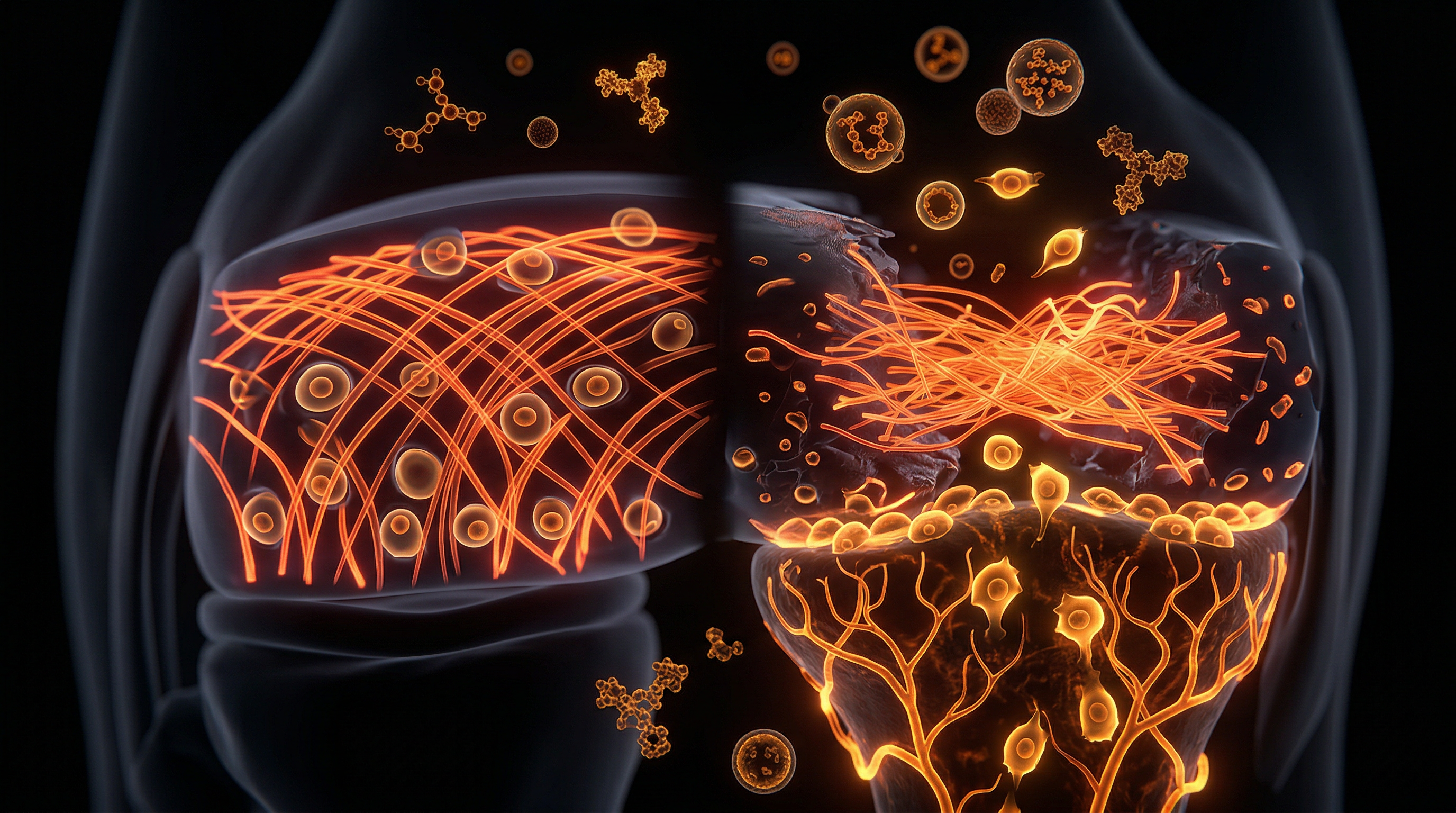
A hydrogel is a polymer network that can hold massive amounts of water—much like cartilage itself. By seeding these gels with Mesenchymal Stem Cells (MSCs)—usually derived from the patient's own bone marrow or adipose (fat) tissue—surgeons can "inject" a liquid scaffold into a cartilage defect.
- —The hydrogel provides immediate structural support.
- —It is infused with growth factors that tell the stem cells: "Become a chondrocyte, not a skin cell."
- —Over time, the hydrogel biodegrades, leaving behind fresh, native hyaline cartilage.
2. Platelet-Rich Plasma (PRP) and Gold-Induced Cytokines
PRP involves concentrating the platelets from a patient's blood and injecting them into the joint. Platelets contain "alpha granules" filled with growth factors (PDGF, TGF-β, VEGF). For UK patients, newer "leukocyte-poor" PRP protocols are showing significant success in reducing inflammation and "awakening" the dormant chondrocytes.
3. Precision Nutrition: The "Building Blocks"
To support any regenerative therapy, the biological terrain must be optimised.
- —Type II Collagen Peptides: Supplementation provides the specific amino acids (proline, glycine) required for matrix synthesis.
- —Glucosamine and Chondroitin Sulphate: While controversial in some mainstream circles, when used in high-purity, pharmaceutical-grade forms, they provide the "raw material" for the proteoglycan "brushes" that hold water.
- —Molecular Hydrogen and Turmeric (Curcumin): These act as potent, non-toxic anti-inflammatories that don't inhibit the healing cascade in the way steroids do.
4. The "Motion is Lotion" Protocol
Because cartilage relies on diffusion, complete rest is the enemy of recovery. Controlled, non-impact movement (such as swimming or professional-grade cycling) is essential to "pump" the nutrients into the hydrogel scaffold. This is mechanotherapy—using physical force to drive biological change.
---
Summary: Key Takeaways
The struggle of joints to regenerate naturally is not a design flaw, but a biological trade-off. In exchange for a tissue that can endure millions of cycles of high-pressure friction without pain, we have a tissue that is isolated from the body's repair systems.
- —Avascularity is the Enemy: The lack of blood supply means cartilage cannot signal for help or receive the nutrients needed for rapid repair.
- —Environment Matters: Microplastics, sugar (AGEs), and endocrine disruptors are "poisoning" the synovial environment, making natural maintenance impossible.
- —Mainstream Limitations: The current UK medical model often relies on "patchwork" solutions or total replacements, ignoring the potential of biological restoration.
- —Hydrogel Hope: The future of joint health lies in biomimetic scaffolds. By combining stem cells with advanced hydrogels, we can finally bypass the biological "lock" and regrow the smooth, hyaline cartilage we were born with.
The transition from "managing" arthritis to "curing" it requires a paradigm shift. We must stop viewing the body as a machine that wears out and start viewing it as a biological system that can be repaired—provided we give it the right instructions and a clean environment. The age of the "hardware" joint is ending; the era of the "biological" joint has begun.
*
"References & Technical Notes:"
*For the researcher seeking deeper technical data, consult the NICE (National Institute for Health and Care Excellence) guidelines on ACI, and the latest publications in 'The Lancet Rheumatology' regarding the efficacy of MSC-based therapies in UK clinical trials.*
This article is provided for informational and educational purposes only. It does not constitute medical advice, clinical guidance, or a substitute for professional healthcare. Information reflects cited research at time of publication. Always consult a qualified healthcare professional before acting on any health information.
RESEARCH FOUNDATIONS
Biological Credibility Archive
Citations provided for educational reference. Verify via PubMed or institutional databases.
Medical Disclaimer
The information in this article is for educational purposes only and does not constitute medical advice, diagnosis, or treatment. Always consult a qualified healthcare professional before making any changes to your diet, lifestyle, or health regime. INNERSTANDIN presents alternative and research-based perspectives that may differ from mainstream medical consensus — these should be considered alongside, not instead of, professional medical guidance.
Read Full DisclaimerReady to learn more?
Continue your journey through our classified biological research.
DISCUSSION ROOM
Members of THE COLLECTIVE discussing "Cartilage Repair: Why Joints Struggle to Regenerate Naturally"
SILENT CHANNEL
Be the first to discuss this article. Your insight could help others understand these biological concepts deeper.
THE ARSENAL
Based on Stem Cell Science & Regenerative Medicine — products curated by our research team for educational relevance and biological support.
Energy Blend Supports

Peptides, one of the secret Russian military health marvels, now available. 40 years research
Albedextrin – Specialist Cyclodextrin Complex
INNERSTANDING may earn a commission on purchases made through these links. All products are selected based on rigorous educational relevance to our biological research.
RABBIT HOLE
Follow the biological thread deeper