Corneal Bio-Engineering: Restoring Sight via Limbal Stem Cells
UK researchers are pioneers in using limbal stem cells to treat blindness caused by corneal damage. This article highlights the success stories of regenerative ophthalmology in the NHS.
# Corneal Bio-Engineering: Restoring Sight via Limbal Stem Cells
Overview
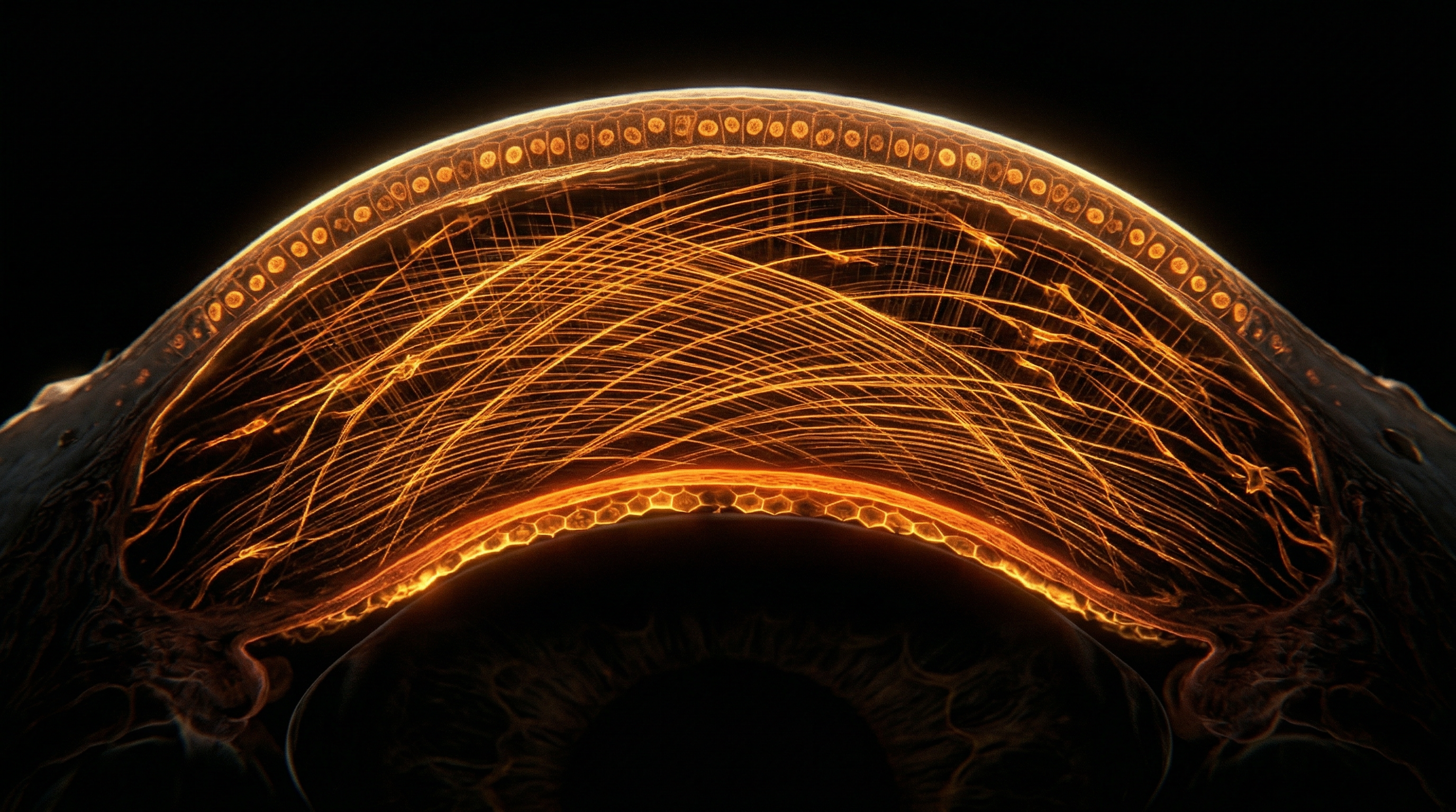
The human eye is often described as a window to the soul, but from a purely biological standpoint, it is a masterwork of optical engineering. At the forefront of this system lies the cornea, a transparent, dome-shaped window that accounts for approximately two-thirds of the eye's total optical power. However, this transparency is not a static state; it is a hard-won equilibrium maintained by a specialised population of regenerative units known as Limbal Stem Cells (LSCs).
In the United Kingdom, we are currently witnessing a revolution in regenerative ophthalmology. For decades, patients suffering from severe corneal scarring—often resulting from chemical burns, thermal injuries, or genetic disorders—were consigned to a life of legal blindness. Traditional corneal transplants (keratoplasty) frequently failed in these patients because the underlying "engine" of corneal renewal was broken.
This article explores the pioneering work of UK researchers and the National Health Service (NHS) in bio-engineering corneal tissue. We are no longer merely replacing tissue; we are restoring the biological capacity for self-repair. By harvesting a tiny biopsy of healthy limbal tissue, scientists can expand these cells in a laboratory and graft them back onto the damaged eye. This process, specifically Cultivated Limbal Epithelial Transplantation (CLET), represents the pinnacle of personalised medicine.
Fact: The cornea is the most densely innervated tissue in the human body, possessing roughly 300 to 600 times the density of pain receptors found in the skin. This sensitivity serves as an evolutionary "early warning system" to protect the visual axis.
As we delve into the mechanics of this breakthrough, we must also acknowledge the barriers that have historically slowed the adoption of such therapies. While the mainstream narrative often focuses on the "miracle" of sight, it frequently ignores the systemic industrial and environmental disruptors that necessitate such interventions, and the regulatory hurdles that keep these life-changing treatments at the periphery of global healthcare.
---
The Biology — How It Works
To understand the restoration of sight, one must first understand the architectural integrity of the ocular surface. The cornea consists of five primary layers: the epithelium, Bowman’s layer, the stroma, Descemet’s membrane, and the endothelium. The epithelium is the outermost protective shield, and it is under constant assault from dust, pathogens, and UV radiation.
The Limbal Niche
The secret to the cornea’s resilience lies at its periphery—a narrow, 1.5mm to 2mm ring known as the limbus. This is the transition zone between the clear cornea and the white sclera. Deep within the limbus, tucked away in microscopic structural undulations called the Palisades of Vogt, resides the reservoir of Limbal Stem Cells.
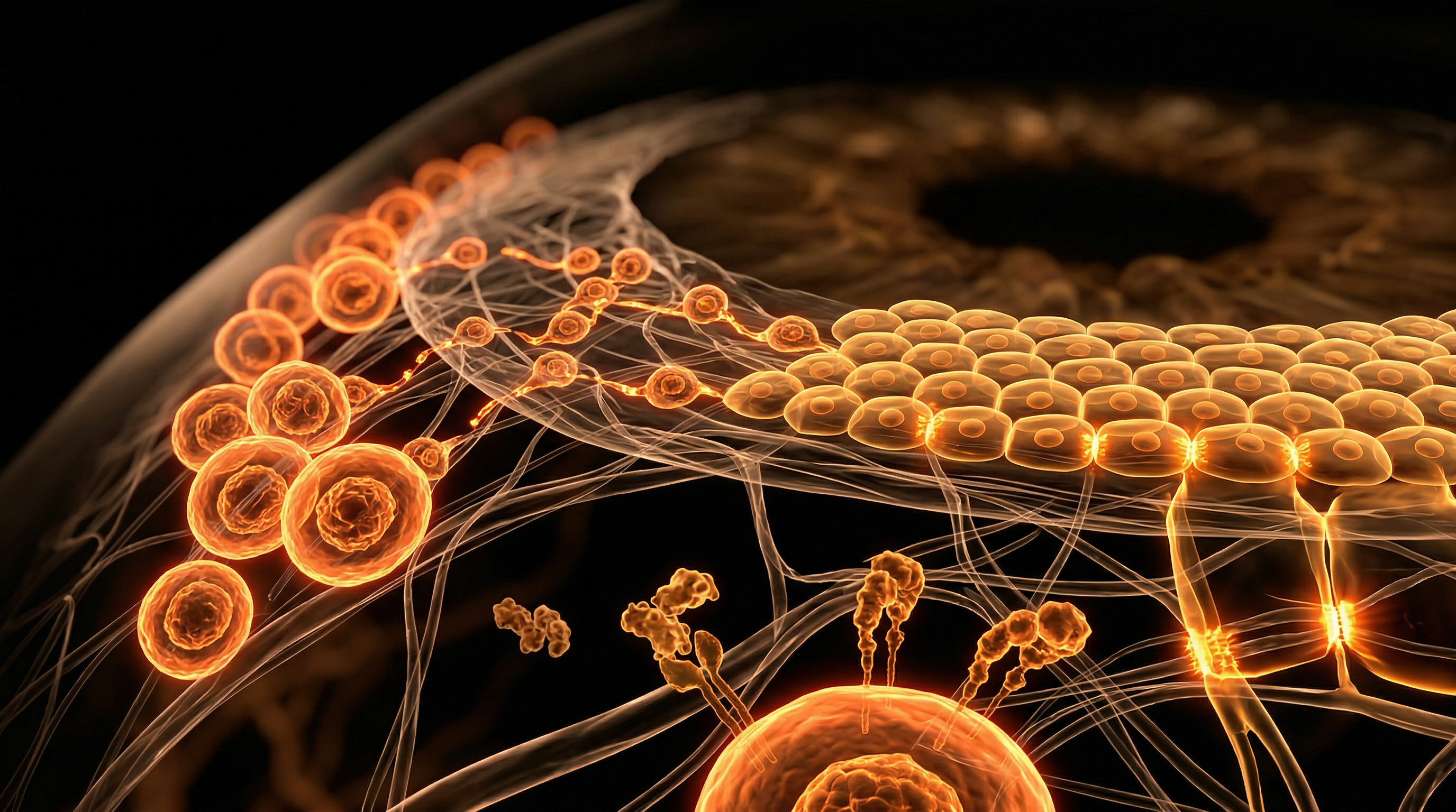
The Regenerative Cycle
In a healthy eye, the corneal epithelium undergoes a complete turnover every 7 to 10 days. This is facilitated by a specific biological pipeline:
- —Quiescence: LSCs remain dormant in the limbal niche, protected from environmental stress.
- —Activation: Upon signaling, LSCs divide. One daughter cell remains a stem cell (self-renewal), while the other becomes a Transit-Amplifying (TA) cell.
- —Migration: These TA cells migrate centripetally (toward the centre of the cornea) and upward toward the surface.
- —Differentiation: As they move, they lose their stem-like qualities and become mature, flattened epithelial cells, eventually sloughing off into the tear film.
When the limbus is destroyed—a condition known as Limbal Stem Cell Deficiency (LSCD)—this pipeline collapses. Without LSCs to replenish the surface, the conjunctiva (the vascularised tissue covering the white of the eye) begins to invade the cornea. This process, called conjunctivalisation, leads to chronic inflammation, neovascularisation (blood vessel growth), and eventual corneal opacification. The window becomes a wall.
---
Mechanisms at the Cellular Level
At the heart of corneal bio-engineering is the manipulation of the cellular phenotype. A stem cell is defined by its potency and its environment. In the lab, UK researchers must replicate the exact conditions of the human eye to ensure that the cultivated cells retain their "stemness."
The Role of p63α
The gold standard biomarker for LSCs is a transcription factor called p63, specifically the ΔNp63α isoform. This protein is essential for the proliferative potential of epithelial stem cells. If a cultivated graft contains fewer than 3% p63α-bright cells, the clinical success rate drops significantly. Researchers use immunohistochemistry to verify that the bio-engineered tissue is biologically "young" enough to survive the transplantation process.
Asymmetric Division and the Microenvironment
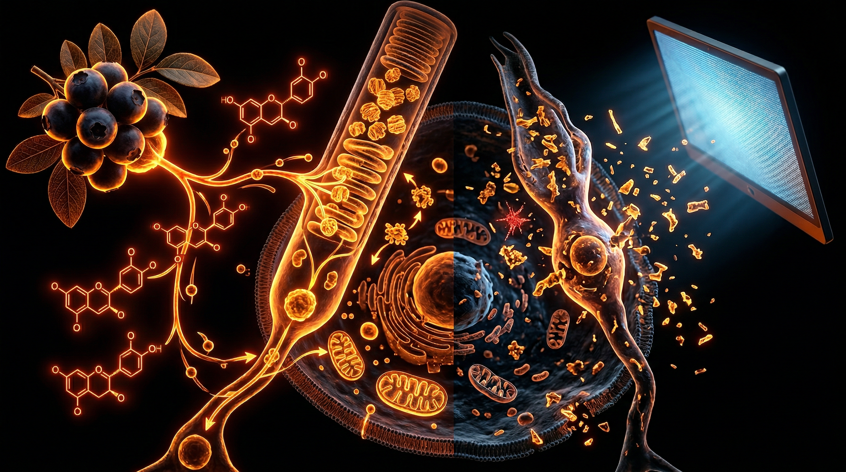
The fate of an LSC is determined by its niche. The niche provides physical support through the extracellular matrix (ECM) and chemical signaling through growth factors like EGF (Epidermal Growth Factor) and FGF (Fibroblast Growth Factor).
- —Mechanical Cues: The stiffness of the substrate matters. Research at Newcastle University has shown that LSCs behave differently on soft versus hard surfaces, a finding that has led to the development of "smart" hydrogels for cell delivery.
- —Wnt Signaling: This pathway is critical for maintaining the balance between stem cell self-renewal and differentiation. Manipulating Wnt signaling allows bio-engineers to "expand" a small biopsy into a large sheet of viable tissue.
Holoclar: The First Stem Cell Product
In the UK and Europe, the first tissue-engineered product to be approved was Holoclar. This involves taking a biopsy of 1-2mm² from the patient’s healthy eye (if the damage is unilateral). The cells are then grown on a scaffold of fibrin, a natural protein involved in blood clotting. Within weeks, a transparent circular sheet of living epithelium is ready for grafting.
---
Environmental Threats and Biological Disruptors
While the science of restoration is advanced, we must address why the incidence of ocular surface disease is shifting. Beyond the obvious traumatic injuries, there is a growing body of evidence suggesting that our modern environment is "antagonistic" to the limbal niche.
Chemical Insults and Industrial Toxicity
The most common cause of LSCD is chemical injury—specifically alkali burns (e.g., lime, ammonia, drain cleaners). Alkalis are more dangerous than acids because they are lipophilic; they saponify the cell membranes and penetrate deep into the limbal niche, effectively "cooking" the stem cells in situ.
The Impact of Glyphosate and Urban Particulates
Recent investigative research (often overlooked by mainstream ophthalmology) has begun to examine the role of environmental toxins in chronic ocular inflammation.
- —Particulate Matter (PM2.5): These ultra-fine particles from traffic and industrial exhaust can lodge in the tear film, inducing oxidative stress that prematurely ages the LSCs.
- —Agricultural Runoff: Emerging studies suggest that chronic exposure to certain organophosphates can interfere with the cholinergic signaling required for corneal nerve maintenance, which in turn destabilises the stem cell niche.
The Contact Lens Paradox
While contact lenses provide vision correction for millions, long-term wear of poorly fitted or low-oxygen-permeable (low Dk/t) lenses can induce hypoxia at the limbus. Chronic oxygen deprivation leads to the exhaustion of the LSC pool. We are seeing an uptick in "Contact Lens-Induced LSCD," a silent epidemic that often goes undiagnosed until the damage is extensive.
Statistic: It is estimated that up to 5% of long-term contact lens wearers show some cytological evidence of limbal stress, though most remain asymptomatic in the early stages.
---
The Cascade: From Exposure to Disease
The progression from a healthy eye to total corneal blindness is a predictable, yet devastating, biological cascade. Understanding this timeline is crucial for timely intervention.
Stage 1: The Primary Insult
The initial trauma (chemical, thermal, or autoimmune) destroys the LSC population. At this point, the eye may appear red and painful, but the cornea might still be clear. This is the "golden window" for intervention that is often missed.
Stage 2: Barrier Failure
As the existing corneal epithelial cells reach the end of their lifespan and slough off, there are no TA cells to replace them. The cornea becomes thin and develops persistent epithelial defects (ulcers).
Stage 3: Conjunctivalisation and Angiogenesis
Nature abhors a vacuum. When the corneal epithelium fails, the surrounding conjunctival cells rush in to cover the wound. However, conjunctival cells are opaque and contain goblet cells that produce mucus. Simultaneously, the body triggers angiogenesis (the growth of new blood vessels). Because the cornea must be avascular to stay clear, these new vessels scatter light and cause "vision whiteout."
Stage 4: Fibrosis and Keratinisation
In the final stages, chronic inflammation leads to the formation of scar tissue (fibrosis). In severe cases, the surface of the eye may even begin to resemble skin (keratinisation), a state from which there is no return without stem cell intervention.
---
What the Mainstream Narrative Omits
In the field of regenerative medicine, the story is often presented as a linear progression of scientific triumph. However, as an "Innerstanding" perspective reveals, there are significant "suppressed truths" regarding why these treatments are not the global standard.
The Profitability of "Maintenance" vs. "Cure"
The pharmaceutical industry derives billions in revenue from chronic treatments for ocular surface disease. Artificial tears, punctal plugs, and immunosuppressive drops like Cyclosporine (Restasis) or Lifitegrast are lifelong commitments for patients. A one-time stem cell graft that permanently restores the ocular surface represents a "threat" to the recurring revenue model of major pharmaceutical firms.
The Regulatory Wall
The classification of bio-engineered cells as "Advanced Therapy Medicinal Products" (ATMPs) has created a regulatory burden that only the largest entities can navigate. While these regulations are intended for safety, they have effectively priced out smaller, non-profit academic labs that were performing these procedures successfully for years before "corporate" versions like Holoclar were patented.
The Bio-Synthetic Bias
There is a heavy push toward synthetic corneas (keratoprostheses like the Boston KPro). While these are useful in end-stage cases, they are mechanical "fixes" that do not integrate with the body's biological systems and carry a high risk of infection and glaucoma. The mainstream narrative often promotes these mechanical devices over biological stem cell restoration because devices are easier to mass-produce and patent than living, patient-derived cells.
The Neglect of Nutritional Co-Factors
Standard ophthalmology rarely discusses the role of Vitamin A, Omega-3 fatty acids, and Zinc in maintaining the limbal niche. Retinoic acid is a key signaling molecule for epithelial differentiation. By ignoring the nutritional status of the patient, mainstream protocols often set the stage for the failure of even the most advanced transplants.
---
The UK Context
The United Kingdom has emerged as a global hub for corneal bio-engineering, largely due to a unique synergy between the NHS, academic institutions, and government funding bodies like the Medical Research Council (MRC).
The London Project to Cure Blindness
Centred at Moorfields Eye Hospital and University College London (UCL), this initiative has been a pioneer in translating stem cell research from the bench to the bedside. While much of their publicised work focuses on the retina (macular degeneration), their expertise in cell delivery has significantly informed corneal treatments.
The Newcastle Legacy
Newcastle University, led by figures such as Professor Francisco Figueiredo and Professor Che Connon, has been instrumental in refining CLET. They have developed techniques to transport cultivated cells across the country in specialised "biocontainers" that maintain cell viability at room temperature, eliminating the need for complex cryopreservation.
The NHS Success Story
The NHS is one of the few healthcare systems in the world to provide a structured pathway for LSC transplantation.
- —Diagnosis: Specialist centres (like those in Newcastle, London, and Nottingham) identify patients with LSCD.
- —Biopsy: A small piece of tissue is taken from the patient's healthy eye.
- —Cultivation: The tissue is sent to a GMP (Good Manufacturing Practice) certified lab.
- —Implantation: The patient receives their own expanded cells, reducing the need for long-term anti-rejection drugs.
In cases of bilateral LSCD (where both eyes are damaged), UK researchers are exploring the use of Allogeneic (donor) stem cells or induced Pluripotent Stem Cells (iPSCs). Another fascinating avenue is COMET (Cultivated Oral Mucosal Epithelial Transplantation), where cells are taken from the lining of the patient's own mouth to rebuild the surface of the eye. This bypasses the need for a healthy eye altogether.
---
Protective Measures and Recovery Protocols
For those seeking to protect their ocular "seed bank" or recovering from injury, a purely surgical focus is insufficient. A holistic, biological approach is required.
1. Niche Protection
To prevent LSC exhaustion, one must mitigate oxidative stress.
- —Blue Light Filtering: Chronic exposure to high-energy visible (HEV) light has been shown to induce mitochondrial stress in corneal cells.
- —Ocular Lubrication: Using preservative-free lubricants is essential. Preservatives like Benzalkonium Chloride (BAK) are known "cytotoxins" that directly damage limbal stem cells.
2. The Anti-Inflammatory Environment
Stem cells cannot thrive in a pro-inflammatory environment.
- —Serum Eye Drops: For patients with severe ocular surface disease, the NHS can produce "autologous serum eye drops" derived from the patient's own blood. These drops contain a cocktail of natural growth factors (TGF-β, Vitamin A, EGF) that mimic the natural tear film better than any synthetic product.
- —Amniotic Membrane Grafts: In the acute phase of an injury, applying a "bandage" made of donated human amniotic membrane can suppress inflammation and provide a temporary scaffold for remaining stem cells to migrate.
3. Post-Operative Bio-Integration
After a stem cell transplant, the "recovery protocol" is rigorous.
- —Steroid Management: Careful modulation of inflammation without inducing high intraocular pressure.
- —Scleral Lenses: These large-diameter gas-permeable lenses create a fluid-filled vault over the new cornea, protecting the fragile bio-engineered graft from the friction of the eyelids.
4. Systemic Support
Biological researchers are increasingly advocating for systemic support to enhance local healing. This includes:
- —High-dose Vitamin C: To support collagen synthesis in the corneal stroma.
- —Epigenetic Support: Compounds like Sulforaphane (from broccoli sprouts) have been shown to upregulate protective enzymes in ocular tissues.
---
Summary: Key Takeaways
The field of corneal bio-engineering is a testament to the power of regenerative medicine when applied with precision and biological respect.
- —The Limbus is Central: Sight is not just about the lens or the retina; it depends on a 2mm ring of stem cells that maintain the cornea's "clarity window."
- —LSCD is a Biological Failure: When stem cells die, the eye loses its identity, becoming "skin-like" and vascularised.
- —UK Leadership: The NHS and UK universities are world leaders in translating lab-grown stem cells into clinical cures, particularly through CLET and COMET procedures.
- —Beyond the Mainstream: We must look critically at environmental disruptors (PM2.5, BAK preservatives) and the pharmaceutical preference for chronic management over permanent cures.
- —The Future is Autologous: The most successful treatments use the patient's own biological material, proving that the body often holds the blueprint for its own repair.
As we move forward, the challenge lies in democratising these "advanced therapies." Sight should not be a luxury dictated by the ability to navigate complex regulatory landscapes or afford proprietary cellular products. By understanding the biology of the limbal niche, we empower ourselves to both protect the vision we have and support the revolutionary science that restores it to those in darkness.
The success stories emerging from the NHS prove that "incurable" blindness is often merely a problem of bio-engineering waiting to be solved. Through the lens of INNERSTANDING, we see that the restoration of sight is more than a medical procedure; it is the reclamation of a fundamental human connection to the world.
This article is provided for informational and educational purposes only. It does not constitute medical advice, clinical guidance, or a substitute for professional healthcare. Information reflects cited research at time of publication. Always consult a qualified healthcare professional before acting on any health information.
RESEARCH FOUNDATIONS
Biological Credibility Archive
Citations provided for educational reference. Verify via PubMed or institutional databases.
Medical Disclaimer
The information in this article is for educational purposes only and does not constitute medical advice, diagnosis, or treatment. Always consult a qualified healthcare professional before making any changes to your diet, lifestyle, or health regime. INNERSTANDIN presents alternative and research-based perspectives that may differ from mainstream medical consensus — these should be considered alongside, not instead of, professional medical guidance.
Read Full DisclaimerReady to learn more?
Continue your journey through our classified biological research.
DISCUSSION ROOM
Members of THE COLLECTIVE discussing "Corneal Bio-Engineering: Restoring Sight via Limbal Stem Cells"
SILENT CHANNEL
Be the first to discuss this article. Your insight could help others understand these biological concepts deeper.
THE ARSENAL
Based on Stem Cell Science & Regenerative Medicine — products curated by our research team for educational relevance and biological support.
Energy Blend Supports

Peptides, one of the secret Russian military health marvels, now available. 40 years research
Albedextrin – Specialist Cyclodextrin Complex
INNERSTANDING may earn a commission on purchases made through these links. All products are selected based on rigorous educational relevance to our biological research.
RABBIT HOLE
Follow the biological thread deeper