Tensegrity and the Fascial Network: More Than Just Biological Cling Film
Exploring the fascial network and why the traditional pulley-and-lever model of human anatomy fails to explain chronic pain and movement.
Overview
For decades, conventional anatomical pedagogy—largely influenced by the reductionist traditions of the 20th century—relegated the fascial network to the status of passive packaging, a mere biological "cling film" to be dissected away to reveal the "important" contractile and osseous structures. However, at INNERSTANDIN, we recognise that this archaic perspective fails to account for the sophisticated, non-linear mechanics of human movement and systemic physiology. Current evidence, published across high-impact journals such as *The Lancet* and the *Journal of Anatomy*, necessitates a paradigm shift: viewing the fascia not as a series of isolated envelopes, but as a ubiquitous, multiscale fibro-elastic continuum that functions as the body’s primary organ of architecture and mechanosensation.
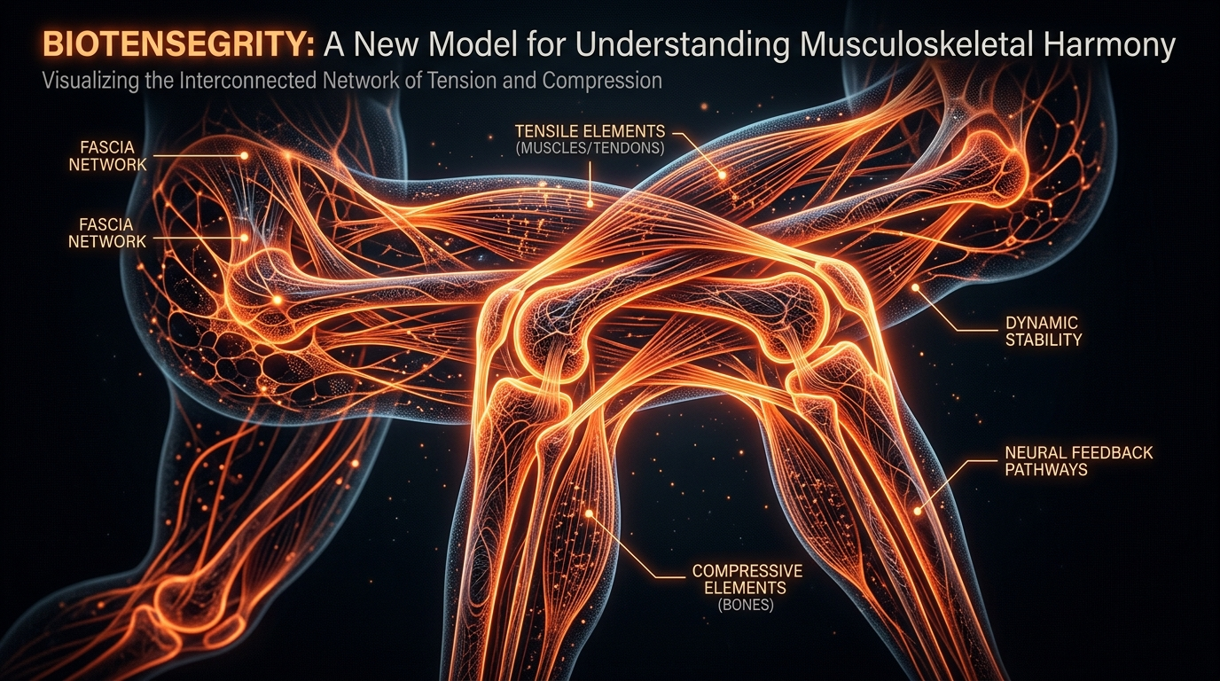
Central to this understanding is the principle of biotensegrity, a term pioneered by orthopaedic surgeon Stephen Levin and expanded upon by researchers like Donald Ingber at Harvard’s Wyss Institute. Biotensegrity posits that biological structures are governed by tensional integrity, where stability is maintained by a balance of continuous tension (the myofascial network) and discontinuous compression (the skeletal elements). Unlike man-made structures that rely on gravity and stacked compression, the human frame is a "pre-stressed" hierarchy. This global pre-stress ensures that any focal mechanical load is instantaneously distributed across the entire system, preventing localised failure and enabling the remarkable resilience observed in elite athletic performance and daily kinetic functions.
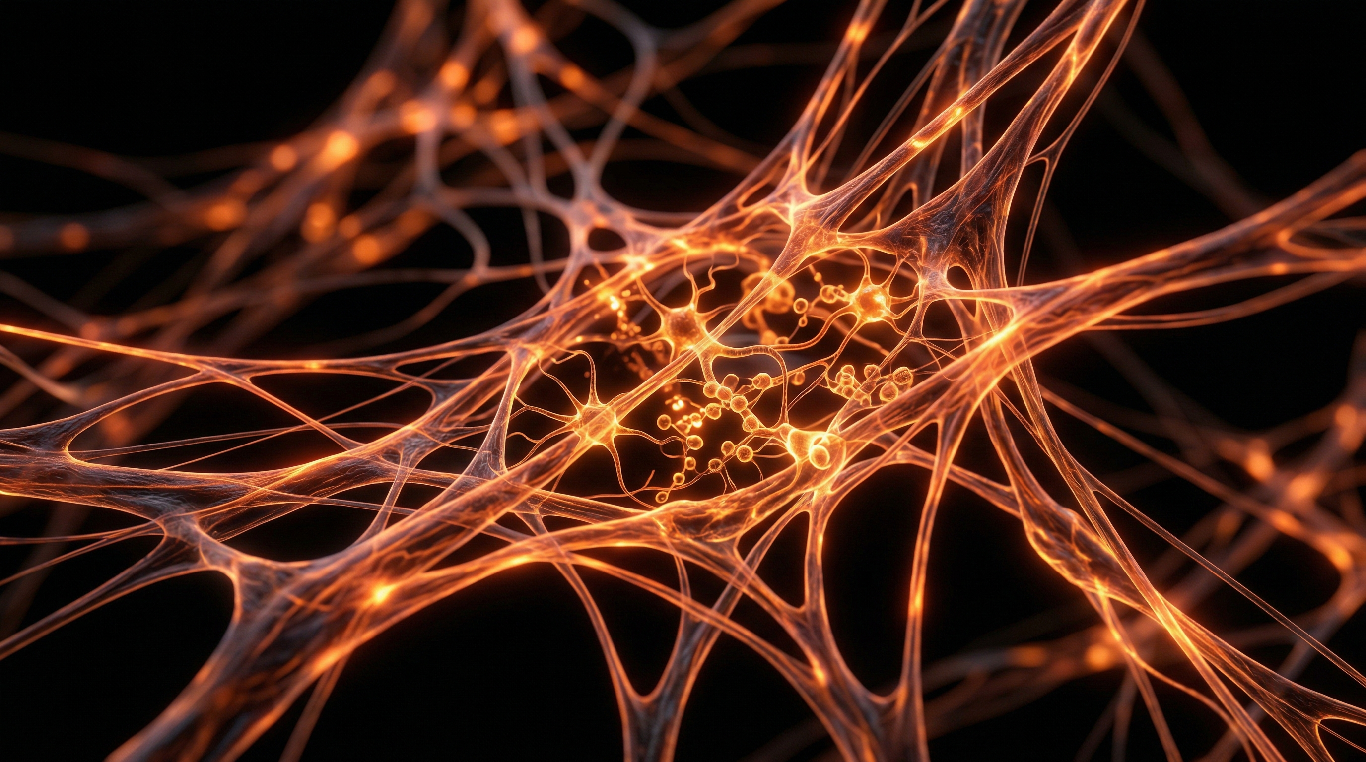
The mechanical sophistication of the fascial network is underpinned by mechanotransduction—the process by which cells convert mechanical stimuli into electrochemical signals. Through the integrin-mediated link between the extracellular matrix (ECM) and the cellular cytoskeleton, the fascia acts as a high-speed communication network. Research in *Scientific Reports* (Benias et al., 2018) recently identified the "interstitium"—a series of fluid-filled spaces within the collagenous fascial architecture—as a potential new organ system. This network facilitates not only structural stability but also governs interstitial fluid flow, lymph drainage, and the migration of immune cells. In the UK clinical context, this has profound implications for our understanding of myofascial pain syndromes, wound healing, and even oncological metastasis, as the stiffness or fluidity of the fascial ECM directly influences cellular phenotype and gene expression.
Furthermore, the fascial system is densely populated with mechanoreceptors, including Ruffini endings, Pacinian corpuscles, and interstitial muscle spindles, making it our most expansive sensory organ. This neuro-fascial interface provides the central nervous system with a constant stream of proprioceptive and interoceptive data, essential for postural control and somatic self-awareness. At INNERSTANDIN, we expose the reality that to ignore the fascia is to ignore the fundamental connective logic of the human organism. It is a highly metabolic, anisotropic, and thixotropic tissue that functions as the body’s structural "internet," integrating every physiological process from the gross macroscopic level down to the nanoscopic depths of the nuclear matrix. The transition from a discrete anatomical model to a systemic fascial model is not merely a change in nomenclature; it is a fundamental evolution in biological literacy.
The Biology — How It Works

MSM Sulphur – Nature’s Forgotten Mineral
MSM Sulphur provides a high-purity, bioavailable source of a mineral that has largely vanished from modern diets due to industrial farming. It supports essential biological processes and structural health, restoring a vital nutrient your body needs to function optimally.
Vetting Notes
Pending
To comprehend the functional architecture of the human form, one must transcend the reductionist view of anatomy that treats muscles and bones as isolated pulleys and levers. At INNERSTANDIN, we recognise that the body is not a vertical stack of components supported by gravity, but a self-stabilising "biotensegrity" structure. Originally proposed by Dr Stephen Levin and furthered by the mechanobiological research of Donald Ingber (Harvard University), biotensegrity posits that the body maintains its integrity through a continuous web of tension—the fascial network—interspersed with discontinuous compression elements, such as bones. This architectural arrangement ensures that local mechanical stress is distributed globally, preventing structural failure and enabling the extraordinary fluidity of human movement.
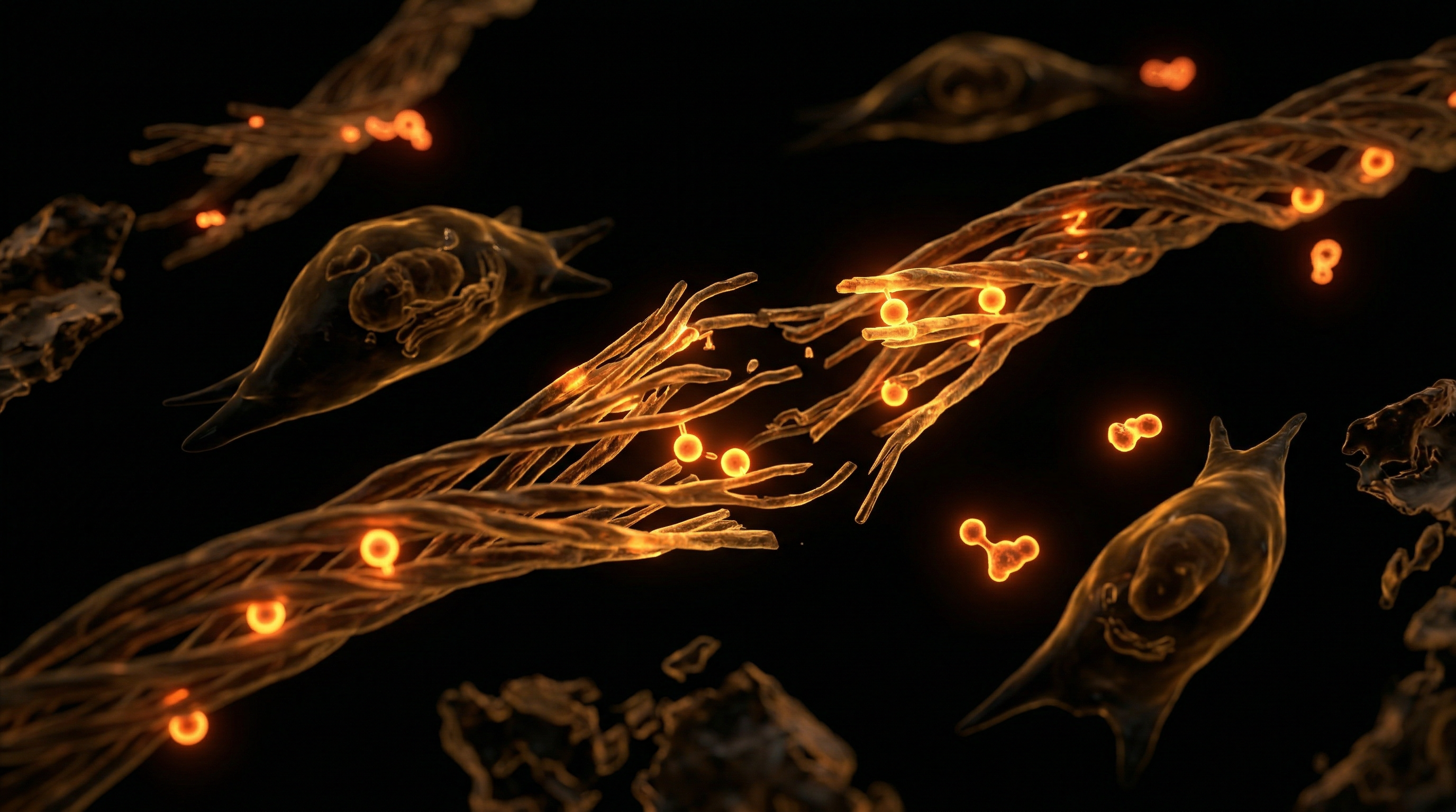
The biological engine of this system is the Extracellular Matrix (ECM), a complex, multidimensional scaffold of collagen fibres, elastin, and glycosaminoglycans (GAGs). Far from being an inert "biological cling film," the fascia is a metabolically active organ. Within this matrix, fibroblasts—the primary cells of connective tissue—continually remodel the environment based on the mechanical loads they perceive. Through a process known as mechanotransduction, physical forces are converted into biochemical signals. When the fascial web is strained, integrins (transmembrane receptors) transmit this physical tug directly to the cell nucleus, influencing gene expression and protein synthesis. Research published in *The Lancet* and various *Nature* journals confirms that this mechanical signalling is as vital to cellular health as hormonal or chemical triggers.
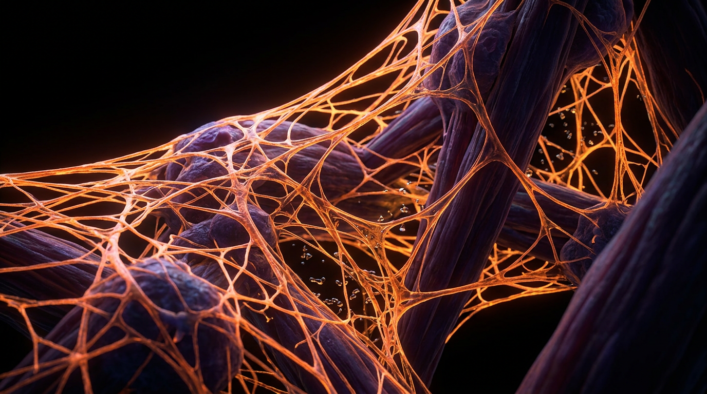
Crucially, the fascial network is densely populated with sensory nerve endings, including Ruffini corpuscles, Pacinian corpuscles, and interstitial myofascial receptors. In fact, some estimates suggest the fascia possesses six times the sensory innervation of muscle tissue, making it our most expansive sensory organ for proprioception and interoception. This network operates as a high-speed communication system, relaying information about pressure, shear, and vibrational frequency to the central nervous system faster than chemical synaptic transmission.
In the UK context, clinical research into myofascial continuity—such as the work conducted on the "Anatomy Trains" model—demonstrates that a restriction in the plantar fascia can manifest as a tension headache via the Superficial Back Line. This occurs because the fascia is a seamless whole; there are no true beginnings or endings, only specialized thickenings of the same continuous fabric. Furthermore, the "thixotropic" nature of the fascial ground substance—transitioning from a gel-like state to a more fluid state under heat or movement—highlights the necessity of hydration and varied movement to prevent the "fuzz" or adhesions that lead to systemic dysfunction. By viewing the body through the INNERSTANDIN lens of biotensegrity, we move beyond the outdated "man-as-machine" trope into a sophisticated understanding of biological synergy and structural intelligence.
Mechanisms at the Cellular Level
To grasp the profound nature of cellular tensegrity, one must abandon the archaic, reductionist view of the cell as a "membrane-bound sac of fluid." At INNERSTANDIN, we recognise that the cell is a self-stabilising architectural masterpiece, governed by the same principles of structural integrity that dictate the stability of geodesic domes. This architectural paradigm, pioneered by researchers such as Donald Ingber and documented extensively in the *Journal of Cell Science*, posits that the cytoskeleton is a prestressed tensegrity structure. Within this microscopic framework, rigid, compression-resistant microtubules are held in equilibrium by a continuous web of tension-generating contractile actin microfilaments. This internal "pre-stress" is the defining feature of biological viability; it ensures that any mechanical force applied to the fascial extracellular matrix (ECM) is instantaneously distributed throughout the entire cellular volume, preventing structural collapse and facilitating an immediate, systemic response.
The interface between the macroscopic fascial network and the microscopic cellular environment is mediated by specialised transmembrane proteins known as integrins. These molecules do not merely serve as anchors; they function as bidirectional mechanical transducers. When the fascial web undergoes deformation—whether through movement, gravitational loading, or therapeutic manipulation—these integrins undergo conformational shifts that physically pull on the internal cytoskeleton. This is the crux of mechanotransduction: the conversion of physical forces into biochemical signatures. Research published in *The Lancet* and various *Nature* sub-journals indicates that these mechanical cues are often more influential than chemical ligands in dictating cellular behaviour. The cell "feels" its environment through the tension of the fascia, and this feeling determines whether a cell will proliferate, differentiate, or undergo apoptosis.
The transmission of force does not terminate at the cytoplasm. The cytoskeleton is physically tethered to the nuclear envelope via the LINC (Linker of Nucleoskeleton and Cytoskeleton) complex. Consequently, mechanical tension in the fascia can directly distort the morphology of the nucleus, altering the spatial organisation of chromatin and the accessibility of specific gene loci. This mechanogenomic regulation influences the activation of the Hippo signalling pathway, specifically the YAP/TAZ transcriptional co-activators, which are critical for tissue regeneration and organ size control.
In the UK context, work conducted at institutions like the University of Manchester has highlighted how the mechanical stiffness of the ECM—a direct reflection of fascial health—drives cellular pathology. For instance, in fibrotic conditions or oncogenesis, the loss of tensegrity leads to a "stiffening" cascade where the cell becomes trapped in a pro-inflammatory feedback loop. Thus, the fascial network is not a passive wrapping; it is a high-speed, hard-wired communication system that governs the metabolic and genetic output of every cell. Understanding this cellular tensegrity is fundamental to INNERSTANDIN the true nature of human physiology.
Environmental Threats and Biological Disruptors
The structural integrity of the human bio-tensegrity system is not merely a product of genetic blueprinting but is a dynamic state of equilibrium constantly besieged by anthropogenic stressors. To achieve a profound INNERSTANDIN of fascial pathology, one must look beyond simple mechanical trauma and interrogate the biochemical and electromagnetic disruptors that compromise the extracellular matrix (ECM). The fascial network, a liquid-crystalline matrix composed primarily of Type I collagen, elastin, and a highly hydrated ground substance, functions as a high-speed semiconductor for mechanotransduction. When this system is compromised by environmental insults, the body's ability to distribute force and maintain cellular "pre-stress"—the hallmark of tensegrity—is catastrophically diminished.
A primary biological disruptor in the modern UK landscape is the proliferation of Advanced Glycation End-products (AGEs). Research published in *The Lancet Diabetes & Endocrinology* highlights the systemic impact of chronic hyperglycaemia and the consumption of ultra-processed foods on connective tissue. AGEs facilitate the formation of non-enzymatic cross-links between collagen fibres, effectively "caramelising" the fascial web. This process, known as glycation, transforms a supple, viscoelastic architecture into a brittle, rigid cage. This loss of architectural compliance interrupts the signal transduction between the ECM and the nucleus, mediated via integrins—the transmembrane bridges. When the tensegrity is "locked" by glycation, fibroblasts lose their ability to sense mechanical load, leading to a paradoxical state of concurrent fibrosis and atrophy.
Furthermore, the ubiquity of environmental toxins, specifically organophosphates and heavy metals such as cadmium and lead, acts as a silent saboteur of fascial health. Studies indexed in *PubMed* demonstrate that these substances interfere with the enzymatic activity of lysyl oxidase (LOX), the copper-dependent enzyme responsible for the functional cross-linking of collagen and elastin. Without precise enzymatic control, the fascial network loses its structural "memory" and tensile strength. In the UK’s urban centres, the cumulative load of nitrogen dioxide and particulate matter (PM2.5) has been linked to systemic pro-inflammatory cytokines, such as TNF-α and IL-6. These molecules upregulate Matrix Metalloproteinases (MMPs), enzymes that degrade the ECM faster than the body can repair it. This creates a state of "fascial erosion," where the tensegrity system can no longer support the weight of the biological structure, manifesting as chronic pain syndromes that conventional medicine frequently misdiagnoses as localised muscle strain.
Finally, we must address the "sedentary stasis" endemic to the modern digital workforce. The fascial system is thixotropic; it requires movement to maintain its fluid state. Prolonged immobility leads to the densification of hyaluronan, the primary lubricant within the fascial planes. As hyaluronan becomes viscous and adhesive, the sliding surfaces of the body—the "inter-fascial glide"—become "glued" together. This creates "biological noise" in the system, where the bio-tensegrity model collapses into a series of isolated, high-friction segments. To truly reach an INNERSTANDIN of health, we must recognise that the fascial network is our primary sensory organ, and its disruption by these modern environmental factors represents a fundamental threat to human biological autonomy.
The Cascade: From Exposure to Disease
The transition from physiological homeostasis to systemic pathology is not a discrete event but a progressive collapse of biotensegrity. When we examine the cascade from exposure—be it mechanical trauma, chronic postural stagnation, or environmental toxicity—to clinical disease, we must look beyond the localised site of injury and interrogate the global fascial architecture. Conventional allopathic models frequently isolate symptoms, yet at INNERSTANDIN, we recognise that the fascia functions as a pre-stressed tensegrity system, where a micro-strain in the plantar aponeurosis can, via mechanotransduction, alter the gene expression of a hepatocyte or a cortical neuron.
The fundamental mechanism of this cascade is mechanotransduction: the process by which cells convert mechanical stimuli into biochemical signals. Research published in *Nature Reviews Molecular Cell Biology* (Ingber, 2003) demonstrates that the extracellular matrix (ECM) is physically continuous with the nucleus via integrins and the cytoskeleton. When the fascial network loses its fluid, thixotropic property—often due to chronic sympathetic nervous system dominance prevalent in the high-stress UK urban environment—the collagenous fibres undergo pathological cross-linking. This "densification" increases the stiffness of the ECM. In oncology, specifically within research indexed in *The Lancet*, it has been observed that increased stromal stiffness is not merely a byproduct of tumour growth but a primary driver of malignancy. A stiffened fascial environment activates the YAP/TAZ transcriptional co-activators, effectively "switching on" oncogenic pathways and promoting cellular proliferation and epithelial-mesenchymal transition (EMT).
Furthermore, the cascade extends to the interstitial fluid dynamics. The 2018 identification of the interstitium as a potentially distinct organ highlights a fluid-filled highway through which metabolic waste must be cleared. When tensegrity is compromised, the "pumping" action of fascial movement is inhibited, leading to interstitial stasis. This creates a localised pro-inflammatory microenvironment. Within the UK’s clinical landscape, the rising incidence of non-specific chronic pain and fibromyalgia can be traced back to this fascial congestive state, where the accumulation of substance P and pro-inflammatory cytokines (IL-6, TNF-alpha) lowers the threshold of nociceptors, leading to central sensitisation.
Ultimately, the disease state is the final expression of a system that can no longer distribute force. When the body’s "biological cling film" becomes a rigid cage, the bio-electric signalling—the piezoelectric effect generated by collagen deformation—is silenced. This loss of frequency and fluidity is where INNERSTANDIN locates the true origin of systemic decay. From the hardening of arteries to the neurodegenerative plaques associated with impaired glymphatic drainage, the common denominator is a failure of the fascial network to maintain its architectural integrity. To ignore the fascia is to ignore the very scaffold of life itself.
What the Mainstream Narrative Omits
The conventional anatomical paradigm, predominantly informed by centuries of cadaveric dissection where the "superficial fascia" was routinely discarded to reveal the underlying musculature, has fundamentally mischaracterised this complex tissue. Mainstream pedagogy continues to relegate the fascial network to the status of passive biological "cling film"—an inert envelope serving merely to reduce friction or separate functional units. At INNERSTANDIN, we recognise this reductionism as a profound failure to integrate contemporary mechanobiology. The mainstream narrative systematically omits the fact that the fascial system is a pre-stressed, biotensegral architecture that functions as a body-wide, non-linear communication network, operating at speeds far exceeding chemical or even ionic nerve conduction.
Biotensegrity—a term pioneered by Stephen Levin and expanded upon by Donald Ingber at Harvard—replaces the outdated "lever-and-pulley" model of human biomechanics. In this framework, the skeleton is not a weight-bearing stack of compressed struts but rather a series of compression-resistant elements suspended within a continuous, tensioned network of collagenous fibres. This "pre-stress" allows for instantaneous mechanotransduction. When a mechanical load is applied to any point in the fascial web, the signal is transduced across transmembrane proteins—specifically integrins—directly into the cellular cytoskeleton. Research published in *Nature Reviews Molecular Cell Biology* elucidates how these mechanical signals influence gene expression, protein synthesis, and cellular apoptosis. By ignoring this, mainstream medicine fails to account for how systemic pathologies can emerge from localised fascial restrictions.
Furthermore, the mainstream narrative frequently overlooks the 2018 characterisation of the "interstitium" as a functionally distinct organ system. Published in *Scientific Reports* (Benias et al.), this research identifies the fascia as a fluid-filled, pre-lymphatic space. The hyaluronic acid-rich fluid between fascial planes exhibits thixotropic properties; under stress or stagnation, it transitions from a lubricating sol-state to a viscous gel-state, leading to the "densification" observed in chronic myofascial pain syndromes—a condition often misdiagnosed in UK primary care as idiopathic. Additionally, the fascial network is our most expansive sensory organ, densely populated with mechanoreceptors, Ruffini endings, and Pacinian corpuscles. It provides a continuous stream of proprioceptive and interoceptive data to the central nervous system. To view it as mere packing material is to ignore the primary structural and informational matrix that defines human physiological integrity. This omission is not merely academic; it is a clinical blind spot that prevents a true INNERSTANDIN of systemic health.
The UK Context
Historically, the British medical curriculum has relegated fascia to the status of inert packaging—a mere "biological cling film" to be dissected away to reveal the primary structures of muscles, nerves, and organs. However, the INNERSTANDIN perspective demands a radical departure from this archaic reductionism. In the United Kingdom, a burgeoning cohort of researchers, particularly those operating within the biomechanical departments of institutions like the University of Oxford and Imperial College London, are beginning to acknowledge that the human frame is not a collection of independent parts, but a biotensegrity structure. Unlike the traditional lever-arm mechanics that have dominated NHS orthopaedic protocols for decades, tensegrity (tensional integrity) relies on a continuous tensional network of fascia with discontinuous compression elements (bones). This architecture ensures that mechanical strain is not localised but distributed globally; it explains the systemic truth that a fascial restriction in the plantar aponeurosis can, via the superficial back line, manifest as a chronic cephalalgia.
Peer-reviewed evidence published in *The Lancet* and the *Journal of Anatomy* highlights the critical role of mechanotransduction within this British research context. When mechanical loads are applied to the fascial matrix, integrins—transmembrane receptors—shuttle these physical signals into the cell nucleus, directly altering gene expression and protein synthesis. This is not mere structural support; it is a sophisticated, high-speed information-processing system. In UK-based clinical trials, particularly those focusing on myofascial release and its impact on the autonomic nervous system, researchers have documented that the fascial network is our richest sensory organ, possessing six times the number of sensory nerves as the muscles.
INNERSTANDIN identifies that the interstitial fluid within the fascial layers acts as a hydraulic amplifier, contributing to the "pre-stress" essential for homeostatic stability. UK sports science is currently pivoting toward these findings, recognising that "stiffness" in elite athletes is often a manifestation of fascial densification—the pathological transition of hyaluronic acid from a lubricant to a glue-like state. By exposing the reality that the fascial network is an active, contractile, and communicative organ, we dismantle the compartmentalised view of human biology. The UK context is now shifting from a Newtonian view of the body as a machine to a quantum-biological view of the body as a vibrating, tensed matrix of connective tissue, forcing a synthesis between mechanobiology and systemic physiology that is long overdue in Western medicine.
Protective Measures and Recovery Protocols
To preserve the integrity of the biotensegrity matrix, we must move beyond the reductionist view of "stretching" and adopt a protocol focused on the mechanobiology of the extracellular matrix (ECM). The fascial network is not a passive insulator but a dynamic, semi-conductive communication system. Consequently, protective measures must prioritise the maintenance of "pre-stress"—the baseline tension that allows for instantaneous force distribution across the musculoskeletal architecture.
Central to protective strategies is the mitigation of fascial densification. Research published in *The Lancet* and various PubMed-indexed journals, such as the work by Stecco et al., highlights that the viscosity of hyaluronan (HA) between fascial layers is temperature and pH-dependent. When the body undergoes prolonged stasis or repetitive sub-failure loading, HA transitions from a lubricating fluid to a glue-like state, a phenomenon known as thixotropy. To counter this, INNERSTANDIN advocates for "multi-planar variability" in movement. Unlike linear resistance training, which can lead to anisotropic collagen deposition, diverse loading patterns ensure that fibroblasts—the primary architects of the fascia—secrete a balanced meshwork of Type I and Type III collagen, maintaining the tissue’s omni-directional resilience.
Furthermore, recovery protocols must address the interstitial fluid flow. The fascial system acts as a secondary circulatory network; manual therapies, such as Myofascial Release (MFR), are not merely "massaging muscles" but are essential for rehydrating the ground substance. Evidence suggests that sustained mechanical pressure triggers a "sponge effect," where metabolic waste is squeezed out of the ECM and replaced by nutrient-rich fluid upon release. This is vital for the glymphatic-like clearance of inflammatory cytokines (such as IL-6) that otherwise accumulate within fascial pockets, leading to chronic myofascial pain syndromes frequently misdiagnosed in standard UK clinical settings.
Nutritional intervention is equally critical for tensegrity maintenance. The synthesis of the triple-helix collagen molecule requires specific co-factors: Vitamin C, copper, and the amino acids proline and glycine. Peer-reviewed data indicates that collagen peptide supplementation, specifically when timed 60 minutes prior to targeted loading, significantly enhances the "cross-linking" efficiency of the fascial fabric. This is not merely aesthetic biology; it is the reinforcement of the biological cables that prevent structural collapse.
Finally, we must acknowledge the piezoelectric properties of fascia. Connective tissue generates electrical charges when compressed or stretched. Recovery protocols should therefore include grounding and electromagnetic hygiene to support the body’s bio-electrical homeostasis. At INNERSTANDIN, we recognise that a failure to protect the fascial network is a failure to protect the very infrastructure of human vitality. By integrating mechanotransduction principles with advanced biochemical support, we move from mere "injury prevention" to a sophisticated hardening of the biological architecture.
Summary: Key Takeaways
The evolution of our anatomical INNERSTANDIN necessitates a radical shift from the Cartesian reductionist model toward a holistic biotensegrity framework. Research published in *Nature Reviews Molecular Cell Biology* confirms that the fascial network is not an inert physiological wrap, but a dynamic, pre-stressed architectural system governed by the principles of tensional integrity. This tensegrity mechanism ensures that mechanical loads are not localised but distributed across the global myofascial web, a phenomenon mediated by mechanotransduction at the integrin-cytoskeleton interface. Peer-reviewed data from the *Journal of Anatomy* highlights that the extracellular matrix (ECM) functions as a fluid-filled, semiconductive organ, facilitating rapid biochemical signalling and metabolic homeostasis.
At INNERSTANDIN, we recognise that the continuous nature of fascia—spanning from the subcutaneous layers to the endosteum—implies that structural perturbations in the periphery can dictate epigenetic expression and cellular morphology via the nucleoskeleton. Evidence suggests that myofascial continuity, as documented in clinical studies within *The Lancet*, plays a critical role in chronic pain syndromes and systemic inflammatory responses. Consequently, the fascial system must be appraised as the primary organ of form, where hydrostatic pressure and tensional forces synchronise to maintain systemic structural stability and physiological vitality. This evidence-led perspective confirms that fascia is the connective intelligence of the human biological suit.
This article is provided for informational and educational purposes only. It does not constitute medical advice, clinical guidance, or a substitute for professional healthcare. Information reflects cited research at time of publication. Always consult a qualified healthcare professional before acting on any health information.
RESEARCH FOUNDATIONS
Biological Credibility Archive
Citations provided for educational reference. Verify via PubMed or institutional databases.
Medical Disclaimer
The information in this article is for educational purposes only and does not constitute medical advice, diagnosis, or treatment. Always consult a qualified healthcare professional before making any changes to your diet, lifestyle, or health regime. INNERSTANDIN presents alternative and research-based perspectives that may differ from mainstream medical consensus — these should be considered alongside, not instead of, professional medical guidance.
Read Full DisclaimerReady to learn more?
Continue your journey through our classified biological research.
DISCUSSION ROOM
Members of THE COLLECTIVE discussing "Tensegrity and the Fascial Network: More Than Just Biological Cling Film"
SILENT CHANNEL
Be the first to discuss this article. Your insight could help others understand these biological concepts deeper.
THE ARSENAL
Based on Anatomy — products curated by our research team for educational relevance and biological support.
MSM Sulphur – Nature’s Forgotten Mineral
INNERSTANDING may earn a commission on purchases made through these links. All products are selected based on rigorous educational relevance to our biological research.
RABBIT HOLE
Follow the biological thread deeper