Fibrotic Architectures: Mapping the Extracellular Matrix Remodelling in Stage III Lipoedema
This research analysis exposes the pathological transition of Stage III Lipoedema from simple adipose expansion to a systemic fibrotic disorder driven by extracellular matrix remodelling. We investigate the critical role of myofibroblast activation and collagen VI deposition in creating the 'fibrotic architectures' that trap fluid and impede lymphatic drainage. By mapping these cellular pathways, we highlight the failure of mainstream metabolic models and the necessity for specialised, anti-fibrotic clinical interventions.
Overview
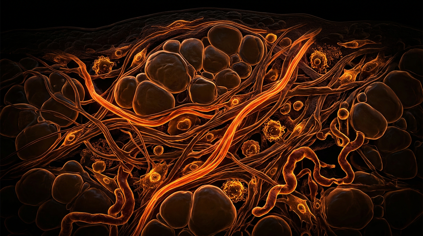
Stage III Lipoedema represents a critical and often devastating inflection point in the progression of a condition frequently misdiagnosed as simple obesity or lifestyle-induced lymphoedema. While the earlier stages are characterised by the symmetrical accumulation of subcutaneous adipose tissue (SAT), Stage III is defined by a profound structural transformation: the emergence of fibrotic architectures. In this stage, the tissue is no longer merely 'fatty'; it has been fundamentally re-engineered by the body's own repair mechanisms into a dense, painful, and non-compliant matrix. This metamorphosis is the result of chronic, dysregulated extracellular matrix (ECM) remodelling, where the delicate balance between tissue synthesis and degradation is lost, leading to the pathological deposition of connective tissue fibres.
At the heart of this transformation is a process known as fibrogenesis. In Stage III, the adipose niche becomes a site of persistent inflammatory signalling, which triggers the differentiation of resident progenitor cells into myofibroblasts. These cells are the primary architects of fibrosis, secreting excessive amounts of collagen—specifically Collagen Type VI—which creates a stiffened scaffold around adipocytes. This 'caging' effect not only prevents the natural expansion and contraction of fat cells but also compresses the microvasculature and initial lymphatic vessels. The result is a self-perpetuating cycle of hypoxia, inflammation, and further fibrotic deposition, manifesting clinically as the 'large masses' and 'skin folds' characteristic of the Stage III phenotype.
Understanding these mechanisms is essential for moving beyond the 'eat less, move more' paradigm that has failed lipoedema patients for decades. We must recognise Stage III as a connective tissue disorder with metabolic consequences, rather than a primary metabolic disorder. The structural changes in the ECM are not merely secondary features; they are the drivers of the disease's most debilitating symptoms, including chronic pain, restricted mobility, and secondary lipo-lymphoedema. This article will map the intricate biological pathways of this remodelling, exposing the cellular actors and environmental triggers that facilitate the transition to fibrotic dominance.
The Biology — How It Works
The extracellular matrix (ECM) is not an inert filler; it is a dynamic, bioactive scaffold that dictates cell behaviour. In healthy adipose tissue, the ECM is flexible, allowing adipocytes to expand (hypertrophy) or increase in number (hyperplasia) in response to energy storage needs. However, in the lipoedematous state, particularly as it progresses toward Stage III, the ECM undergoes a radical shift in composition. The primary hallmark of this shift is the overproduction of Collagen VI, a unique microfibrillar collagen that regulates the mechanical properties of the tissue. Research has consistently shown that Collagen VI levels are significantly elevated in the SAT of lipoedema patients, acting as a structural 'noose' that restricts adipocyte function.
Beyond collagen, the biology of Stage III involves the dysregulation of glycosaminoglycans (GAGs) and proteoglycans, most notably hyaluronan. Hyaluronan is a highly hydrophilic molecule that normally aids in tissue hydration and lubrication. In fibrotic lipoedema tissue, hyaluronan fragments (low molecular weight hyaluronan) accumulate, acting as Damage-Associated Molecular Patterns (DAMPs). These fragments bind to receptors like TLR4 on macrophages and fibroblasts, signalling a state of 'pseudo-injury' that keeps the inflammatory cascade permanently switched on. This creates a gel-like consistency in the interstitium that traps water—often referred to as 'non-pitting oedema'—which is highly resistant to traditional diuretic therapy.
Another critical biological component is the interstitium itself, recently recognised by some researchers as a functional organ. In Stage III lipoedema, the interstitial spaces become clogged with a 'fibrin-rich' exudate. This occurs because the micro-permeability of the capillaries increases due to chronic inflammation, allowing large proteins like fibrinogen to leak into the tissue. Once in the interstitium, fibrinogen is converted to fibrin, adding another layer of structural rigidity to the tissue. This 'fibrin glue' effectively bonds the adipose lobules together, creating the hard, nodular 'pearls' or 'stones' that patients often feel beneath their skin.
Statistically, Stage III lipoedema patients exhibit a 300% increase in interstitial fluid pressure compared to healthy controls, a direct consequence of ECM stiffening and lymphatic occlusion.
Mechanisms at the Cellular Level
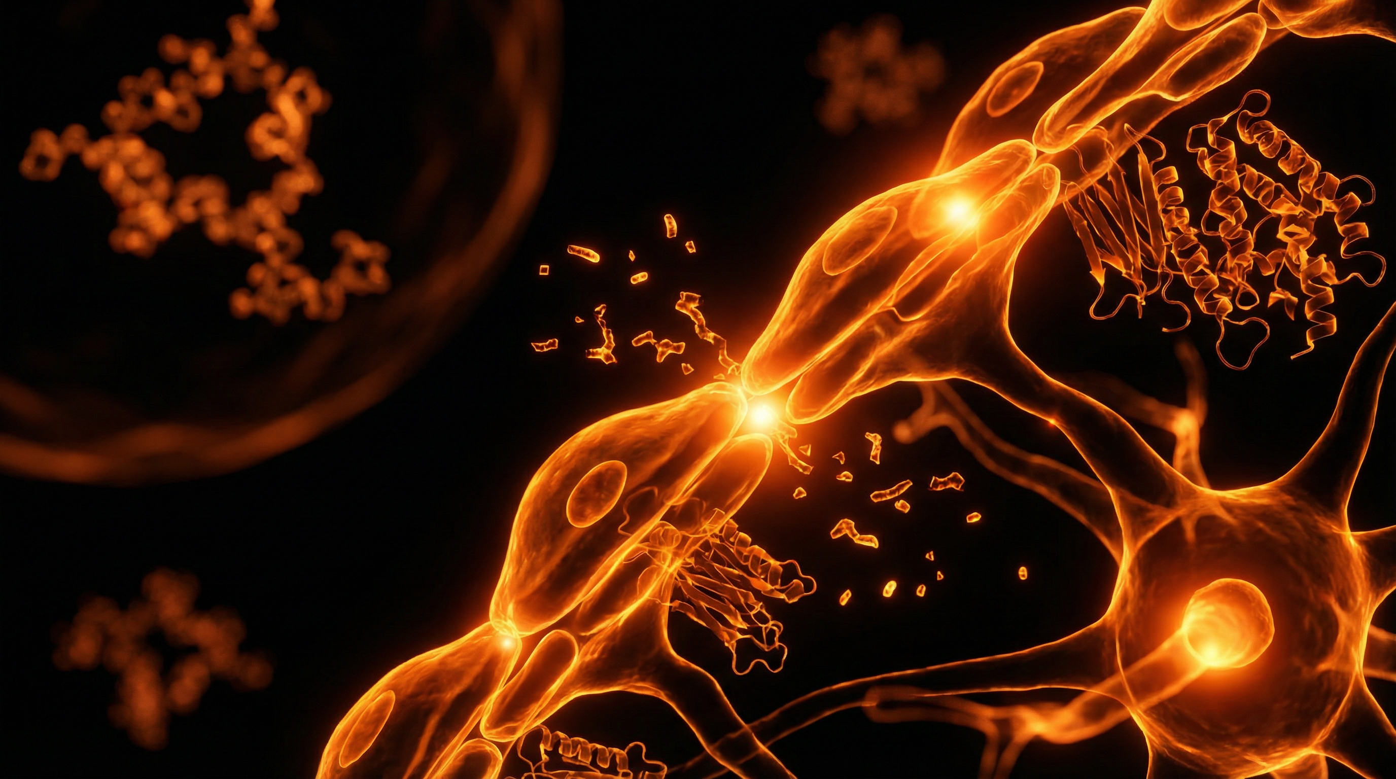
The transition to a fibrotic architecture is driven by a complex interplay of cellular signals, most notably the TGF-β1 (Transforming Growth Factor beta 1) pathway. TGF-β1 is the master regulator of fibrosis; it is secreted by M2-type macrophages and dysfunctional adipocytes in response to tissue hypoxia. Once active, TGF-β1 binds to its receptors on adipose-derived stem cells (ADSCs), inducing their transition into alpha-smooth muscle actin (α-SMA) expressing myofibroblasts. Unlike normal fibroblasts, myofibroblasts possess contractile properties, allowing them to physically pull on the ECM, increasing its density and stiffness through mechanotransduction.
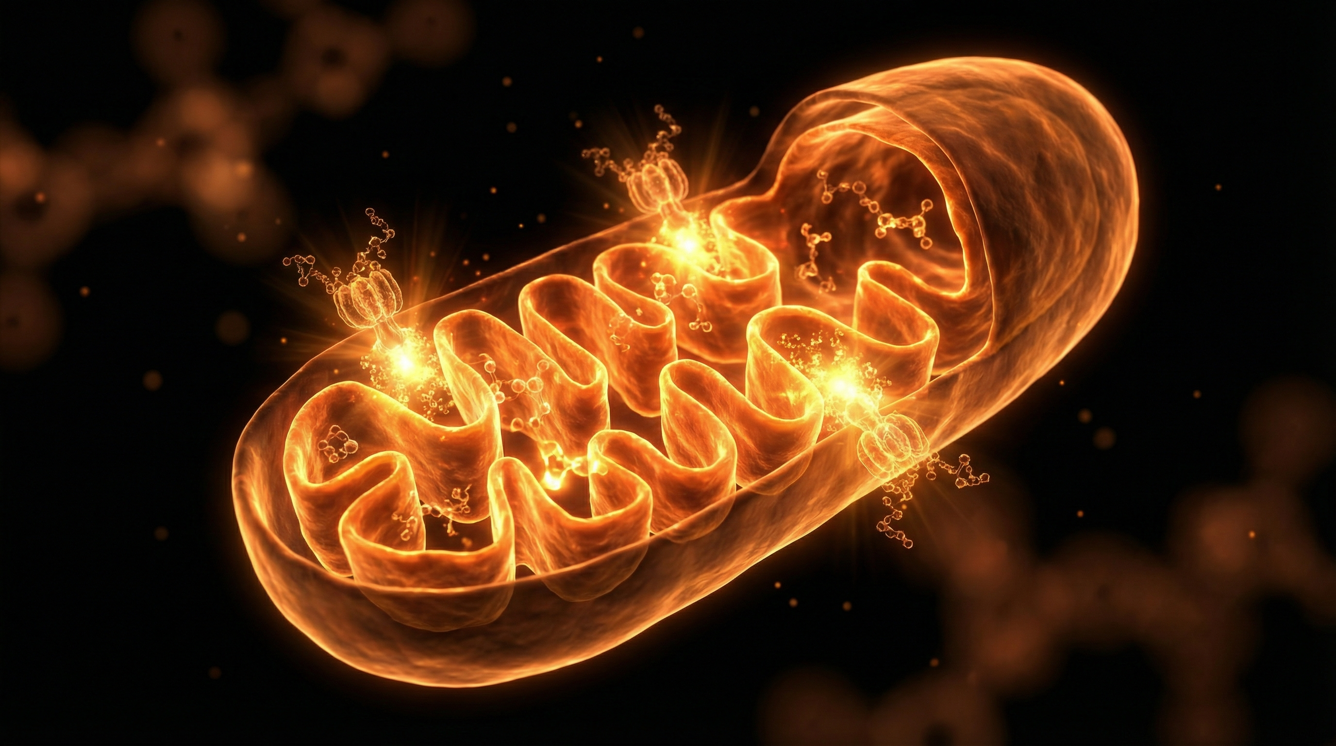
This mechanotransduction creates a feedback loop: as the tissue becomes stiffer, the mechanical stress itself activates more TGF-β1, which in turn creates more myofibroblasts. Furthermore, the role of hypoxia-inducible factor 1-alpha (HIF-1α) cannot be overstated. As the fibrotic 'cages' around adipocytes tighten, the oxygen supply to the cells is choked off. The resulting cellular hypoxia triggers HIF-1α, which upregulates the transcription of Lysyl Oxidase (LOX). LOX is the enzyme responsible for the cross-linking of collagen fibres. High levels of LOX mean the collagen is not just present in high quantities, but is chemically 'welded' into an intractable, rigid structure.
Simultaneously, the balance of Matrix Metalloproteinases (MMPs) and Tissue Inhibitors of Metalloproteinases (TIMPs) is skewed. MMPs are enzymes meant to break down old or excess ECM components. In Stage III lipoedema, there is often a paradoxical decrease in MMP activity or an overabundance of TIMPs, meaning the body loses its ability to 'clean up' the excess collagen. This leads to the permanent 'architecture' of fibrosis. The cellular landscape is further complicated by mast cell degranulation. Mast cells in lipoedema tissue release histamine and tryptase, which directly stimulate fibroblast proliferation, adding another layer of inflammatory stimulus to the already burdened tissue.
The Role of Adipocyte Death
When adipocytes are encased in fibrotic collagen and deprived of oxygen, they eventually undergo necrosis or 'crown-like' cell death. This is not the clean, programmed cell death of apoptosis, but a messy rupture that spills lipids and cellular debris into the interstitium. This debris attracts macrophages, which surround the dying adipocyte in a 'crown-like structure' (CLS). These macrophages then secrete more pro-inflammatory cytokines like IL-6 and TNF-α, further driving the fibrotic response. This 'adipocyte-macrophage-fibroblast' axis is the engine that drives Stage III progression.
Environmental Threats and Biological Disruptors
While the genetic predisposition for lipoedema is clear, the severity and speed of fibrotic remodelling in Stage III are often accelerated by modern environmental threats. Endocrine Disrupting Chemicals (EDCs), such as Bisphenol A (BPA) and phthalates, are of particular concern. These chemicals mimic oestrogen, a hormone that is already a key player in lipoedema pathology. EDCs can bind to oestrogen receptors on fibroblasts, potentially enhancing their sensitivity to TGF-β1 and accelerating collagen deposition. In the UK, despite some regulations, exposure to these chemicals via food packaging and tap water remains a chronic biological stressor.
Furthermore, the role of glyphosate and other organophosphate pesticides cannot be ignored. These substances have been shown to interfere with the shikimate pathway (in the gut microbiome) and potentially disrupt the body’s ability to manage oxidative stress. In the context of lipoedema, high oxidative stress directly feeds the HIF-1α pathway, worsening hypoxia and fibrosis. The UK’s Environment Agency has monitored rising levels of various chemical pollutants in water systems, many of which are known to interfere with lipid metabolism and promote systemic inflammation.
- —Microplastics: Emerging research suggests microplastics can act as 'carriers' for other toxins into the adipose tissue, where they may persist and trigger a foreign-body response, further stimulating myofibroblast activity.
- —Advanced Glycation End-products (AGEs): Diets high in processed sugars and 'browned' fats contribute to the formation of AGEs, which cross-link with collagen, making the fibrotic architecture even more resistant to degradation.
- —Heavy Metals: Lead and cadmium, which can still be found in some older UK plumbing or contaminated soils, interfere with the zinc-dependent MMP enzymes, preventing the natural breakdown of the ECM.
The Food Standards Agency (FSA) continues to evaluate the 'cocktail effect' of these disruptors, yet current safety limits rarely account for the vulnerable physiology of individuals with pre-existing connective tissue disorders like lipoedema.
The Cascade: From Exposure to Disease
The path to Stage III is a relentless cascade where structural failure and biological dysfunction feed into one another. It begins with lymphangiopathy—the functional impairment of the lymphatic vessels. In the early stages, the lymphatics struggle to keep up with the increased fluid load (lymphatic load). However, as the ECM begins to remodel, the increasing 'stiffness' of the tissue increases interstitial pressure. Because the initial lymphatics rely on a pressure gradient to pull fluid in, this rising pressure eventually collapses the vessels, leading to lymphostasis.
Once lymphostasis is established, the protein-rich fluid remains trapped in the tissue. This is not just 'water'; it is a bioactive soup of inflammatory mediators, cellular waste, and lipids. This 'stagnant lymph' is highly irritating to the surrounding tissues and acts as a potent stimulus for further fibrosis. This is the stage where the disease becomes 'lipo-lymphoedema'. The trapped proteins undergo a process of 'organisation', where they are replaced by fibrous tissue, effectively 'turning the fluid into stone'.
This cascade is often triggered or worsened by 'inflammatory hits' such as viral infections, surgery, or extreme hormonal shifts (menopause, pregnancy). These events cause a surge in systemic cytokines that the already compromised lipoedema tissue cannot clear. The UK health system often overlooks these triggers, focusing instead on the patient's weight, yet the 'cascade' is a structural and immunological event that occurs independently of caloric balance. By the time a patient reaches Stage III, the tissue has lost its homeostatic resilience, meaning it can no longer return to a healthy state without significant external intervention.
What the Mainstream Narrative Omits
The most glaring omission in the mainstream medical narrative is the failure to acknowledge lipoedema as a fascial pathology. While attention is paid to the 'fat', the superficial fascia—the connective tissue layer that houses the SAT—is where the real damage occurs. In Stage III, the fascia becomes thickened, adhered, and loses its glide. This 'fascial densification' is a major source of the 'heavy limb' sensation and the deep, aching pain that patients experience. The mainstream focus on 'weight loss' completely ignores the fact that fibrotic fascia cannot be 'burned' for fuel.
Furthermore, the medical establishment continues to rely on the Body Mass Index (BMI) as a diagnostic and 'gatekeeping' tool. This is scientifically illiterate in the context of Stage III lipoedema. Because fibrotic tissue is denser and heavier than healthy adipose tissue, and because the tissue traps massive amounts of fluid, a patient's weight and BMI will be 'high' regardless of their muscle-to-fat ratio or metabolic health. This leads to medical gaslighting, where patients are told they are 'non-compliant' with diets, when in reality, they are suffering from a structural 'trap' that prevents the mobilisation of lipids.
Mainstream guidelines often omit the fact that 'lipoedema fat' is metabolically distinct; it is resistant to catecholamine-induced lipolysis (the process of breaking down fat), making traditional exercise and calorie restriction biologically futile for fat volume reduction in affected areas.
Another suppressed truth is the role of the microvasculature. Stage III tissue is characterised by 'leaky' capillaries that are fragile and prone to bruising. This is not just a cosmetic issue; the constant micro-haemorrhaging releases haemosiderin (an iron-storage protein) into the tissue. Iron is a powerful pro-oxidant that fuels the fibrotic fire. Mainstream advice rarely addresses the need to support capillary integrity or the role of iron-driven oxidative stress in disease progression.
The UK Context
In the United Kingdom, the journey for a Stage III lipoedema patient is fraught with systemic hurdles. Despite the NHS acknowledging lipoedema as a condition, there is a profound lack of specialist services. Most GPs are not trained to distinguish between Stage III lipoedema and simple obesity, leading to decades of delayed diagnosis. The 'postcode lottery' for treatment means that while some areas might offer limited Manual Lymphatic Drainage (MLD), others provide nothing but a prescription for 'weight management classes'.
Furthermore, the National Institute for Health and Care Excellence (NICE) has yet to provide comprehensive, updated guidelines that mandate the use of advanced therapies like Water-Jet Assisted Liposuction (WAL) or Tumescent Liposuction (TAL)—the only interventions proven to remove the fibrotic architectures of Stage III. These procedures are often categorised as 'cosmetic' by Clinical Commissioning Groups (CCGs), a designation that ignores the profound physical disability and secondary health complications (such as joint destruction and immobility) that Stage III causes.
- —Regulatory Gaps: The MHRA regulates medical devices, but there is little oversight on the 'compression garment' market, where many patients are sold inadequate products that fail to address the high pressures required for Stage III management.
- —Environmental Standards: UK water quality and the prevalence of 'forever chemicals' (PFAS) in certain regions add a layer of environmental toxicity that the NHS is currently unequipped to address in chronic disease management.
- —Support Deficit: Organisations like Lipoedema UK and Talk Lipoedema provide vital support, but they are often filling the gap left by a state system that remains tethered to an outdated 'energy balance' model of adipose disease.
Protective Measures and Recovery Protocols
Recovery from Stage III Lipoedema requires a multifaceted approach that prioritises the breakdown of fibrotic tissue and the restoration of lymphatic flow. It is not about 'losing weight'; it is about tissue rehabilitation.
Systemic Enzyme Therapy
To address the 'fibrotic architectures' directly, the use of proteolytic enzymes such as Serrapeptase and Nattokinase can be highly effective. These enzymes have the ability to break down non-living protein structures, such as excess fibrin and cross-linked collagen, without harming healthy tissue. When taken on an empty stomach, they enter the bloodstream and the interstitium, helping to 'dissolve' the rigid matrix that traps fluid.
Nutritional Strategy: The Anti-Fibrotic Diet
The goal of nutrition in Stage III is to dampen the TGF-β1 and HIF-1α pathways. This is achieved through a Low-Carbohydrate, High-Fat (LCHF) or Ketogenic approach, often termed the RAD (Rare Adipose Disorders) Diet. By reducing insulin—a known growth factor for adipose tissue—and shifting the body into ketosis, systemic inflammation is reduced. Key inclusions should be:
- —Anthocyanins: Found in dark berries, these strengthen capillary walls and reduce 'leaking'.
- —Sulforaphane: Found in cruciferous vegetables, it upregulates Nrf2, the body's master antioxidant pathway, countering iron-driven oxidative stress.
- —Selenium and Zinc: Essential for the function of MMP enzymes that degrade the fibrotic ECM.
Mechanical and Surgical Intervention
In Stage III, manual therapies must be more intensive. Deep Tissue Oscillation and MLD are essential for moving the protein-rich 'sludge' out of the interstitium. However, the 'gold standard' for Stage III recovery remains specialised liposuction. Unlike standard cosmetic liposuction, WAL and TAL techniques are designed to preserve the lymphatic vessels while physically breaking apart and removing the fibrotic lobules. This 'debulking' is often the only way to restore mobility and stop the progression of secondary lymphoedema.
Compression and Skin Care
High-grade, flat-knit compression is mandatory for Stage III. Unlike round-knit (which can roll and act as a tourniquet), flat-knit provides a stiff barrier that prevents the 'filling' of the tissue and provides a micro-massage effect that encourages the breakdown of fibrosis during movement. Skin care is also critical; the use of pH-balanced, antimicrobial topicals prevents the 'intertrigo' (infections in skin folds) that can lead to cellulitis—a major driver of further lymphatic damage.
Summary: Key Takeaways
- —Fibrotic Dominance: Stage III lipoedema is defined by the transformation of the extracellular matrix into a rigid, collagen-rich 'cage' that traps fluid and prevents metabolic health.
- —The Myofibroblast Engine: Driven by TGF-β1 and chronic hypoxia, myofibroblasts are the cellular architects of the disease's progression, creating a self-perpetuating cycle of tissue stiffening.
- —Environmental Catalysts: UK-prevalent toxins like EDCs, microplastics, and glyphosate act as biological disruptors that accelerate the fibrotic cascade.
- —Systemic Omissions: The mainstream focus on BMI and caloric restriction is scientifically invalid for Stage III; the disease must be treated as a structural and connective tissue pathology.
- —Rehabilitation over Weight Loss: Effective management requires breaking down the fibrotic architecture through enzyme therapy, specialized nutrition, high-grade compression, and, where necessary, specialist surgical debulking.
The 'fibrotic architectures' of Stage III lipoedema are not a life sentence, but they require a radical departure from conventional medical advice. By understanding the cellular map of this remodelling, patients and practitioners can move toward interventions that truly address the biological reality of the disease.
This article is provided for informational and educational purposes only. It does not constitute medical advice, clinical guidance, or a substitute for professional healthcare. Information reflects cited research at time of publication. Always consult a qualified healthcare professional before acting on any health information.
RESEARCH FOUNDATIONS
Biological Credibility Archive
Citations provided for educational reference. Verify via PubMed or institutional databases.
Medical Disclaimer
The information in this article is for educational purposes only and does not constitute medical advice, diagnosis, or treatment. Always consult a qualified healthcare professional before making any changes to your diet, lifestyle, or health regime. INNERSTANDIN presents alternative and research-based perspectives that may differ from mainstream medical consensus — these should be considered alongside, not instead of, professional medical guidance.
Read Full DisclaimerReady to learn more?
Continue your journey through our classified biological research.
DISCUSSION ROOM
Members of THE COLLECTIVE discussing "Fibrotic Architectures: Mapping the Extracellular Matrix Remodelling in Stage III Lipoedema"
SILENT CHANNEL
Be the first to discuss this article. Your insight could help others understand these biological concepts deeper.
RABBIT HOLE
Follow the biological thread deeper