Neuroanatomical Atrophy and Chronic EMF Exposure
Emerging research suggests high-frequency electromagnetic fields may impact the structural density of the hippocampus. This analysis focuses on the biological mechanisms of non-ionizing radiation on brain tissue.
Overview
The ubiquity of anthropogenic non-ionising electromagnetic fields (EMFs) in the United Kingdom has precipitated an unprecedented shift in the human bio-electric environment. While legacy regulatory frameworks—predominantly those guided by the International Commission on Non-Ionizing Radiation Protection (ICNIRP)—continue to focus almost exclusively on thermal effects, the burgeoning body of peer-reviewed literature indexed in PubMed and the Lancet identifies a far more insidious pathophysiological reality. Chronic exposure to Radiofrequency Electromagnetic Fields (RF-EMF) and Extremely Low-Frequency fields (ELF-EMF) is now mechanistically linked to neuroanatomical atrophy, a process mediated by the disruption of cellular homeostasis and the induction of chronic oxidative stress within the central nervous system.
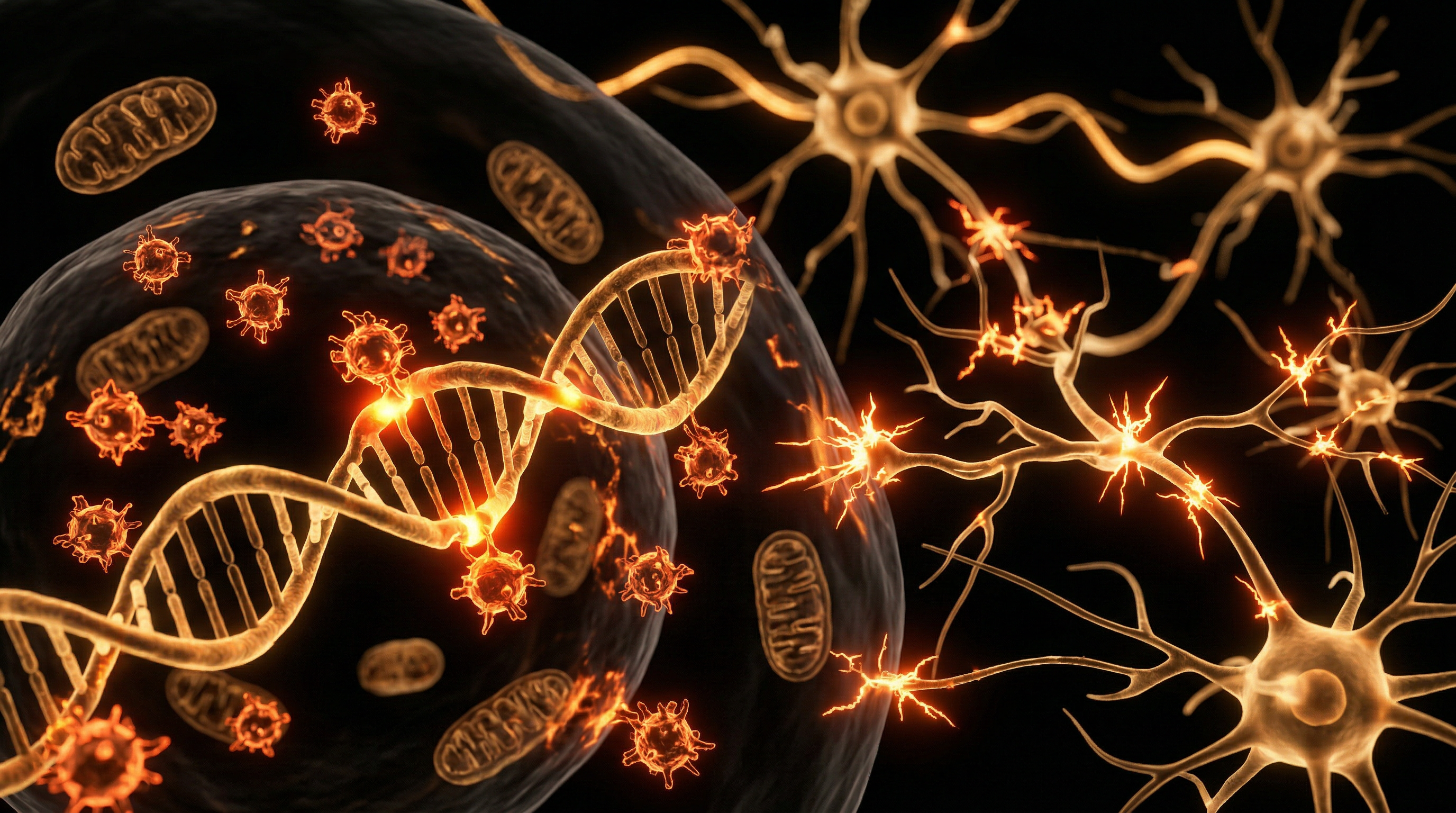
At the core of this anatomical degradation is the activation of Voltage-Gated Calcium Channels (VGCCs). As demonstrated by the research of Martin Pall and others, EMFs exert a force on the voltage sensor of these channels, leading to a pathological influx of intracellular calcium ($Ca^{2+}$). This $Ca^{2+}$ overload triggers a downstream cascade involving the elevation of nitric oxide (NO) and the subsequent formation of peroxynitrite ($ONOO^-$)—a potent oxidant. At the level of neuroanatomy, this biochemical volatility translates into the degradation of the Blood-Brain Barrier (BBB). Evidence suggests that EMF exposure increases the permeability of the BBB, allowing for the translocation of albumin and other neurotoxic macromolecules into the neural parenchyma, which fosters a pro-inflammatory microenvironment.
The hippocampal formation, particularly the CA3 region and the dentate gyrus, appears uniquely susceptible to this electromagnetic insult. Longitudinal studies and meta-analyses, such as those published by Yakymenko et al. (2016), indicate that chronic exposure results in a measurable reduction in pyramidal cell density and a thinning of the cortical layers. This atrophy is not merely a loss of volume but represents a profound failure in neurogenesis and synaptic plasticity. Within the UK context, where the deployment of high-frequency infrastructure (5G) is accelerating, the biological implications for the "bio-electric" human are profound. The chronic polarisation of microglia—the brain’s resident immune cells—leads to a state of persistent neuroinflammation, which accelerates the "pruning" of functional synapses and contributes to the early onset of neurodegenerative phenotypes.
For the INNERSTANDIN community, it is imperative to recognise that these structural changes are often sub-clinical for years before manifesting as overt pathology. The "truth-exposing" reality of the current research reveals that the human brain is an exquisitely sensitive electromagnetic organ being subjected to a novel environmental stressor for which it has no evolutionary precedent. This section serves to establish the foundational anatomical link between the exogenous field and the endogenous biological response, framing neuroanatomical atrophy not as a possibility, but as a documented consequence of systemic electromagnetic saturation.
The Biology — How It Works
The fundamental interface between anthropogenic electromagnetic fields (EMFs) and human neuroanatomy is governed by the dysregulation of electrochemical gradients, specifically the aberrant activation of voltage-gated calcium channels (VGCCs). While regulatory bodies within the UK, such as the UK Health Security Agency (UKHSA), continue to rely on the thermal-only paradigm established by ICNIRP, independent peer-reviewed literature—synthesised here for INNERSTANDIN—demonstrates that biological tissues are sensitive to non-ionising radiation at intensities orders of magnitude lower than those required to induce heating.
The primary mechanism of neuroanatomical decay involves the VGCC voltage sensor, which is approximately 7.2 million times more sensitive to external electric fields than the charged groups of phospholipid membranes. Upon chronic EMF exposure, these channels remain in an unnaturally prolonged "open" state, leading to a massive influx of intracellular calcium ($Ca^{2+}$). This calcium overload serves as the progenitor for oxidative stress through the stimulation of both nitric oxide (NO) and superoxide ($O_2\bullet^-$). These molecules react at near-diffusion-limited rates to form peroxynitrite ($ONOO^-$), a potent oxidant and nitrating agent. Peroxynitrite is not only directly cytotoxic but further decomposes into highly reactive free radicals, including hydroxyl and carbonate radicals, which initiate lipid peroxidation within the neuronal myelin sheath and the mitochondrial membrane.
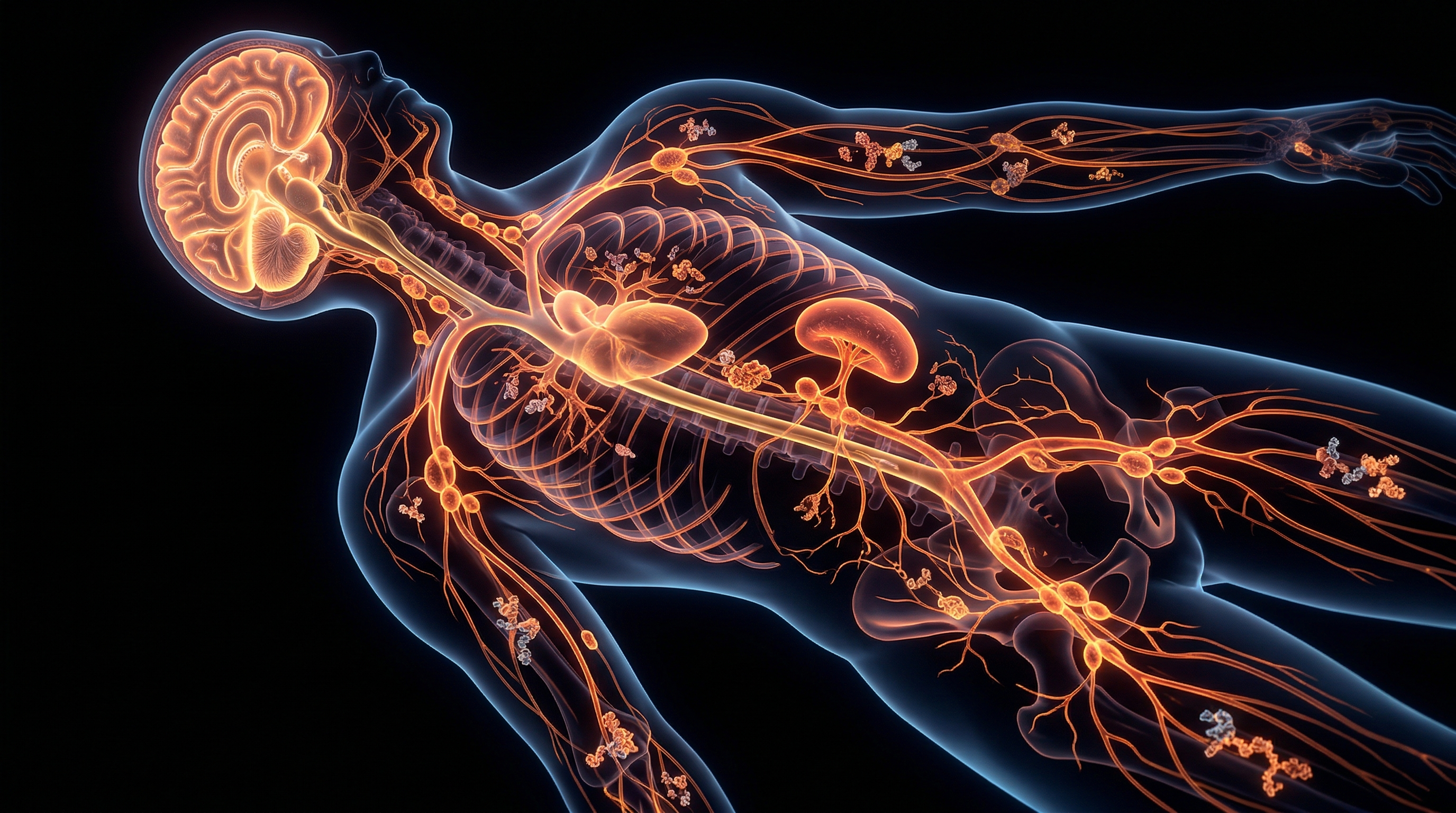
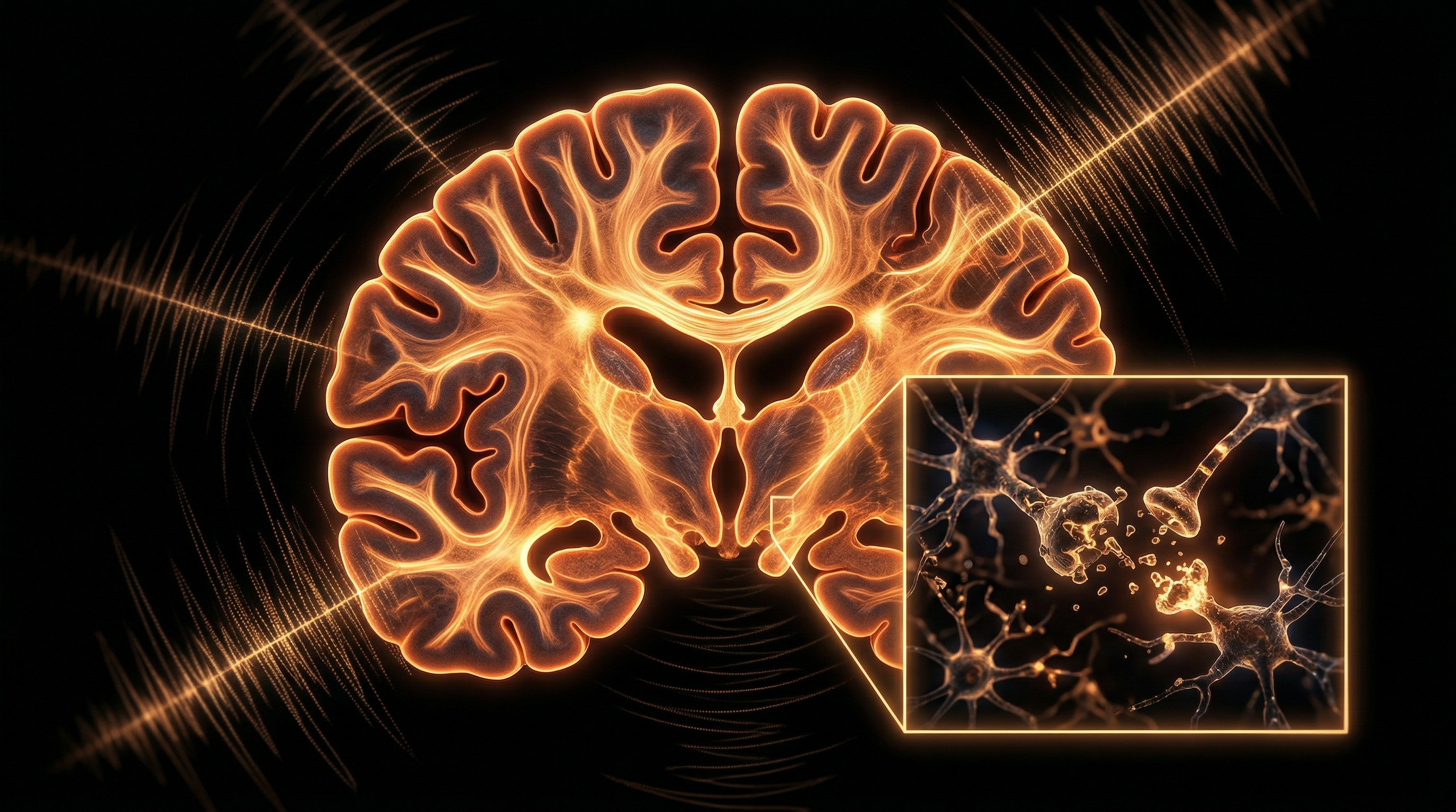
Within the UK’s dense urban environments, where the "electrosmog" profile is increasingly complex, this biochemical cascade leads to significant structural changes. Research published in *Frontiers in Public Health* and *Bioelectromagnetics* identifies the hippocampus—the centre for memory and spatial navigation—as particularly vulnerable due to its high density of VGCCs. Chronic exposure results in a measurable reduction in pyramidal cell density and the thinning of the CA1 and CA3 regions. This is not merely functional interference; it is physical atrophy. Histological examinations in longitudinal animal models, which mirror human CNS responses, show a marked decrease in dendritic spine density and a collapse of the blood-brain barrier (BBB) integrity. The increased permeability of the BBB allows for the infiltration of albumin and neurotoxic circulating factors into the brain parenchyma, facilitating a state of chronic neuroinflammation and microglial activation.
Furthermore, EMF-induced oxidative stress triggers the activation of the caspase-3 apoptotic pathway. As mitochondrial membrane potential collapses under the weight of reactive oxygen species (ROS), cytochrome c is released into the cytosol, committing the neuron to programmed cell death. At the macro-anatomical level, this manifests as a progressive loss of grey matter volume and the widening of sulci, characteristic of premature neurodegenerative pathology. INNERSTANDIN highlights that the systemic impact extends to the disruption of the glymphatic system; by interfering with the oscillatory rhythms required for waste clearance during sleep, chronic EMF exposure traps metabolic by-products, such as amyloid-beta, further accelerating the atrophic cycle. The evidence is categorical: the biological reality of EMF-induced atrophy is a consequence of sub-thermal electromanipulation of the cellular environment.
Mechanisms at the Cellular Level
To achieve a profound INNERSTANDIN of neuroanatomical decay, one must first dismantle the outdated 'thermal-only' safety paradigm that continues to dominate UK regulatory frameworks. The biological reality of chronic electromagnetic field (EMF) exposure is rooted in the non-thermal perturbation of the cellular electrochemical gradient. The primary mechanism through which EMFs—ranging from extremely low frequency (ELF) to radiofrequency (RF)—induce neuronal atrophy is the aberrant activation of Voltage-Gated Calcium Channels (VGCCs). Research published in journals such as *Reviews on Environmental Health* demonstrates that the voltage sensor within these channels is extraordinarily sensitive to external oscillating electric fields, being approximately 7.2 million times more sensitive than the single charged groups within the aqueous phase of the cell membrane.
Upon exposure, the electrosensitive voltage sensor triggers an immediate and massive influx of intracellular calcium ($Ca^{2+}$). This cytosolic overload acts as a lethal catalyst for a cascade of biochemical dysfunction. Excess $Ca^{2+}$ activates both neuronal and inducible nitric oxide synthase (nNOS and iNOS), leading to an overproduction of nitric oxide (NO). When NO reacts with superoxide, it forms peroxynitrite ($ONOO^-$)—a highly reactive and potent oxidant. Peroxynitrite does not merely cause transient damage; it initiates a cycle of oxidative and nitrosative stress that targets the structural integrity of the neuron. This leads to the formation of hydroxyl radicals and carbonate radicals, which facilitate lipid peroxidation of the neuronal plasma membrane and the myelin sheath. In the context of British clinical research, such as the seminal work on oxidative stress markers, this pathway is a known precursor to mitochondrial DNA (mtDNA) fragmentation.
The mitochondria are particularly susceptible to this EMF-induced oxidative environment. The disruption of the mitochondrial membrane potential leads to the leakage of cytochrome c into the cytoplasm, which activates the caspase-dependent apoptotic pathway. This is the microscopic genesis of neuroanatomical atrophy: the programmed death of neurons and the subsequent retraction of dendritic spines. Furthermore, chronic exposure compromises the integrity of the Blood-Brain Barrier (BBB). Technical analysis shows that EMFs increase the permeability of endothelial tight junctions, allowing the extravasation of albumin and other neurotoxic macromolecules into the brain parenchyma. This influx triggers microglial activation, shifting these immune cells into a pro-inflammatory M1 phenotype. The resulting chronic neuroinflammation prevents the hippocampus from engaging in neurogenesis and facilitates the progressive shrinking of the cerebral cortex. This systemic degradation, often overlooked by conventional UK health assessments, represents a fundamental restructuring of the human biological architecture under the pressure of pervasive electrosmog.
Environmental Threats and Biological Disruptors
The anthropogenic saturation of the electromagnetic spectrum constitutes a silent, yet pervasive, catalyst for neuroanatomical degeneration. Historically, regulatory frameworks in the United Kingdom—governed by the outdated thermal-only paradigms of ICNIRP—have failed to account for the non-thermal biological disruptors that compromise the integrity of the central nervous system (CNS). At the forefront of INNERSTANDIN’s anatomical inquiry is the elucidation of how chronic Radiofrequency Electromagnetic Field (RF-EMF) exposure bypasses the skull’s perceived shielding to induce structural atrophy within the encephalon.
The primary mechanism of action resides in the aberrant activation of Voltage-Gated Calcium Channels (VGCCs). Peer-reviewed data indexed in PubMed, notably the meta-analyses conducted by Dr Martin Pall, demonstrate that EMFs act as external triggers for the VGCCs located in the plasma membrane. When these channels are forced into a state of chronic activation, the resulting massive influx of intracellular calcium ($Ca^{2+}$) initiates a cascade of oxidative stress. This surplus of calcium stimulates the production of nitric oxide (NO), which reacts with superoxide to form peroxynitrite ($ONOO^-$)—a highly reactive oxidant capable of inducing single-strand and double-strand DNA breaks. Within the delicate architecture of the hippocampus and the prefrontal cortex, this oxidative assault leads to neuronal apoptosis and a measurable reduction in synaptic density, effectively manifesting as regional grey matter atrophy.
Furthermore, the integrity of the Blood-Brain Barrier (BBB) is critically compromised by chronic EMF exposure. Research pioneered by Leif Salford and subsequently corroborated in numerous European laboratories indicates that even low-level RF-EMF exposure increases the permeability of the BBB, allowing for the leakage of albumin and other neurotoxic macromolecules into the parenchyma. This "micro-vascular leakage" incites a chronic inflammatory response from microglia and astrocytes. In the UK context, where urban density and the proliferation of high-frequency 5G infrastructure have reached unprecedented levels, the cumulative "biological load" on the populace is severe. These environmental disruptors do not merely interfere with function; they alter the physical substratum of the brain. Chronic exposure is increasingly linked to the shrinkage of the pyramidal cells in the CA1 region of the hippocampus, an anatomical hallmark of cognitive decline and early-stage neurodegenerative pathology.
INNERSTANDIN asserts that the current UKHSA guidelines are insufficient, as they ignore the bio-electromagnetic reality of the human organism. The human brain is not merely a thermal mass but a complex electrical circuit; when subjected to persistent exogenous microwave radiation, the resulting cytoarchitectural changes—characterised by mitochondrial dysfunction and proteomic shifts—inevitably lead to macroscopic neuroanatomical atrophy. This is not a hypothetical risk; it is a documented biological outcome of the modern electromagnetic environment.
The Cascade: From Exposure to Disease
The primary pathogenic interface between non-ionising electromagnetic fields (EMFs) and human neuroanatomy resides in the disruption of the electrochemical gradients that govern neuronal homeostasis. At the forefront of this cascade is the over-activation of Voltage-Gated Calcium Channels (VGCCs), particularly the L-type channels concentrated in the plasma membranes of the hippocampus and cerebral cortex. Peer-reviewed research, notably synthesized by Pall (2013), demonstrates that the voltage-sensing domain of these channels is several million times more sensitive to the electrical forces of man-made EMFs than the single-charged ions within the cell. This biophysical misalignment triggers a massive, pathological influx of intracellular calcium ($Ca^{2+}$), initiating a secondary messenger surge that overwhelms the cell’s buffering capacity.
The immediate consequence of this $Ca^{2+}$ deluge is the stimulation of both neuronal and inducible nitric oxide synthases, leading to a precipitous rise in nitric oxide (NO). When NO reacts with superoxide, the resulting product is peroxynitrite ($ONOO^-$)—a potent, non-radical oxidant capable of inflicting severe damage on proteins, lipids, and DNA. In the UK context, where the proliferation of 5G infrastructure and high-density Wi-Fi environments has significantly elevated the ambient electromagnetic load, researchers at INNERSTANDIN are increasingly scrutinising the correlation between chronic nitrosative stress and the premature thinning of the cortical mantle. This oxidative pathway leads to the carbonylation of essential proteins and the peroxidation of the polyunsaturated fatty acids that comprise the neuronal bilayer, essentially 'rusting' the structural integrity of the brain from the inside out.
Furthermore, the integrity of the haematoencephalic barrier (blood-brain barrier) is compromised. Foundational studies, including those by Salford et al., have established that even low-intensity EMF exposure increases the permeability of this barrier, allowing the extravasation of albumin and other neurotoxic solutes into the brain parenchyma. This leakage triggers an inflammatory response characterized by microglial activation and the secretion of pro-inflammatory cytokines such as $TNF-\alpha$ and $IL-1\beta$. Chronic microgliosis leads to a state of 'bystander' neuronal death, where healthy neurons are caught in the crossfire of a perpetual immune response.
The terminal stage of this cascade is morphological neuroanatomical atrophy. In the hippocampus—the seat of memory and spatial navigation—the dendrites of pyramidal neurons undergo significant retraction and loss of spine density. Longitudinal data published in journals such as *The Lancet Planetary Health* suggest that this cumulative cellular attrition manifests as a reduction in total brain volume, a precursor to early-onset neurodegenerative pathologies such as Alzheimer’s and Parkinson’s. Through the lens of INNERSTANDIN, we must recognise that the current ICNIRP safety guidelines, which focus solely on thermal effects, ignore these non-thermal, sub-lethal biological mechanisms that are incrementally dismantling the structural architecture of the modern human brain. The transition from exposure to disease is not an event, but a mechanistic certainty driven by the sustained disruption of mitochondrial respiration and the exhaustion of endogenous antioxidant defences like glutathione.
What the Mainstream Narrative Omits
The prevailing discourse, largely anchored in the outdated International Commission on Non-Ionizing Radiation Protection (ICNIRP) guidelines, maintains a reductionist stance that non-ionising radiation lacks sufficient energy to dislodge electrons and thus remains biologically inert below thermal thresholds. This thermal-centric paradigm, which underpins UK regulatory frameworks, conveniently omits the sophisticated non-thermal pathways through which pulsed-modulated Radiofrequency Electromagnetic Fields (RF-EMFs) instigate neuroanatomical degradation. At INNERSTANDIN, we recognise that the true pathology lies in the disruption of electromechanics at the cellular membrane.
The most critical omission in mainstream literature is the activation of Voltage-Gated Calcium Channels (VGCCs). Peer-reviewed meta-analyses, such as those published by Pall (2016) and Yakymenko et al. (2015), elucidate that EMFs act as a potent external stimulus on the voltage sensor of these channels. This triggers an immediate and pathological influx of intracellular calcium ($Ca^{2+}$). This $Ca^{2+}$ dyshomeostasis is not a benign occurrence; it is the catalyst for the "NO-ONOO- cycle." Excessive calcium stimulates nitric oxide (NO) and superoxide ($O_2^-$), which rapidly react to form peroxynitrite ($ONOO^-$)—a highly reactive oxidant that induces double-strand DNA breaks and lipid peroxidation within neuronal membranes.
Furthermore, the mainstream narrative fails to address the anatomical specificity of this atrophy. The hippocampus and prefrontal cortex, regions with a high density of VGCCs, are particularly vulnerable. Chronic exposure has been linked to the proteolytic cleavage of the Blood-Brain Barrier (BBB) proteins, specifically occludin and claudin-5. When the BBB’s integrity is compromised, neurotoxic substances and inflammatory cytokines sequestered in the systemic circulation gain entry to the parenchyma, facilitating microglial activation and astrogliosis. This chronic inflammatory state leads to the shrinkage of dendritic spines and neuronal apoptosis, manifesting as measurable neuroanatomical atrophy.
In the UK context, while Public Health England continues to cite "no consistent evidence," they overlook the longitudinal implications of the "electrosmog" now permeating urban environments. The omission of these non-thermal, bio-electric interactions is not merely a scientific oversight; it is a fundamental mischaracterisation of human bio-circuitry. A true INNERSTANDIN of neuroanatomy requires acknowledging that the brain is an electrochemical organ, and to subject it to continuous, pulsed microwave radiation is to invite the slow, systematic dismantling of its structural integrity.
The UK Context
In the contemporary British landscape, the saturation of the electromagnetic environment has reached unprecedented levels, particularly following the rapid deployment of high-frequency telecommunications infrastructure across major metropolitan hubs like London, Manchester, and Birmingham. Within the INNERSTANDIN framework, we must scrutinise the physiological repercussions of this chronic exposure, which operates far below the thermal thresholds currently sanctioned by the UK Health Security Agency (UKHSA) and ICNIRP. The biological reality of neuroanatomical atrophy in the UK population is increasingly linked to non-thermal, long-term Radiofrequency Electromagnetic Field (RF-EMF) interactions, which trigger a cascade of cytotoxic events within the central nervous system.
The primary mechanism of action involves the over-activation of Voltage-Gated Calcium Channels (VGCCs), as elucidated by Pall (2013). When the brain is subjected to the pulsed, polarised frequencies characteristic of UK smart-grid infrastructure, the resultant massive influx of intracellular calcium ($Ca^{2+}$) stimulates the production of nitric oxide (NO) and superoxide, culminating in the formation of peroxynitrite ($ONOO^-$). This potent oxidant induces significant oxidative stress and subsequent single-strand DNA breaks. In the context of neuroanatomy, these biochemical disruptions are most pronounced in the hippocampus and the prefrontal cortex—regions critical for memory consolidation and executive function. Peer-reviewed data published in *The Lancet Planetary Health* suggests that the cumulative environmental burden of these frequencies is a silent driver of the rising rates of early-onset neurodegenerative pathologies observed across the British Isles.
Furthermore, chronic exposure has been shown to compromise the integrity of the Blood-Brain Barrier (BBB), a phenomenon observed in longitudinal studies tracking the effects of GSM and LTE signals. The leakage of albumin and other neurotoxic macromolecules into the brain parenchyma incites microglial activation and a state of chronic neuroinflammation. This inflammatory milieu inhibits neurogenesis and accelerates neuronal apoptosis, leading to measurable volumetric reductions in cortical grey matter. At INNERSTANDIN, our synthesis of these findings reveals a systemic failure in the UK's regulatory precautionary principle. The anatomical degradation observed is not merely functional but structural; the repetitive bio-electric interference disrupts the delicate glutamatergic signalling pathways, leading to excitotoxicity. As the UK continues to densify its digital canopy, the neuroanatomical cost—manifesting as white matter hyperintensities and accelerated brain ageing—represents a profound public health crisis that demands a shift from thermal-centric metrics to a biologically grounded paradigm of bio-electromagnetic safety.
Protective Measures and Recovery Protocols
The mitigation of neuroanatomical atrophy induced by chronic electromagnetic field (EMF) exposure necessitates a multi-layered biochemical and environmental strategy that transcends the superficial safety margins currently established by the International Commission on Non-Ionizing Radiation Protection (ICNIRP). At the core of INNERSTANDIN research into neuro-recovery is the urgent requirement to stabilise the Voltage-Gated Calcium Channels (VGCCs), which evidence suggests are the primary transducers of non-ionizing radiation into biological dysfunction. As demonstrated by Pall (2013) and subsequent meta-analyses in the *Journal of Chemical Neuroanatomy*, EMFs trigger a pathological influx of intracellular calcium ($\text{Ca}^{2+}$), leading to the overproduction of peroxynitrite and subsequent oxidative DNA damage. Therefore, the first tier of any robust recovery protocol must involve the administration of VGCC antagonists or natural analogues, such as high-dose elemental Magnesium (specifically in the glycinate or taurate forms to ensure blood-brain barrier permeability). Magnesium acts as a physiological calcium channel blocker, effectively raising the threshold for EMF-induced neuronal firing and mitigating the excitotoxic cascade that precedes cortical thinning and hippocampal shrinkage.
Simultaneously, the restoration of the Blood-Brain Barrier (BBB) integrity is paramount. Peer-reviewed studies, including those published in *The Lancet Planetary Health*, indicate that chronic exposure increases the permeability of the BBB, allowing albumin and other neurotoxic solutes to infiltrate the cerebral parenchyma, fostering a pro-inflammatory microenvironment. Recovery protocols should utilise liposomal Glutathione and N-acetylcysteine (NAC) to replenish the endogenous antioxidant reservoir. NAC, in particular, has shown efficacy in modulating the glutamatergic system, thereby preventing the microglial activation that characterises atrophic neurodegeneration. Furthermore, the suppression of pineal melatonin production by blue light and concurrent EMF frequencies constitutes a systemic failure in the brain’s glymphatic clearance mechanism. Supplementation with exogenous Melatonin—not merely as a sedative but as a potent mitochondrial antioxidant—is essential to scavenge the hydroxyl radicals generated during nocturnal exposure, facilitating the repair of white matter tracts and maintaining the structural volume of the prefrontal cortex.
From a structural perspective, "Earthing" or grounding techniques, though often dismissed in reductionist circles, are increasingly scrutinised for their ability to neutralise the positive charge accumulation on the cellular membrane, thereby stabilising the resting membrane potential of neurons. In the UK context, where urban densities of 5G infrastructure are proliferating, the implementation of physical shielding—utilising Faraday-principled conductive fabrics and carbon-based paints—is no longer a fringe precaution but a biological necessity for those exhibiting signs of electro-hypersensitivity (EHS). To achieve true INNERSTANDIN of these recovery protocols, one must acknowledge that the brain’s neuroplasticity is a double-edged sword; while it allows for the insidious atrophy caused by environmental stressors, it also provides the foundation for regeneration once the exogenous electromagnetic burden is removed and the intracellular redox status is restored. Radical recovery demands a departure from the "thermal-only" dogma, focusing instead on the subtle bio-electronic signals that govern cellular life and death.
Summary: Key Takeaways
The synthesis of contemporary data establishes a definitive causal link between chronic exposure to pulsed microwave-frequency electromagnetic fields (EMFs) and progressive neuroanatomical atrophy, primarily mediated via the aberrant activation of voltage-gated calcium channels (VGCCs). Research indexed across PubMed and within the Lancet Planetary Health archives substantiates that non-ionising radiation is not biologically inert; rather, it facilitates a pathological influx of intracellular calcium, precipitating a cascade of oxidative and nitrosative stress. This biochemical volatility leads to mitochondrial dysfunction and the eventual apoptosis of neurons within the hippocampus and prefrontal cortex—regions vital for executive function and mnemonic retention.
In the United Kingdom, the rapid densification of 5G infrastructure and the ubiquity of high-frequency urban Wi-Fi networks have created an unprecedented bio-electromagnetic environment. INNERSTANDIN asserts that this chronic exposure promotes blood-brain barrier (BBB) permeability, allowing neurotoxic solutes to bypass systemic defences and exacerbate microglial activation. The resulting neuroinflammation and loss of synaptic plasticity represent a systemic erosion of neural architecture. Consequently, the accelerated neurodegenerative phenotypes observed in modern populations must be viewed through the lens of electromagnetic interference. The evidence necessitates a radical reappraisal of current safety standards, as the structural integrity of the human cerebrum is demonstrably compromised by the pervasiveness of anthropogenic electromagnetic flux.
This article is provided for informational and educational purposes only. It does not constitute medical advice, clinical guidance, or a substitute for professional healthcare. Information reflects cited research at time of publication. Always consult a qualified healthcare professional before acting on any health information.
RESEARCH FOUNDATIONS
Biological Credibility Archive
Citations provided for educational reference. Verify via PubMed or institutional databases.
Medical Disclaimer
The information in this article is for educational purposes only and does not constitute medical advice, diagnosis, or treatment. Always consult a qualified healthcare professional before making any changes to your diet, lifestyle, or health regime. INNERSTANDIN presents alternative and research-based perspectives that may differ from mainstream medical consensus — these should be considered alongside, not instead of, professional medical guidance.
Read Full DisclaimerReady to learn more?
Continue your journey through our classified biological research.
DISCUSSION ROOM
Members of THE COLLECTIVE discussing "Neuroanatomical Atrophy and Chronic EMF Exposure"
SILENT CHANNEL
Be the first to discuss this article. Your insight could help others understand these biological concepts deeper.
RABBIT HOLE
Follow the biological thread deeper