Spike Protein Pathogenicity: Examining the Toxicity of S-Protein Expression
A deep dive into the biological activity of the spike protein produced by genetic vaccines. It explores interactions with ACE2 receptors and potential vascular implications.
Overview
The shift in global health paradigms over the last four years has pivoted largely on a single biological entity: the Spike Protein (S-protein). Originally identified as the structural anchor by which the SARS-CoV-2 virus gains entry into human cells, the spike protein has since transcended its role as a mere viral component. Through the advent of mass-scale genetic vaccination programmes, this protein has been introduced into the human body not via the natural respiratory route, but through direct intramuscular injection of synthetic genetic instructions.
As a senior biological researcher, it is my duty to scrutinise the distinction between a viral infection and the systemic production of a truncated, modified, and highly stable version of this protein. The narrative maintained by public health authorities has long suggested that the spike protein produced by mRNA and viral vector platforms is a benign "antigen," serving only to train the immune system before being rapidly degraded. However, an accumulating body of peer-reviewed evidence and clinical observation suggests a more harrowing reality.
The spike protein is not merely a passive marker; it is a biologically active pathotoxin. Its interaction with the human body is multifaceted, involving the disruption of the Renin-Angiotensin System (RAS), the induction of endothelial inflammation, and the potential for long-term persistence within vital organs. This article serves as a comprehensive examination of Spike Protein Pathogenicity, exploring the molecular mechanisms of its toxicity and the systemic implications of its uncontrolled expression within the human host. We are moving beyond the simplistic "lock and key" model of immunity into the realm of complex toxicology and synthetic biology.
The Spike Protein is increasingly recognised not just as a viral component, but as a bioactive protein capable of inducing systemic vascular and neurological pathology independent of the virus itself.
---
##
The Biology — How It Works
To understand the toxicity of the spike protein, one must first understand how the genetic "blueprints" function within the cellular machinery. In the context of mRNA therapeutics, the natural uridine bases of the genetic code are replaced with N1-methylpseudouridine. This modification was intended to prevent the innate immune system from immediately destroying the injected mRNA, thereby increasing the half-life of the template and the volume of protein produced.
The Genetic Instruction Set
Unlike natural infection, where the virus is primarily sequestered in the upper respiratory tract, the Lipid Nanoparticles (LNPs) used to deliver mRNA are designed for systemic distribution. Once injected into the deltoid, these LNPs travel through the lymphatic system and can enter the bloodstream, reaching the liver, spleen, heart, and even crossing the Blood-Brain Barrier (BBB).
Synthetic Modifications
The spike protein produced by the body following vaccination is not an exact replica of the viral version. It has been engineered for stability:
- —Proline Substitution (2P): Two proline molecules are inserted into the S2 subunit to lock the protein in its "pre-fusion" state. This was intended to make it more recognisable to antibodies.
- —Furin Cleavage Site: The junction between the S1 and S2 subunits is a critical point of biological activity. In some contexts, the S1 subunit (which contains the Receptor Binding Domain or RBD) can be cleaved and released into the systemic circulation.
- —Longevity: While natural viral proteins are degraded quickly, the "synthetic" spike has been found in the blood for weeks, and in some cases months, after the initial injection.
Bypassing Innate Immunity
When a virus enters through the nose or throat, it triggers a robust IgA mucosal response. By bypassing this through injection, the body is forced into a systemic response that prioritises IgG antibodies. This shift changes the landscape of how the body interacts with the protein, potentially leading to "immune exhaustion" or IgG4 class switching, where the body begins to tolerate the protein rather than neutralise it, allowing the toxin to persist longer in the tissues.
---
##
Mechanisms at the Cellular Level
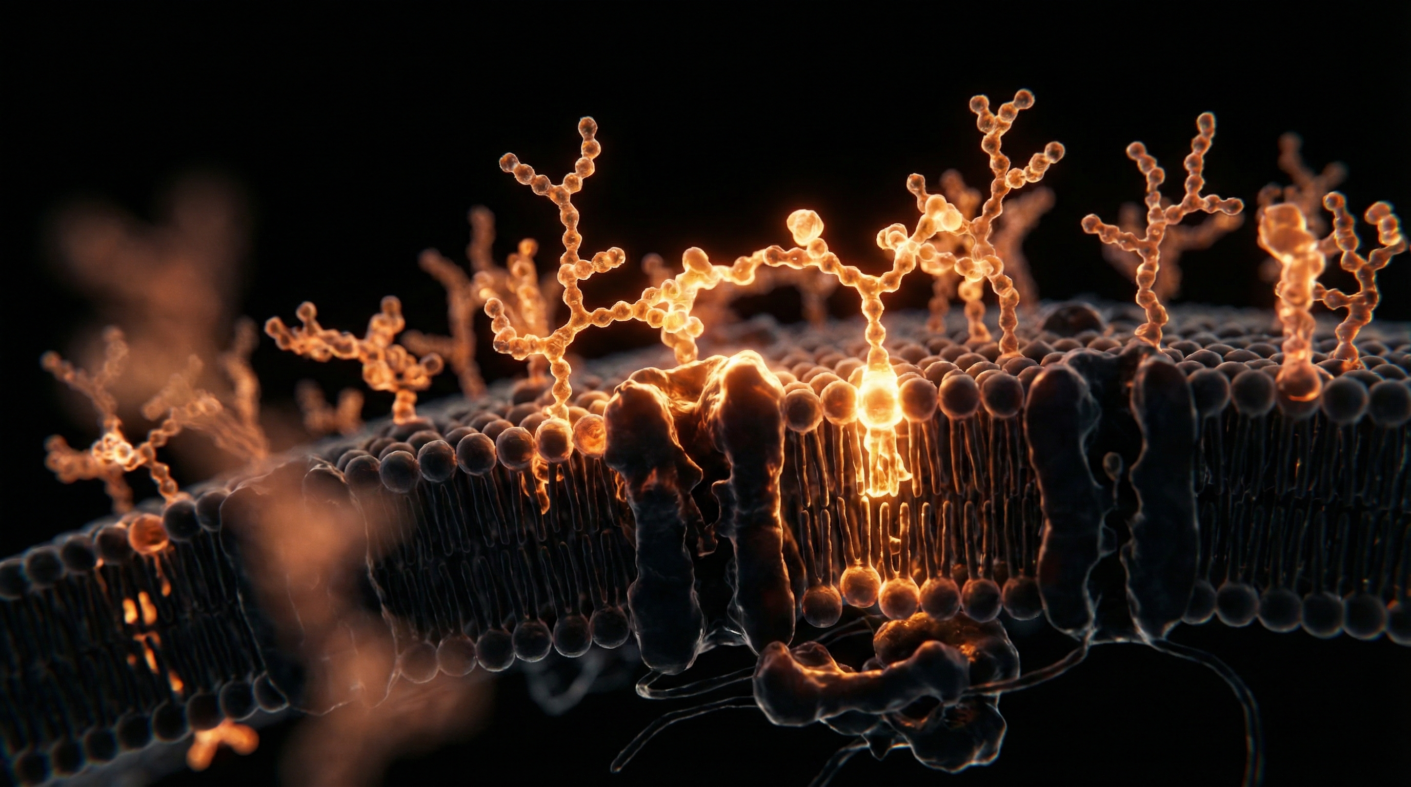
The primary gateway for the spike protein into the human cell is the Angiotensin-Converting Enzyme 2 (ACE2) receptor. While the mainstream narrative focuses on this as an "entry point" for the virus, the biological consequence of this binding is far more sinister.
ACE2 Downregulation and the RAS Imbalance
ACE2 is a vital regulatory enzyme. Its primary role is to convert Angiotensin II (a potent vasoconstrictor and pro-inflammatory agent) into Angiotensin (1-7) (a vasodilator and anti-inflammatory agent).
- —When the spike protein binds to ACE2, it causes the receptor to be internalised and degraded.
- —This leads to a systemic deficiency of ACE2.
- —The result is an uncontrolled accumulation of Angiotensin II.
This "RAS imbalance" is a primary driver of hypertension, oxidative stress, and endothelialitis (inflammation of the blood vessel lining). By depleting the body's natural "brakes" on inflammation, the spike protein creates a hyper-inflammatory environment at the cellular level.
Mitochondrial Dysfunction
Recent studies have highlighted the spike protein’s ability to interfere with mitochondrial function. The mitochondria are the "powerhouses" of the cell, but they also regulate cell death (apoptosis) and signalling. The S-protein has been shown to:
- —Disrupt the Electron Transport Chain, leading to reduced ATP production (causing the profound fatigue seen in many patients).
- —Increase the production of Reactive Oxygen Species (ROS), which damages cellular DNA and membranes.
- —Induce mitochondrial "fragmentation," a state often seen in degenerative diseases.
The Protein-Protein Interaction (PPI) Web
Beyond ACE2, the spike protein has been found to interact with a variety of other receptors, including CD147 (on red blood cells and neurons) and TLR4 (Toll-like Receptor 4), which is a key initiator of the innate immune system's inflammatory "alarm." By triggering TLR4, the spike protein directly mimics the effects of bacterial endotoxins, leading to a "cytokine storm" on a micro-scale.
---
##
Environmental Threats and Biological Disruptors
In the study of toxicology, we must look at the Bio-persistence and Bio-distribution of the substance in question. The spike protein is not just a "threat" because of its shape, but because of its persistence in an environment that was never designed to handle it.
The Role of Lipid Nanoparticles (LNPs)
LNPs are not inert delivery vehicles. They are highly inflammatory in their own right. When combined with the production of the spike protein, they create a "synergistic toxicity." LNPs allow the spike-producing instructions to enter cells that would never naturally encounter a respiratory virus, such as:
- —Hepatocytes (Liver cells): Leading to altered metabolic function.
- —Cardiomyocytes (Heart muscle cells): Leading to the well-documented risk of myocarditis.
- —Endothelial Cells: The cells lining every blood vessel in the body.
Exosomal Shedding and Micro-vesicles
One of the most overlooked aspects of spike protein pathogenicity is the production of Exosomes. Cells that have been transfected by mRNA can package the spike protein into small vesicles and "shed" them into the extracellular space. These exosomes can travel throughout the body, delivering the spike protein to distant organs that were never directly "hit" by the initial injection. This mechanism explains why spike protein has been found in breast milk and why some individuals report secondary exposure symptoms.
The Question of Reverse Transcription
While the "central dogma" of biology suggests that RNA cannot be converted back into DNA, recent in vitro studies (such as the Lund University study in Sweden) have suggested that the BNT162b2 mRNA can be reverse-transcribed into DNA within human liver cells (Huh7) via endogenous LINE-1 retrotransposons. If this occurs in vivo, it raises the terrifying prospect of permanent or long-term spike protein expression, effectively turning the human body into a permanent "spike protein factory."
---
##
The Cascade: From Exposure to Disease
The path from spike protein expression to clinical disease is a "cascade" of events that often begins in the vascular system.
Endothelialitis and Micro-clotting
The most profound impact of the spike protein is on the vascular endothelium. Because ACE2 receptors are highly expressed on the surface of blood vessel walls, the spike protein binds there, causing inflammation.
- —Vascular Permeability: The "tight junctions" between cells break down, allowing fluids to leak into tissues.
- —Platelet Activation: The spike protein can directly activate platelets, the cells responsible for clotting.
- —Fibrinoid Micro-clots: Research by Dr Resia Pretorius has identified "amyloid-like" micro-clots in the blood of those suffering from post-vaccination syndromes. These clots are resistant to the body’s natural breakdown processes (fibrinolysis) and can block the smallest capillaries, starving organs of oxygen.
"The spike protein is a 'clotting machine.' It doesn't just trigger the cascade; it structurally changes the fibrinogen into a form that the body cannot easily dissolve."
Myocarditis and Pericarditis
The heart is particularly susceptible to spike-mediated damage. Cardiomyocytes have a high density of ACE2 receptors. When the spike is expressed in the heart muscle, the immune system may attack those cells, leading to inflammation (Myocarditis). Furthermore, the spike protein can damage the delicate pericytes that support the heart's capillaries, leading to micro-ischaemia (small areas of tissue death).
Neurological Impact
The spike protein’s ability to cross the Blood-Brain Barrier is a matter of grave concern. Once in the central nervous system, it can:
- —Activate Microglia (the brain's immune cells), leading to neuro-inflammation.
- —Bind to the S1 receptor in the brain, potentially contributing to "brain fog" and cognitive decline.
- —Behave like a Prion-like protein. Computational models have suggested that the spike protein contains "prion domains," which could lead to the misfolding of other proteins (like amyloid-beta or tau), potentially accelerating neurodegenerative diseases like Alzheimer's or CJD.
---
##
What the Mainstream Narrative Omits
The suppression of scientific data regarding spike protein toxicity is perhaps the greatest academic scandal of the 21st century. Several key facts have been systematically downplayed or ignored by regulatory bodies.
The "Stay at the Injection Site" Myth
Early in the rollout, the public was told the vaccine stays in the arm. This was demonstrably false. The Pharmacokinetic data submitted by Pfizer to the Japanese regulator (PMDA) showed that LNPs distribute throughout the body within 48 hours, with significant concentrations in the ovaries, liver, and adrenal glands.
The Fallacy of "Short-lived" mRNA
We were told the mRNA disappears within days. However, research published in *Cell* (Röltgen et al., 2022) found that the vaccine mRNA and the spike protein persisted in the germinal centres of lymph nodes for at least 60 days. This prolonged presence means the body is subjected to a constant "drip-feed" of a toxic protein, rather than a short-lived "training" session.
Antibody-Dependent Enhancement (ADE) and Original Antigenic Sin
The narrative omits the risk that high levels of non-neutralising antibodies could actually facilitate viral entry into cells or that the immune system becomes "tethered" to an obsolete version of the spike protein (the original Wuhan strain), making it less effective against new variants.
The Presence of DNA Contamination
Recent independent laboratory analyses (led by researchers like Kevin McKernan) have discovered significant plasmid DNA contamination in the mRNA vials. This DNA includes SV40 promoter sequences. This contamination could potentially lead to genomic integration or prolonged spike protein production, a risk that was never disclosed to the public or properly evaluated by the MHRA or FDA.
---
##
The UK Context
In the United Kingdom, the deployment of genetic vaccines was hailed as a "triumph of British science." However, the data emerging from the Office for National Statistics (ONS) and the MHRA Yellow Card reporting system tells a more complex story.
The MHRA and the "Yellow Card" Data
The Yellow Card system has recorded hundreds of thousands of adverse event reports, including thousands of deaths. While the MHRA maintains that "the benefits outweigh the risks," the sheer volume of vascular and haematological reports is unprecedented.
- —Excess Mortality: The UK has seen sustained "unexplained" excess mortality since 2021, particularly in non-COVID categories such as cardiovascular disease.
- —The "Gold Standard" Burden: British researchers are finding it increasingly difficult to secure funding for spike-protein specific toxicity studies, as the funding landscape is dominated by the very organisations that promoted the "safe and effective" mantra.
The NHS Crisis: A Symptom of Systemic Injury?
The NHS is currently buckling under a wave of chronic illness. While "long-wait times" are blamed on logistics, clinicians are seeing a rise in complex, multi-system inflammatory conditions that do not fit traditional diagnostic boxes. Many of these cases involve the very "cascade" of spike protein toxicity described earlier: micro-clotting, POTS (Postural Orthostatic Tachycardia Syndrome), and chronic fatigue.
---
##
Protective Measures and Recovery Protocols
For those concerned about the persistence of the spike protein in their systems, the focus must shift from "prevention" to "degradation" and "neutralisation." Biological science offers several pathways for mitigating the impact of the S-protein.
Fibrinolytic Therapy
Since the spike protein induces resistant micro-clots, using enzymes that can break down fibrin is crucial.
- —Nattokinase: An enzyme derived from fermented soy (Natto). It has been shown in vitro to degrade the spike protein directly and to break down the amyloid-like clots.
- —Lumbrokinase: A more potent fibrinolytic enzyme derived from earthworms, often used in complex clotting disorders.
Proteolytic Enzymes
- —Bromelain: Derived from pineapple stems, bromelain has the capacity to cleave the spike protein, particularly when used in combination with N-Acetylcysteine (NAC), which breaks the disulphide bonds that hold the protein structure together.
Anti-inflammatory and Autophagy Support
- —Autophagy: This is the body's natural "recycling" process. Intermittent fasting and certain compounds like Spermidine or Resveratrol can trigger autophagy, potentially helping the body clear out the spike-producing cells or the proteins themselves.
- —Vitamin D and Zinc: Essential for maintaining the integrity of the immune system and preventing the "over-activation" of the inflammatory response.
- —Quercetin: A zinc ionophore that also has anti-inflammatory properties and can inhibit the binding of the spike protein to the ACE2 receptor.
Detoxification of LNPs
Supporting the liver is paramount for clearing the synthetic lipid nanoparticles. Milk Thistle (Silymarin) and Glutathione precursors (like NAC) are essential for maintaining the liver's phase II detoxification pathways.
Recovery is not a single event but a process of reducing the "total toxic load" and restoring the balance of the Renin-Angiotensin System.
---
##
Summary: Key Takeaways
The examination of Spike Protein Pathogenicity reveals a biological landscape that is far more perilous than the public has been led to believe. The "synthetic" spike protein produced by genetic vaccines is a multi-modal toxin that interacts with the body on vascular, neurological, and immunological levels.
- —Direct Toxicity: The spike protein is not an inert antigen; it is a pathotoxin that can cause disease independently of the virus.
- —Vascular Damage: Through ACE2 downregulation and the formation of resistant micro-clots, the spike protein poses a significant threat to cardiovascular health.
- —Bio-persistence: Modified mRNA and LNPs allow for the prolonged and systemic production of the spike protein, far exceeding the duration of a natural infection.
- —Regulatory Failure: There has been a systematic failure to acknowledge the biodistribution and toxicity data, leading to a lack of informed consent.
- —Pathway to Recovery: Mitigation strategies involving nattokinase, bromelain, and autophagy-promoting protocols offer hope for those suffering from spike-mediated injuries.
As we continue to peel back the layers of this unprecedented biological experiment, it becomes clear that the spike protein is the "smoking gun" in the surge of post-2021 chronic illnesses. Only through rigorous, independent science—free from corporate influence—can we fully understand the scale of this toxicity and develop the protocols necessary to heal those affected. The era of "blind faith" in genetic products must end; the era of biological accountability must begin.
This article is provided for informational and educational purposes only. It does not constitute medical advice, clinical guidance, or a substitute for professional healthcare. Information reflects cited research at time of publication. Always consult a qualified healthcare professional before acting on any health information.
RESEARCH FOUNDATIONS
Biological Credibility Archive
The SARS-CoV-2 spike protein alone can damage vascular endothelial cells by downregulating ACE2 expression and inhibiting mitochondrial function.
Exposure to the spike protein subunit 1 induces a pro-inflammatory response in human brain microvascular endothelial cells via TLR4 signaling.
The S1 subunit of the spike protein was detected in the plasma of individuals following mRNA vaccination, indicating systemic distribution of the expressed antigen.
Intracellular expression of the spike protein in human cells can lead to syncytia formation and subsequent cellular dysfunction or death through membrane fusion mechanisms.
The spike protein directly interacts with platelet receptors and promotes thrombosis through the activation of vascular signaling pathways independently of viral replication.
Citations provided for educational reference. Verify via PubMed or institutional databases.
Medical Disclaimer
The information in this article is for educational purposes only and does not constitute medical advice, diagnosis, or treatment. Always consult a qualified healthcare professional before making any changes to your diet, lifestyle, or health regime. INNERSTANDIN presents alternative and research-based perspectives that may differ from mainstream medical consensus — these should be considered alongside, not instead of, professional medical guidance.
Read Full DisclaimerReady to learn more?
Continue your journey through our classified biological research.
DISCUSSION ROOM
Members of THE COLLECTIVE discussing "Spike Protein Pathogenicity: Examining the Toxicity of S-Protein Expression"
SILENT CHANNEL
Be the first to discuss this article. Your insight could help others understand these biological concepts deeper.
RABBIT HOLE
Follow the biological thread deeper