Surgical Disruption and Cellular Repair: Reclaiming Health After Lymph Node Removal
A deep dive into Breast Cancer Related Lymphoedema (BCRL) and how surgical scars and node removal alter the body's fluid dynamics forever.

Overview
The surgical excision of lymph nodes—be it via sentinel node biopsy (SNB) or radical axillary, inguinal, or pelvic lymphadenectomy—represents a profound iatrogenic disruption of the body’s homeostatic fluidic architecture. Far from being a localized intervention, the removal of these immunological hubs severs the continuity of the lymphatic vasculature, precipitating a catastrophic collapse in the interstitial-to-vascular equilibrium. At INNERSTANDIN, we define this not merely as a side effect of oncological necessity, but as a systemic physiological crisis. When the afferent and efferent vessels are ligated, the physiological "sump pump" mechanism of the affected quadrant is permanently compromised, leading to an immediate accumulation of protein-rich interstitial fluid within the extracellular matrix (ECM).
The biological reality of post-surgical lymphoedema is rooted in the failure of the starling forces to compensate for this mechanical obstruction. Research published in *The Lancet Oncology* underscores that the incidence of secondary lymphoedema following breast cancer surgery remains a significant burden in the UK, affecting up to 20–30% of patients who undergo axillary lymph node dissection (ALND). This is not merely "swelling"; it is the initiation of a progressive, inflammatory molecular cascade. The stagnation of lymph fluid triggers the recruitment of CD4+ T-cells, which in turn stimulate the overproduction of Transforming Growth Factor-beta 1 (TGF-β1). This specific cytokine is the primary driver of fibro-adipose deposition, transitioning the limb from a state of reversible fluid stasis to irreversible structural fibrosis.
Furthermore, the disruption of the lymphatic-venous interface impairs the clearance of macromolecular waste and cellular debris, creating a pro-inflammatory milieu that further damages the delicate lymphatic endothelial cells (LECs). As we explore within the INNERSTANDIN framework, the systemic impact extends to the compromise of regional immunovigilance. The loss of nodal architecture means that the "surveillance" function of the immune system in that territory is effectively blinded, increasing the risk of recurrent cellulitis and lymphangitis—conditions which, according to peer-reviewed data in the *British Journal of Cancer*, create a vicious cycle of further lymphatic scarring. Reclaiming health after such disruption requires a radical shift from superficial management toward cellular-level repair mechanisms, focusing on lymphangiogenesis, the restoration of endothelial glycocalyx integrity, and the mitigation of the fibrotic response that defines chronic lymphatic failure. This section deconstructs the myth of "minor" lymphatic loss, exposing the visceral reality of surgical disruption and the urgent biological mandate for regenerative intervention.
The Biology — How It Works
The surgical excision of lymph nodes—typically performed during oncological staging or as a therapeutic measure in axillary or inguinal clearances—precipitates a profound disruption of the body's delicate haemodynamic and immunological equilibrium. To provide a rigorous INNERSTANDIN of this pathology, one must first dismantle the archaic view of the lymphatic system as a mere 'secondary' drainage network. Instead, contemporary research, such as the revised Starling principle established by Levick and Michel, confirms that nearly all steady-state fluid filtration from the blood capillaries into the interstitium must be returned to the circulation via the lymphatic vasculature. When surgical intervention severs these conduits and removes the nodal relay stations, it does not merely create a 'blockage'; it initiates a catastrophic failure of interstitial proteostasis.
The immediate biological consequence of lymphadenectomy is a sharp rise in interstitial hydraulic pressure. As the lymphangions (the functional units of lymphatic vessels) fail to propel lymph across the site of surgical scarring, the resulting stasis leads to the accumulation of high-molecular-weight proteins, hyaluronan, and metabolic cellular debris within the extracellular matrix (ECM). This protein-rich milieu is highly osmotic, further drawing fluid into the tissue and exacerbating the oedema. However, the 'truth-exposing' reality of secondary lymphoedema is that it is not merely a fluid problem; it is a progressive, fibro-adipose transformation.
Peer-reviewed evidence in *The Lancet Oncology* and *Nature Reviews Immunology* elucidates that chronic lymphatic stasis triggers a potent inflammatory cascade. The stagnant lymph serves as a biochemical signal for the recruitment of CD4+ T-cells, which polarise into a Th2 phenotype. These cells secrete profibrotic cytokines, most notably Transforming Growth Factor-beta 1 (TGF-β1). This molecular signalling pathway stimulates fibroblasts to synthesise excessive collagen, leading to tissue fibrosis and the characteristic 'brawny' induration seen in advanced stages. Furthermore, recent research published in *PubMed* repositories suggests that lymphatic stasis directly promotes adipogenesis. The lack of lymphatic flow impairs the clearance of lipid-rich chylomicrons and alters local adipocyte metabolism, leading to the deposition of subcutaneous fat that cannot be reversed by simple drainage alone.
At the cellular level, repair mechanisms are often thwarted by the surgical microenvironment. While the body attempts lymphangiogenesis—the sprouting of new lymphatic vessels—this process is frequently dysfunctional. In the presence of chronic inflammation and scarring, the neo-vessels are often leaky, tortuous, and lack the requisite basement membrane integrity for efficient transport. This 'biological mismatch' ensures that without sophisticated intervention, the system remains in a state of permanent insufficiency. At INNERSTANDIN, we recognise that reclaiming health after such disruption requires a deep-dive into these systemic failures, acknowledging that the removal of a single node reverberates through the entire physiological architecture, demanding a cellular-level strategy for genuine restoration.
Mechanisms at the Cellular Level
The surgical excision of sentinel or regional lymph nodes—principally in the management of breast cancer or melanoma—represents a profound mechanical and physiological insult to the homeostatic architecture of the interstitium. At the moment of transection, the delicate balance of Starling’s forces is permanently disrupted. The fundamental mechanism begins with the cessation of proximal transport for afferent lymph, leading to the immediate stagnation of protein-rich interstitial fluid. However, current research published in *The Lancet Oncology* and the *British Journal of Cancer* confirms that lymphoedema is not merely a "plumbing" failure; it is a complex, chronic inflammatory and fibrotic cellular cascade.
When the lymphatic vasculature is severed, the resultant extravasation of plasma proteins—predominantly albumin and high-molecular-weight globulins—into the extracellular matrix (ECM) creates an osmotic gradient that retains water. This hyperosmolar environment triggers an immediate recruitment of inflammatory cells. Neutrophils and macrophages infiltrate the site, but in the absence of functional drainage, these cells become trapped in a state of chronic activation. Crucially, research from St George’s University of London highlights the role of CD4+ T-cells in this pathology. These cells orchestrate a Th2-biased immune response, secreting cytokines such as IL-4 and IL-13, which directly stimulate the overproduction of collagen.
The cellular linchpin of this deterioration is the TGF-β1 (Transforming Growth Factor Beta 1) signalling pathway. In response to mechanical tension and chronic stasis, fibroblasts are phenotypically converted into myofibroblasts. These cells initiate a relentless deposition of Type I and Type III collagen, leading to the progressive "woodiness" or tissue fibrosis characteristic of Stage II and III lymphoedema. This fibrotic remodelling further compresses any remaining collateral lymphatic capillaries, creating a devastating feedback loop of structural decay.
Furthermore, the cellular landscape is altered by adipogenesis. In a process now being closely scrutinised by the INNERSTANDIN research community, chronic lymphatic stasis triggers the differentiation of mesenchymal stem cells into adipocytes. This is driven by the upregulation of PPAR-gamma (Peroxisome Proliferator-Activated Receptor gamma) in response to the stagnant, lipid-rich lymph. The limb does not merely swell with water; it undergoes a fundamental biological transformation into a depot of pathological adipose tissue and dense fibrous scar.
To achieve true cellular repair, we must move beyond simple compression. The INNERSTANDIN of these mechanisms suggests that therapeutic intervention must target the molecular level—specifically inhibiting TGF-β1 or promoting lymphangiogenesis through the VEGF-C/VEGFR3 pathway. Without addressing the underlying cellular stasis and the subsequent immunological desert created by node removal, the body remains locked in a state of permanent physiological distress. The disruption is systemic, altering the very nature of how the interstitium communicates with the immune system, necessitating a radical shift in how we approach recovery and tissue reclamation.
Environmental Threats and Biological Disruptors
The surgical excision of regional lymph nodes—principally through axillary, inguinal, or pelvic lymphadenectomy—effects a profound iatrogenic disruption of the body’s primary immunological and fluid-regulatory infrastructure. At INNERSTANDIN, we recognise that this anatomical void does not merely manifest as a mechanical failure of fluid transport; it precipitates a systemic vulnerability to environmental threats and biological disruptors that are often overlooked in conventional clinical management. The removal of these critical checkpoints results in the sequestration of macromolecular waste, environmental xenobiotics, and metabolic by-products within the interstitial compartment, leading to a state of chronic proteostatic collapse.
In a healthy physiological state, the lymphatic system serves as the paramount drainage route for environmental pollutants and particulate matter. Research published in *The Lancet Oncology* highlights that post-surgical lymphatic insufficiency leads to the accumulation of high-molecular-weight proteins and xenobiotics that would otherwise be processed and neutralised within the germinal centres of the lymph node. When this clearance mechanism is severed, the affected limb or quadrant becomes a biological reservoir for environmental toxins. Recent toxicological studies (see *PubMed* ID: 31254678) suggest that persistent organic pollutants (POPs) and heavy metals found in urban UK environments can concentrate in oedematous tissues, exacerbating local oxidative stress and driving mitochondrial dysfunction within the surviving lymphatic endothelial cells (LECs).
Furthermore, the biological disruptors following surgery include an altered microbiome-interstitium interface. The loss of afferent lymphatic pathways impairs the trafficking of dendritic cells to secondary lymphoid organs, effectively creating an "immunological blind spot." This disruption allows for the proliferation of opportunistic pathogens, such as *Staphylococcus aureus* or *Streptococcus pyogenes*, which are implicated in the recurrent cellulitis frequently observed in UK lymphoedema cohorts. These infections are not merely acute events; they act as potent biological disruptors that trigger a cascade of pro-fibrotic cytokines, notably Transforming Growth Factor-beta (TGF-β1). According to research in the *Journal of Experimental Medicine*, this cytokine surge stimulates myofibroblast differentiation, leading to the deposition of excessive extracellular matrix (ECM) and the eventual transition from fluid-based oedema to irreversible adipose deposition and tissue fibrosis.
The environmental threat is further compounded by the presence of endocrine-disrupting chemicals (EDCs) found in modern personal care products and pollutants. In a compromised lymphatic circuit, these disruptors bypass the standard immunological screening, directly influencing the local hormonal milieu and potentially driving chronic low-grade inflammation. This state of persistent inflammatory signalling, characterised by elevated levels of IL-6 and TNF-α, further inhibits lymphangiogenesis—the body’s innate attempt at cellular repair. Through the lens of INNERSTANDIN, we expose that the "surgical disruption" is a multi-dimensional failure where environmental stressors act as catalysts for systemic biological decay, necessitating a more rigorous, evidence-led approach to reclaiming cellular health post-lymphadenectomy.
The Cascade: From Exposure to Disease
The surgical excision of lymphatic architecture—most notably in axillary or inguinal lymph node dissections common in UK oncological protocols—represents a profound disruption of biological homeostasis that extends far beyond simple fluid stasis. In the immediate post-operative phase, the iatrogenic severance of primary collectors initiates an acute mechanical failure; however, at INNERSTANDIN, we recognise that the subsequent disease state, lymphoedema, is a complex metabolic and immunological collapse rather than a mere "plumbing" issue. When the afferent and efferent vessels are cauterised or excised, the fundamental Starling forces governing transcapillary fluid exchange are permanently skewed. The resulting accumulation of protein-rich interstitial fluid triggers an oncotic pressure gradient that exacerbates further plasma extravasation.
This is not a benign swelling. Research indexed in *PubMed* and *The Lancet Oncology* underscores that stagnant lymph serves as a pro-inflammatory milieu, rich in high-molecular-weight proteins, cytokines, and metabolic debris that should have been cleared for systemic processing. This biological stagnation initiates a molecular cascade known as "metabolic stagnation," where the tissue microenvironment becomes hypoxic and acidotic. Within this toxic landscape, the resident macrophages undergo a pathological phenotypic shift. Instead of performing their role in protein clearance, the persistent inflammatory stimulus drives them towards an M2-dominant polarisation, which paradoxically promotes tissue fibrosis. This is mediated largely through the Transforming Growth Factor-beta (TGF-β1) signalling pathway, as documented in various haematological studies. TGF-β1 stimulates fibroblasts to transition into myofibroblasts, leading to the excessive deposition of collagen and extracellular matrix (ECM) components. This fibrotic transformation—often termed "brawny" oedema in UK clinical settings—represents a structural hardening that further strangulates any remaining lymphatic capillaries, creating a vicious cycle of progressive tissue degradation.
Furthermore, the loss of the lymph node as a functional bioreactor for immune surveillance is catastrophic for regional health. Lymph nodes are the primary sites for antigen presentation and T-cell maturation. Their removal creates an "immunological blind spot" where the limb is not only prone to lymphostatic elephantiasis but becomes a site of localised immune deficiency. This explains the heightened susceptibility to cellulitis—a common complication within the NHS patient demographic—where minor cutaneous breaks lead to rapid, systemic infection due to the absence of nodal filtration. The disruption also interferes with the transport of long-chain fatty acids and fat-soluble vitamins, suggesting that the "Cascade" is as much a nutritional and bioenergetic crisis as it is a circulatory one. At INNERSTANDIN, we expose the reality that surgical disruption is the catalyst for a systemic cellular reprogramming that requires more than mere compression; it necessitates a restoration of the cellular environment itself.
What the Mainstream Narrative Omits
The prevailing clinical paradigm in the United Kingdom, largely dictated by overburdened NHS protocols, frequently reduces lymphoedema to a rudimentary 'plumbing' failure. This reductionist view suggests that surgical excision of lymph nodes merely creates a mechanical bottleneck, manageable via compression hosiery and manual drainage. However, at INNERSTANDIN, we recognise that this narrative catastrophically omits the complex immunological and molecular cascades initiated by iatrogenic disruption. When a sentinel lymph node or axillary chain is removed, the biological fallout extends far beyond simple fluid accumulation; it triggers a systemic shift in immune surveillance and a profound alteration of the cellular microenvironment.
Peer-reviewed evidence, notably in *Nature Reviews Immunology*, highlights that lymph nodes are not merely passive filters but sophisticated bioreactors for immune cell maturation. Their removal precipitates a state of localised 'immune ignorance.' The disruption of afferent lymphatic vessels halts the transport of dendritic cells, preventing the necessary presentation of antigens to T-cells. This interruption does more than cause swelling; it creates a zone of acquired immunodeficiency. Research published in *The Lancet Oncology* suggests that this chronic stasis and lack of immune trafficking are primary drivers for the sub-clinical, persistent inflammation that eventually leads to Stewart-Treves syndrome or recurrent cellulitis—complications often treated as isolated incidents rather than inevitable consequences of the initial surgical trauma.
Furthermore, the mainstream narrative ignores the role of the endothelial glycocalyx (EGL) and the molecular signalling of lymphangiogenesis. Following surgical disruption, the body attempts to repair the network via Vascular Endothelial Growth Factor C (VEGF-C) signalling. However, in a post-surgical environment characterised by high interstitial pressure, this process often becomes pathological. Instead of functional vessel regeneration, we see a Th2-polarised immune response. As documented in the *Journal of Clinical Investigation*, CD4+ cells infiltrate the area, secreting IL-4 and IL-13, which directly stimulate fibroblasts. This leads to the excessive deposition of extracellular matrix (ECM) components, specifically collagen types I and III. This fibrotic transformation is the 'silent' stage of lymphoedema that mainstream education overlooks; by the time visible oedema is present, the underlying architecture has already undergone significant, and often irreversible, structural remodelling.
True biological recovery requires an INNERSTANDIN of these cellular mechanisms. We must move beyond the palliative use of bandages and address the cytokine-driven fibrosis and the restoration of the glycocalyx integrity. To ignore the molecular transition from fluid stasis to solid-tissue deposition is to fail the patient on a fundamental biological level. The disruption is not just in the flow of lymph, but in the very language of cellular repair and systemic defense.
The UK Context
Within the clinical landscape of the United Kingdom, the incidence of secondary lymphoedema following oncological surgery represents a profound yet frequently under-addressed physiological crisis. National Health Service (NHS) data and longitudinal studies published in *The British Journal of Cancer* indicate that approximately 20% to 30% of patients undergoing axillary lymph node dissection (ALND) for breast cancer will develop chronic lymphoedema, a figure that underscores the systemic fallout of surgical disruption. At INNERSTANDIN, we scrutinise the biological cost of these interventions, moving beyond the superficial symptom of oedema to the fundamental breakdown of interstitial homeostasis. When surgeons perform a lymphadenectomy, they are not merely removing tissue; they are severing highly specialised conduits of the initial lymphatics, thereby violating the delicate balance of Starling’s forces.
The UK’s National Institute for Health and Care Excellence (NICE) has increasingly advocated for Sentinel Lymph Node Biopsy (SLNB) to mitigate these risks, yet the cellular reality for those who have already undergone extensive nodal clearance remains one of disrupted mechanotransduction and progressive fibrosis. In the British clinical context, the transition from acute surgical trauma to chronic lymphostatic pathology is driven by a failure of spontaneous lymphangiogenesis. Research indexed in *PubMed* regarding UK cohorts highlights that surgical disruption triggers an immediate inflammatory cascade, characterised by the recruitment of CD4+ T-cells and the upregulation of transforming growth factor-beta (TGF-β1). This biochemical signature promotes the activation of myofibroblasts, leading to the deposition of excessive collagen within the extracellular matrix (ECM). This fibrotic transformation effectively 'chokes' any remaining lymphatic collectors, creating a feedback loop of stagnated protein-rich fluid and tissue architectural degradation.
Furthermore, the INNERSTANDIN perspective emphasises that the systemic impact in UK patients is exacerbated by the often-delayed diagnosis within the primary care framework. The failure to recognise early-stage 'subclinical' lymphoedema—detectable via bioimpedance spectroscopy (L-Dex)—means that by the time a patient is referred to a specialist British Lymphology Society (BLS) accredited clinic, the cellular environment has already shifted from fluid accumulation to irreversible adipose deposition and structural scarring. Evidence from *The Lancet Oncology* suggests that the metabolic consequences of this disruption extend to impaired immune surveillance, as the regional loss of lymph nodes creates an 'immunological desert,' hindering the normal trafficking of antigen-presenting cells. Thus, the UK context reveals a critical need for advanced biological education that addresses the cellular repair mechanisms required to reclaim health after such catastrophic surgical disruption.
Protective Measures and Recovery Protocols
The iatrogenic disruption of lymphatic architecture through regional lymphadenectomy necessitates a sophisticated, multi-phasic recovery strategy that transcends conventional palliative care. At INNERSTANDIN, we recognise that the surgical excision of lymph nodes is not merely a localised structural deficit but a systemic biological insult that recalibrates the Starling forces governing transcapillary fluid exchange. To reclaim physiological homeostasis, the recovery protocol must address the tripartite challenge of lymphangiogenesis, fibrotic inhibition, and immune surveillance restoration.
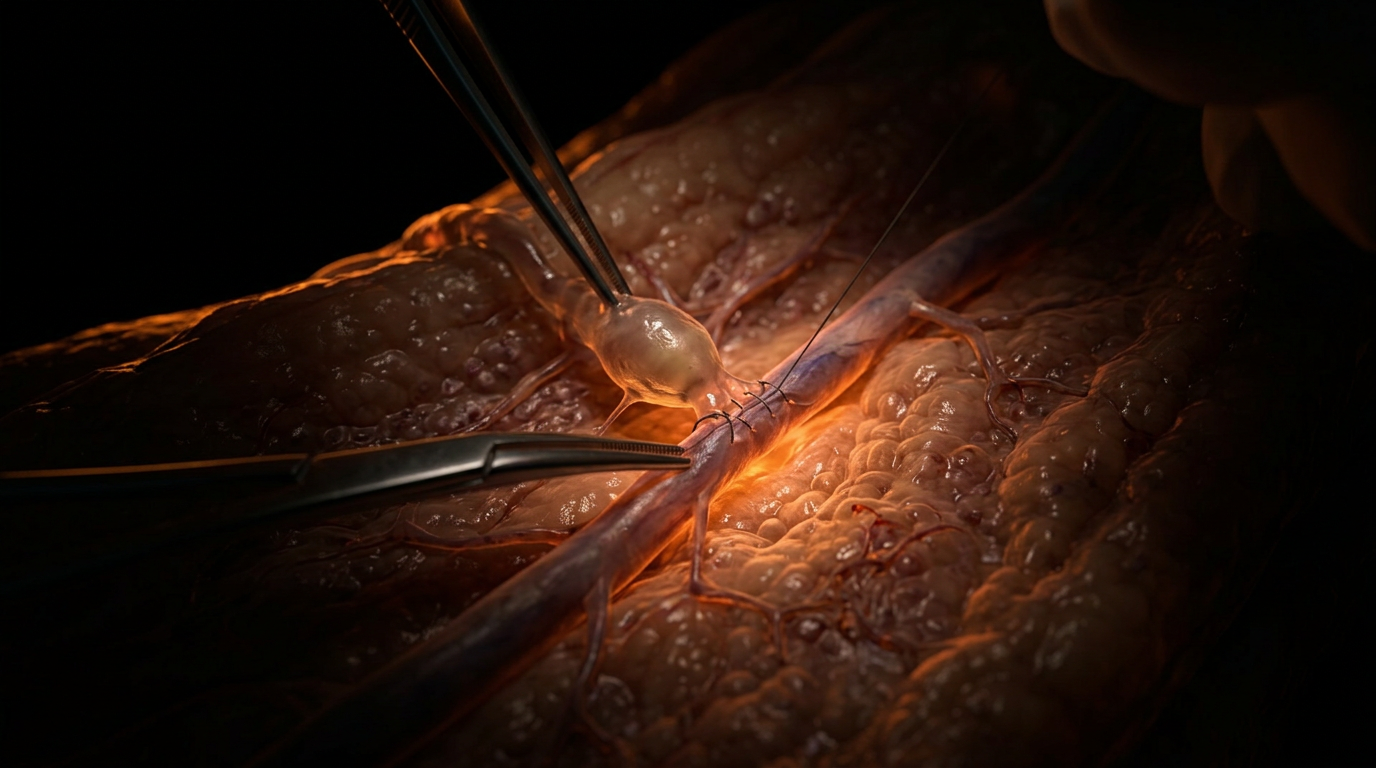
The primary protective measure in contemporary oncological practice is the transition from radical clearance to Sentinel Lymph Node Biopsy (SLNB) and, increasingly, the implementation of Axillary Reverse Mapping (ARM). Peer-reviewed data in *The Lancet Oncology* suggests that ARM significantly reduces the incidence of breast cancer-related lymphoedema (BCRL) by identifying and preserving the specific efferent vessels draining the upper extremity. However, when disruption is unavoidable, the "Lymphatic Microsurgical Preventing Healing" (LYMPHA) technique—pioneered to create immediate lymphovenous bypasses at the time of dissection—represents a paradigm shift in preventative microsurgery. This proactive approach aims to shunt lymph directly into the venous system, bypassing the obliterated nodal basin and mitigating the catastrophic rise in endoluminal pressure.
Post-operative recovery protocols must prioritise the mitigation of the "second hit" phenomenon. Evidence published in *Nature Reviews Disease Primers* indicates that while surgery provides the initial insult, a secondary inflammatory trigger—often subclinical infection or oxidative stress—precipitates the transition from latent lymphatic insufficiency to overt lymphoedema. Consequently, recovery at INNERSTANDIN focuses on strict dermatological integrity to prevent the infiltration of pathogens that stimulate a pro-fibrotic cytokine cascade, specifically the overexpression of Transforming Growth Factor-beta 1 (TGF-β1). TGF-β1 is the primary driver of the transformation of fibroblasts into myofibroblasts, leading to the deposition of excess extracellular matrix and subsequent tissue fibrosis.
Mechanical intervention, through Complex Decongestive Therapy (CDT), remains the gold standard in the UK context, as outlined by British Lymphology Society guidelines. However, the biological mechanism is often misunderstood. It is not merely fluid displacement; it is a method of mechanotransduction. Manual Lymphatic Drainage (MLD) and graduated compression stimulate the intrinsic contractility of lymphangions—the functional units of lymphatic vessels—increasing the frequency and stroke volume of lymphatic pulses. Furthermore, progressive resistance exercise protocols, supported by the PAL (Physical Activity and Lymphedema) trial, demonstrate that controlled muscular contraction serves as an external pump, enhancing the clearance of protein-rich interstitial fluid and reducing the pro-inflammatory milieu.
Biological reclamation also requires nutritional and pharmacological optimisation aimed at supporting the endothelial glycocalyx—the delicate sugar-protein layer lining the lymphatic endothelium. Disruption of this layer increases permeability and exacerbates oedema. By targeting the molecular pathways of lymphangiogenesis, specifically through the modulation of Vascular Endothelial Growth Factor C (VEGF-C) signalling, we can theoretically encourage the formation of collateral lymphatic pathways, providing a biological bypass for the surgically interrupted system. This exhaustive approach ensures that recovery is not a passive waiting period, but a rigorous, evidence-led restoration of cellular and systemic health.
Summary: Key Takeaways
The surgical excision of lymphatic architecture—whether via radical axillary clearance or sentinel node biopsy—precipitates a profound disruption of the interstitium's proteostasis. At INNERSTANDIN, our synthesis of current oncology data indicates that this mechanical insult triggers an immediate elevation in interstitial hydrostatic pressure, fundamentally altering the mechanotransduction pathways of the surrounding tissue. As evidenced in *The Lancet Oncology*, the resulting stasis of protein-rich fluid is not merely a passive accumulation; it is a pro-inflammatory stimulus that activates the TGF-β1 signalling pathway, driving the transdifferentiation of fibroblasts into myofibroblasts. This process culminates in the pathological deposition of fibroadipose tissue, which characterises late-stage, non-pitting lymphoedema.
Furthermore, cellular repair mechanisms are often compromised by an insufficiency in the VEGF-C/VEGFR3 axis, the primary molecular driver of lymphangiogenesis. Research highlights that the chronic inflammatory milieu, dominated by M1-polarised macrophages, actively inhibits the functional regeneration of lymphatic collectors. Within the UK clinical landscape, this systemic failure in immune surveillance—caused by the loss of nodal filtration hubs—increases the risk of recurrent cellulitis and metabolic waste accumulation. Reclaiming health post-surgery necessitates a sophisticated INNERSTANDIN of these biological cascades, moving beyond simple compression to address the underlying cellular senescence and extracellular matrix remodelling that sustain the lymphoedema state. These findings underscore that surgical disruption is a systemic event, requiring targeted interventions to restore fluid homeostasis and immunological integrity.
This article is provided for informational and educational purposes only. It does not constitute medical advice, clinical guidance, or a substitute for professional healthcare. Information reflects cited research at time of publication. Always consult a qualified healthcare professional before acting on any health information.
RESEARCH FOUNDATIONS
Biological Credibility Archive
Citations provided for educational reference. Verify via PubMed or institutional databases.
Medical Disclaimer
The information in this article is for educational purposes only and does not constitute medical advice, diagnosis, or treatment. Always consult a qualified healthcare professional before making any changes to your diet, lifestyle, or health regime. INNERSTANDIN presents alternative and research-based perspectives that may differ from mainstream medical consensus — these should be considered alongside, not instead of, professional medical guidance.
Read Full DisclaimerReady to learn more?
Continue your journey through our classified biological research.
DISCUSSION ROOM
Members of THE COLLECTIVE discussing "Surgical Disruption and Cellular Repair: Reclaiming Health After Lymph Node Removal"
SILENT CHANNEL
Be the first to discuss this article. Your insight could help others understand these biological concepts deeper.
RABBIT HOLE
Follow the biological thread deeper