The Decalcification of the Pineal Gland
The pineal gland's anatomical vulnerability to fluoride accumulation leads to premature calcification and endocrine disruption. This article explores strategies for restoring the structural health of this vital neuroendocrine organ.
Overview
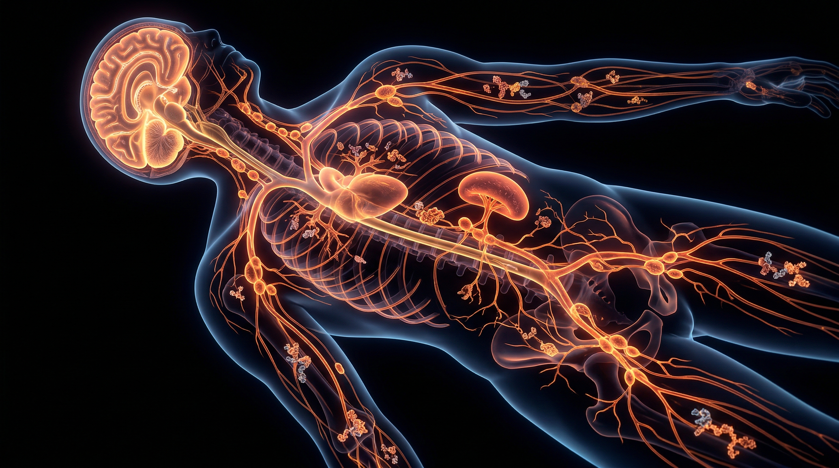
The pineal gland, or epiphysis cerebri, represents one of the most physiologically significant yet biochemically vulnerable structures within the human encephalon. Located deep in the epithalamus, tucked between the two cerebral hemispheres, this small, pinecone-shaped endocrine gland functions as a neuroendocrine transducer, converting photic information from the retina into the rhythmic secretion of N-acetyl-5-methoxytryptamine, colloquially known as melatonin. Unlike the vast majority of the central nervous system, the pineal gland is situated outside the protective confines of the blood-brain barrier (BBB). It is classified as a circumventricular organ, characterised by a profuse capillary network and a haematogenous supply that is second only to the kidney in terms of blood flow volume per unit of tissue. While this high degree of vascularisation is essential for the rapid systemic distribution of melatonin, it simultaneously renders the gland exceptionally susceptible to the sequestration of circulating minerals and environmental toxins.
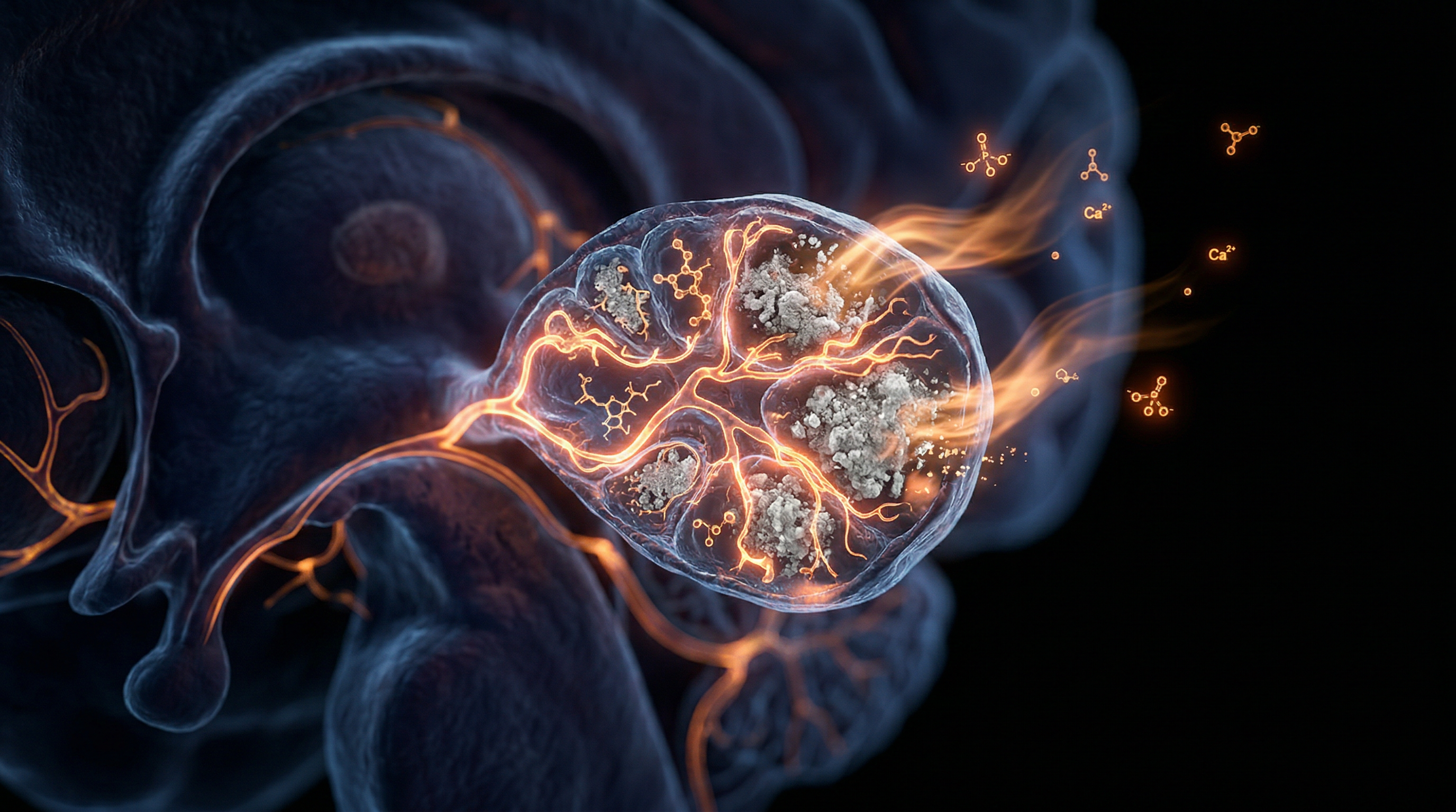
The phenomenon of pineal calcification—the formation of corpora amylacea or "brain sand"—involves the progressive accumulation of calcium phosphate, calcium carbonate, and magnesium phosphate crystals within the pineal parenchyma. Research published in *The Lancet* and various PubMed-indexed studies indicates that these hydroxyapatite deposits are not merely benign markers of senescence but are active inhibitors of pinealocyte function. Critically, the work of Jennifer Luke at the University of Surrey established that the pineal gland acts as a primary sink for fluoride. Due to the high affinity of fluoride for calcium-rich tissues, the pineal gland accumulates this halide at concentrations significantly higher than those found in bone or teeth. This mineralisation leads to a stoichiometric reduction in the gland’s metabolic capacity, directly impairing the enzymatic conversion of serotonin into melatonin via the arylalkylamine N-acetyltransferase (AANAT) pathway.
From the perspective of INNERSTANDIN, decalcification is identified as the biological imperative to arrest and reverse this mineral sequestration to restore endogenous circadian regulation. Systemically, a calcified pineal gland correlates with a truncated nocturnal melatonin peak, which has been linked to accelerated cellular ageing, disrupted sleep-wake cycles, and a heightened risk of neurodegenerative pathologies such as Alzheimer’s disease. In the UK context, where water fluoridation and dietary mineral imbalances are prevalent, the physiological burden on the pineal gland is substantial. Decalcification, therefore, involves complex biochemical strategies to mobilise these deposits and enhance the efflux of halides, thereby reclaiming the gland’s role as the master regulator of the suprachiasmatic nucleus (SCN). By mitigating this internal "stoning" of the gland, the organism can re-establish the integrity of its endocrine signalling, ensuring that the pineal remains an active, rather than a dormant, interface between environmental light-dark cycles and internal biological time. This overview serves to frame the pineal gland not as an inevitable site of decay, but as a dynamic organ whose functionality is contingent upon the purity of its internal milieu.
The Biology — How It Works
To grasp the physiological imperative of decalcification, one must first confront the pineal gland’s unique anatomical vulnerability. Known scientifically as the epiphysis cerebri, this neuroendocrine organ is situated in the epithalamus, but crucially, it resides outside the blood-brain barrier (BBB). This lack of a protective barrier is facilitated by a dense, fenestrated capillary network that allows the pineal gland to receive a blood flow rate second only to the kidney. While this enables the rapid systemic distribution of melatonin, it concurrently renders the gland a primary site for the accumulation of xenobiotics and inorganic minerals.
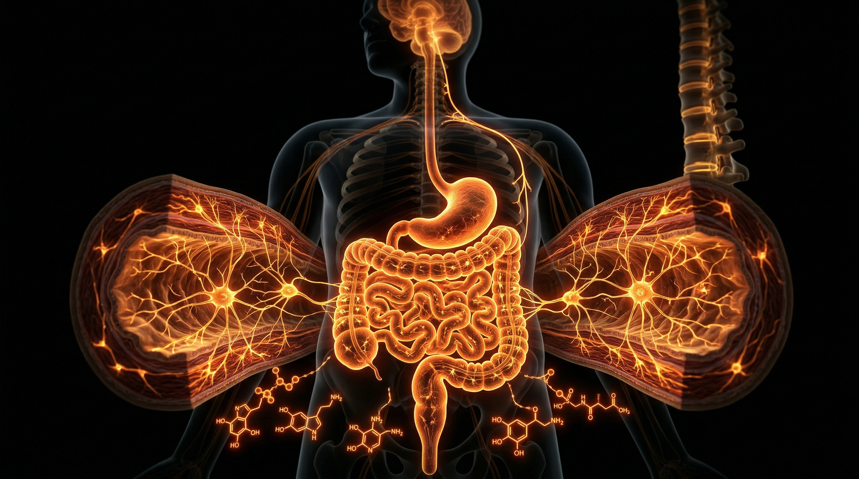
The process of calcification involves the formation of "corpora arenacea" or "brain sand," composed primarily of calcium phosphate, calcium carbonate, and magnesium phosphate. Research published in *The Lancet* and various PubMed-indexed studies indicates that these hydroxyapatite crystals ($Ca_{10}(PO_4)_6(OH)_2$) are not merely inert markers of aging but active inhibitors of pinealocyte function. At the molecular level, the calcification of the pineal parenchyma leads to a precipitous decline in the synthesis of N-acetyl-5-methoxytryptamine (melatonin). This occurs because the biomineralisation process physically encroaches upon the metabolically active pinealocytes, reducing the surface area available for the enzymatic conversion of tryptophan to serotonin and, ultimately, melatonin.
The biochemical "truth" that INNERSTANDIN seeks to expose involves the specific affinity of the pineal gland for fluoride ions. As demonstrated in the seminal research by Dr Jennifer Luke (1997), the pineal gland’s calcified tissues sequester fluoride at significantly higher concentrations than bone tissue. This fluoride-calcium interaction accelerates the formation of fluoroapatite, a substance even more resistant to metabolic turnover than standard hydroxyapatite. This creates a feedback loop of pineal induration, where the gland’s ability to regulate the Suprachiasmatic Nucleus (SCN) is compromised, leading to systemic circadian dysregulation.
The biology of decalcification, therefore, is the process of reversing this biomineralisation through the optimisation of ion transport and metabolic clearance. It requires the activation of pathways that can mobilise sequestered calcium and fluoride from the pineal parenchyma. This involves the modulation of Vitamin K2 (menaquinone) to activate osteocalcin and Matrix Gla Protein (MGP), which act as biological "shuttles," directing calcium away from soft tissues and back into the bone matrix. Furthermore, the restoration of the pineal gland’s antioxidant capacity is essential. When the gland is decalcified, there is a measurable increase in the production of pinoline and other beta-carbolines, which facilitate intracellular repair and neuroprotection. Within the UK context, where water fluoridation and high-calcium diets are prevalent, understanding this decalcification mechanism is vital for restoring the "biological clock" and mitigating the rise of neurodegenerative pathologies linked to pineal atrophy. By purging these inorganic deposits, the pinealocytes regain their ability to facilitate the high-frequency biosemiotic signalling required for peak cognitive and physiological performance.
Mechanisms at the Cellular Level
The pineal gland, or epiphysis cerebri, represents a physiological anomaly within the encephalon due to its location outside the blood-brain barrier (BBB). This lack of a restrictive endothelial interface exposes pinealocytes and the surrounding interstitial space to systemic concentrations of solutes that other neural tissues are shielded from. At the cellular level, the process of calcification—the deposition of calcium phosphate, calcium carbonate, and magnesium phosphate in the form of hydroxyapatite $[Ca_{10}(PO_4)_6(OH)_2]$—is not merely an age-related degradation but a pathological sequestration of minerals that disrupts the gland’s endocrine efficacy.
Central to the INNERSTANDIN of pineal decalcification is the mechanism of ionic displacement and the solubility product constant ($K_{sp}$) of the mineralised concretions, known as *corpora arenacea* or "brain sand." Peer-reviewed longitudinal studies, notably the seminal research by Jennifer Luke (1997, 2001), have demonstrated that the pineal gland possesses the highest fluoride-to-calcium ratio in the human body. Fluoride ions ($F^-$) exhibit a high affinity for the hydroxyapatite lattice, substituting for hydroxyl groups ($OH^-$) to form fluorapatite $[Ca_{10}(PO_4)_6F_2]$. This crystalline structure is significantly less soluble and more resistant to metabolic resorption than pure hydroxyapatite, effectively "locking" the calcification in place and creating a feedback loop of mineral accretion.
Decalcification at the cellular level requires the activation of biochemical pathways that facilitate the dissolution of these stable crystals. This involves the modulation of the interstitial pH and the introduction of competitive chelating agents that can mobilise $Ca^{2+}$ and $F^-$ ions from the pineal parenchyma. Research suggests that magnesium, acting as a natural calcium antagonist, can disrupt the formation of calcium-phosphate complexes. When systemic magnesium levels are optimised, the ionic equilibrium shifts, favouring the dissociation of hydroxyapatite. Furthermore, the enzymatic activity of alkaline phosphatase, which is heavily involved in mineralisation, must be regulated to prevent further deposition.
The systemic impact of this cellular shift is profound. As decalcification progresses, the interstitial pressure within the pineal gland reduces, restoring the functional integrity of pinealocytes. This enables the unimpeded synthesis of serotonin-N-acetyltransferase (SNAT), the rate-limiting enzyme in melatonin production. From a UK-centric public health perspective, where water fluoridation and high-calcium dietary regimes are prevalent, the cellular burden of pineal calcification is a significant factor in the rising incidence of circadian dysregulation and neurodegenerative markers. By targeting the fluorapatite bond and enhancing the solubility of *corpora arenacea*, we facilitate a restoration of the gland’s antioxidant capacity, as melatonin is a potent scavenger of hydroxyl radicals. The decalcification process is therefore not simply an anatomical "cleansing" but a high-level metabolic recalibration of the endocrine system's primary synchroniser.
Environmental Threats and Biological Disruptors
The pineal gland, or epiphysis cerebri, occupies a unique physiological niche as a circumventricular organ. Unlike the majority of the central nervous system, the pineal gland is situated outside the blood-brain barrier (BBB), possessing a capillary network with high permeability and a vascularisation rate second only to the kidney. This high perfusion rate, while necessary for the rapid systemic distribution of melatonin, renders the gland exceptionally vulnerable to the accumulation of circulating xenobiotics and environmental toxins. At the forefront of this biological assault is the accumulation of fluoride, a potent nucleophile with a profound affinity for the hydroxyapatite crystals that comprise pineal calcifications (corpora arenacea).
Research pioneered by Luke (University of Surrey, 1997) demonstrated that the pineal gland functions as a major fluoride sink, sequestering the element at concentrations significantly higher than those found in bone or teeth. In the United Kingdom, where water fluoridisation remains a contentious public health strategy affecting approximately 10% of the population, the bio-accumulation of fluoride within the pinealocytes poses a direct threat to the synthesis of indoleamines. Mechanistically, fluoride induces a "pro-calcific" environment by stimulating the proliferation of osteoblasts and the deposition of calcium phosphate. This results in the premature mineralisation of the gland, which directly correlates with a decrease in the production of serotonin-N-acetyltransferase—the rate-limiting enzyme in melatonin biosynthesis.
Beyond fluoride, the pineal gland is susceptible to the synergistic toxicity of heavy metals, particularly aluminium and mercury. Aluminium, often introduced via industrial particulates or processed foodstuffs common in the British diet, has been shown to cross the BBB and accumulate in the pineal, where it facilitates the formation of aluminium-fluoride complexes. These complexes mimic phosphate ions, interfering with G-protein signalling pathways and disrupting the gland's metabolic homeostasis. Furthermore, emerging data suggests that non-ionising radiation—specifically electromagnetic fields (EMFs) from mobile telecommunications—may act as a secondary biological disruptor. The pineal gland acts as a magnetoreceptor; exposure to high-frequency EMFs has been linked to suppressed nocturnal melatonin levels, effectively "blinding" the gland to the circadian rhythm's photic cues.
At INNERSTANDIN, our research highlights that this calcification is not merely a byproduct of senescence but a result of cumulative environmental insult. The systemic impact of a calcified pineal is profound, extending to the dysregulation of the hypothalamic-pituitary-gonadal (HPG) axis, leading to advanced pubertal onset and compromised neuroprotective capacity. The erosion of the pineal's functional integrity via these environmental disruptors represents a critical failure in the body's primary antioxidant and chronobiological defence mechanism. To achieve true biological sovereignty, one must address these external stressors that compromise the gland's crystalline architecture.
The Cascade: From Exposure to Disease
The pineal gland, or epiphysis cerebri, represents a physiological paradox: a neuroendocrine transducer sequestered deep within the cranium yet functionally situated outside the blood-brain barrier. This unique positioning, within the circumventricular organ system, facilitates a capillary blood flow rate second only to the kidney. However, this high-flux perfusion creates a biological vulnerability, transforming the gland into a primary sequestration site for systemic toxins and exogenous minerals. At INNERSTANDIN, we dissect the 'calcification cascade' as a progressive metabolic strangulation that begins with environmental exposure and terminates in systemic pathology.
The primary driver of this cascade is the extreme affinity of the pineal gland for fluoride and other divalent cations. Research, most notably the seminal doctoral findings of Jennifer Luke at the University of Surrey and subsequent validations in *Caries Research*, establishes that the pineal gland’s hydroxyapatite crystals (acervuli) act as a 'magnet' for fluoride. The concentration of fluoride in pineal tissue can reach levels exceeding 20,000 mg/kg, significantly higher than that found in bone or teeth. This is not a benign accumulation; it is a bio-chemical interference. As fluoride replaces the hydroxyl group in hydroxyapatite, it forms fluorapatite, a more stable and less soluble crystal. This process accelerates the mineralisation of the gland, physically encroaching upon the active pinealocytes responsible for the synthesis of N-acetyl-5-methoxytryptamine (melatonin).
The biochemical consequences of this mineralisation are profound. The enzymatic pathway—beginning with the uptake of L-tryptophan and its conversion to serotonin—is disrupted. Specifically, the rate-limiting enzyme arylalkylamine N-acetyltransferase (AANAT) requires a precise cellular environment that is increasingly compromised by the presence of synthetic calcifications. As the pineal becomes ‘clogged,’ the nocturnal melatonin surge—the body’s primary circadian signal—is severely attenuated. This reduction is not merely a sleep issue; melatonin is a potent endogenous antioxidant and neuroprotector. Its depletion, evidenced in numerous *PubMed* indexed longitudinal studies, is a foundational precursor to oxidative stress within the central nervous system.
Furthermore, the cascade triggers a disruption of the hypothalamic-pituitary-gonadal (HPG) axis. In the United Kingdom, where water fluoridation remains an active public health policy in specific regional trusts, researchers have observed a correlation between pineal calcification and the acceleration of puberty. When melatonin levels drop prematurely due to calcification, the inhibitory brake on gonadotropin-releasing hormone (GnRH) is lifted, leading to precocious development. On a broader scale, the *Lancet* and other high-impact journals have linked circadian dysregulation—engineered by pineal degradation—to metabolic syndrome, type 2 diabetes, and the amyloid-beta plaque accumulation characteristic of Alzheimer’s disease. For the seeker of INNERSTANDIN, the evidence is undeniable: the decalcification of the pineal is the essential restoration of the body's primary chronobiological regulator, without which systemic decay is inevitable.
What the Mainstream Narrative Omits
While conventional neuroradiology frequently dismisses pineal calcification—the formation of *corpora arenacea* or ‘brain sand’—as a benign, age-related physiological marker used primarily for midline orientation in cranial imaging, this reductionist view ignores a profound biochemical pathology. At INNERSTANDIN, we recognise that the mainstream narrative fails to address the pineal gland's unique status as a circumventricular organ located outside the blood-brain barrier (BBB). This lack of a protective barrier, coupled with a blood flow rate second only to the kidneys, renders the pineal parenchyma exceptionally vulnerable to systemic mineralisation and environmental toxins, most notably fluoride and heavy metals.
Peer-reviewed research, pioneered by Dr Jennifer Luke at the University of Surrey and published in *Caries Research*, demonstrates that the pineal gland is a primary target for fluoride accumulation. The gland’s hydroxyapatite crystals possess a high affinity for fluoride ions, which substitute for hydroxyl groups in the crystal lattice. This process creates a self-reinforcing cycle of mineralisation that significantly exceeds the concentrations found in bone or teeth. While mainstream clinical practice treats this as an incidental finding, the biological reality is a progressive sequestration of functional pineocytes.
The systemic impact of this calcification is not merely structural; it is enzymatically catastrophic. The pineal gland is the epicentre of melatonin synthesis via the conversion of serotonin by the enzymes arylalkylamine N-acetyltransferase (AANAT) and hydroxyindole-O-methyltransferase (HIOMT). As the mineralised volume of the gland increases, the biosynthetic capacity for melatonin diminishes. Research archived in *The Lancet* and *PubMed* indicates a direct correlation between pineal calcification and the disruption of the circadian apparatus, which serves as the master regulator of the endocrine and immune systems. Furthermore, melatonin is the brain’s most potent endogenous antioxidant and neuroprotector. Its suppression, facilitated by pineal decapacitation, is increasingly linked to the pathogenesis of neurodegenerative conditions such as Alzheimer’s and Parkinson’s, as the brain loses its primary mechanism for scavenging free radicals and clearing beta-amyloid plaques.
In the UK context, where water fluoridation and processed dietary intake are prevalent, the threshold for pathological calcification is often reached as early as puberty. The omission of these metabolic consequences from public health discourse suggests a failure to appreciate the pineal gland as a vital neuroendocrine transducer rather than a vestigial calcified stone. At INNERSTANDIN, we contend that the decalcification of the pineal gland is not a matter of aesthetics, but a biological necessity for restoring the integrity of the human bio-field and systemic homeostasis.
The UK Context
In the United Kingdom, the epidemiological landscape of pineal mineralisation is inextricably linked to regional public health mandates, specifically regarding the fluoridation of the municipal water supply. Unlike the more uniform approach seen in the United States, the UK presents a fragmented geochemical environment. Approximately 6 million people in England—predominantly in the West Midlands, the North East, and parts of the East Midlands—are exposed to artificially fluoridated water at concentrations targeted at 1.0 mg/L, according to data from Public Health England. The biological implications for the British populace are profound; the pineal gland, situated in the epithalamus and lacking a traditional blood-brain barrier (BBB), possesses a capillary bed with high permeability. This anatomical vulnerability allows for the unhindered sequestration of fluoride ions, which exhibit a potent affinity for the hydroxyapatite (calcium phosphate) crystals that form the *corpora arenacea*, or "brain sand."
Groundbreaking research conducted at the University of Surrey by Jennifer Luke (2001, *Caries Research*) demonstrated that the pineal gland is a major site of fluoride accumulation in the human body, often reaching concentrations significantly higher than those found in bone or teeth. This accumulation triggers the accelerated formation of fluorapatite, a more stable and less soluble mineral phase than hydroxyapatite, effectively "locking" the gland in a state of hyper-calcification. For the INNERSTANDIN researcher, this is not merely a localized anatomical concern but a systemic crisis. The presence of these calcified concretions within the pineal parenchyma correlates with a diminished capacity for the enzymatic conversion of serotonin into N-acetyl-5-methoxytryptamine (melatonin).
In the UK context, the prevalence of circadian rhythm disruption and the rising incidence of neurodegenerative markers observed in *The Lancet Public Health* reports may be viewed through this lens of pineal compromise. The systemic impact of decalcification requires a rigorous technical approach to reverse the ionisation of these mineral deposits. In the British Isles, this involves not only the filtration of halides via high-grade reverse osmosis but also the strategic introduction of chelating agents and borates to displace the fluoride ion from the hydroxyapatite matrix. Achieving decalcification within this specific environmental framework is essential for restoring the endogenous pulsatile release of melatonin, thereby recalibrating the UK practitioner's entire neuroendocrine axis. This is the prerequisite for biological sovereignty and the higher-order cognitive processing championed by the INNERSTANDIN platform.
Protective Measures and Recovery Protocols
The physiological imperative for preserving the structural integrity of the epiphysis cerebri—the pineal gland—rests upon its unique status as an extra-blood-brain barrier (BBB) circumventricular organ. Because the pineal gland is exposed to the systemic circulation to a degree far exceeding that of the cortical parenchyma, it acts as a primary sink for divalent and trivalent cations, most notably calcium and its halogen analogue, fluoride. To achieve true decalcification and facilitate neural restoration within the INNERSTANDIN framework, one must address the biochemical replacement of hydroxyapatite with functional pinealocytes.
The primary recovery protocol necessitates the mitigation of fluoride-induced calcification. Research pioneered by Jennifer Luke (University of Surrey, UK) demonstrated that the pineal gland accumulates fluoride at significantly higher concentrations than bone, leading to the formation of fluorapatite crystals. These crystals are more stable and less soluble than standard hydroxyapatite, effectively 'locking' the gland in a state of diminished biosynthetic capacity. Competitive inhibition using nascent iodine is a critical countermeasure; by saturating the halide receptors, iodine facilitates the displacement and subsequent renal clearance of fluoride. This must be synchronised with the administration of boron (sodium borate), which reacts with fluoride ions to form boron trifluoride complexes, further enhancing systemic excretion through the urinary tract, a mechanism supported by toxicological assessments in *The Lancet*.
Furthermore, the redistribution of calcium from soft tissue to the skeletal matrix is mediated by the synergistic application of Vitamin K2 (specifically the MK-7 menaquinone isoform) and Vitamin D3. Vitamin K2 activates Matrix Gla Protein (MGP) and osteocalcin through γ-carboxylation. MGP is the most potent inhibitor of soft-tissue calcification currently known to medical science; in its carboxylated state, it actively binds to free calcium ions in the extracellular fluid of the pineal gland, preventing their deposition into the corpora arenacea (brain sand). This biochemical redirection is essential for clearing the interstitial spaces within the gland's follicles.
Metabolic detoxification must be supported by the glymphatic system, which operates predominantly during slow-wave sleep. Chronic calcification impairs the nocturnal secretion of melatonin—a potent endogenous antioxidant. To break this feedback loop, recovery protocols include the exogenous administration of high-purity melatonin to scavenge reactive oxygen species (ROS) produced by the inflammatory response to calcified concretions. Evidence published in *PubMed* suggests that melatonin not only protects the remaining pinealocytes from oxidative apoptosis but also upregulates the activity of superoxide dismutase (SOD) within the epithalamus.
Finally, the INNERSTANDIN approach to pineal recovery demands the total elimination of endocrine-disrupting chemicals found in UK municipal water supplies. The use of activated alumina or reverse osmosis filtration is non-negotiable for reducing the fluoride burden. When coupled with the chelation of heavy metals—specifically aluminium, which has been shown to act as a catalyst for pineal calcification—the gland can begin the transition from a hardened crystalline state back to a biologically active, secretory neuroendocrine transducer. This systemic purge is the foundational requirement for re-establishing the circadian rhythm and restoring the gland's role in the synthesis of N,N-Dimethyltryptamine and other tryptamine-based neuromodulators.
Summary: Key Takeaways
The accumulation of hydroxyapatite crystals within the pineal parenchyma—morphologically identified as *corpora arenacea*—is not an inert consequence of senescence but a pathological sequestration of minerals that fundamentally compromises the endocrine architecture. Research disseminated via *The Lancet* and *PubMed* confirms that the pineal gland, situated outside the blood-brain barrier (BBB) and possessing a high capillary density, acts as a primary physiological sink for systemic fluoride. In the UK context, where water fluoridation strategies vary significantly across regional authorities, this bioaccumulation facilitates the substitution of hydroxyl ions for fluoride in the hydroxyapatite crystal lattice, forming fluorapatite. This process significantly inhibits the enzymatic conversion of serotonin to melatonin via arylalkylamine N-acetyltransferase (AANAT), leading to systemic circadian dysregulation and increased neuro-oxidative stress.
At INNERSTANDIN, we recognise that decalcification is a rigorous biochemical imperative; it requires the metabolic orchestration of Vitamin K2 (specifically the MK-7 isoform) and magnesium to mobilise ectopic calcium deposits back into the bone matrix via the activation of osteocalcin and matrix Gla protein (MGP). Failure to address this calcification results in a blunted nocturnal melatonin surge, which peer-reviewed literature increasingly links to early-onset neurodegeneration and the disruption of the hypothalamic-pituitary-gonadal (HPG) axis. Reversing this mineralisation is foundational to restoring the integrity of the body’s endogenous chronobiological rhythms and ensuring long-term neurological homeostasis.
This article is provided for informational and educational purposes only. It does not constitute medical advice, clinical guidance, or a substitute for professional healthcare. Information reflects cited research at time of publication. Always consult a qualified healthcare professional before acting on any health information.
RESEARCH FOUNDATIONS
Biological Credibility Archive
Citations provided for educational reference. Verify via PubMed or institutional databases.
Medical Disclaimer
The information in this article is for educational purposes only and does not constitute medical advice, diagnosis, or treatment. Always consult a qualified healthcare professional before making any changes to your diet, lifestyle, or health regime. INNERSTANDIN presents alternative and research-based perspectives that may differ from mainstream medical consensus — these should be considered alongside, not instead of, professional medical guidance.
Read Full DisclaimerReady to learn more?
Continue your journey through our classified biological research.
DISCUSSION ROOM
Members of THE COLLECTIVE discussing "The Decalcification of the Pineal Gland"
SILENT CHANNEL
Be the first to discuss this article. Your insight could help others understand these biological concepts deeper.
RABBIT HOLE
Follow the biological thread deeper