Advanced Glycation End-Products and the Impairment of Claudin-Dependent Barrier Function in Diabetic Nephropathy
Discuss this resourceUpdated August 2026
An exploration of how metabolic dysfunction leads to the degradation of renal tight junctions, focusing on the role of AGEs in disrupting claudin-mediated barrier integrity in diabetic kidney disease.
Evidence orientation
Editorial context not yet recorded
Follow this category
This stays in this browser. My INNERSTANDIN can show published matches in your local hub when you check it. It does not send email, push, or alert notifications.
Local learning review
A private browser aid for revisiting ideas. It is not an alert or a health recommendation.
Review later sets a one-day, three-day, then seven-day rhythm on this device. Choose it only when you want to revisit this article.
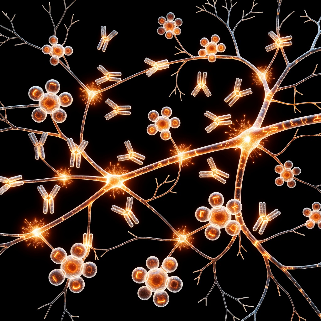
In the landscape of chronic metabolic disease, Diabetic Nephropathy (DN) remains a leading cause of end-stage renal failure. While systemic blood glucose control is often the primary focus, the cellular root cause involves a profound collapse of the renal glomerular and tubular barrier. At the heart of this collapse lies the intersection of Advanced Glycation End-Products (AGEs) and the specialized protein complexes known as Tight Junctions (TJs).
The Role of Claudins in Renal Homeostasis
Tight junctions are the gatekeepers of the kidney. Among their most critical components are the claudin family of transmembrane proteins. These proteins regulate the paracellular permeability pathway—the "spaces" between cells through which ions and water must pass. In a healthy kidney, specific claudins, such as claudin-1, -2, and -16, maintain a selective barrier, ensuring that essential solutes are reabsorbed while harmful metabolic waste is excreted. When these claudin bridges are compromised, the barrier becomes 'leaky,' leading to proteinuria and eventual fibrosis.
AGEs: The Biochemical Saboteurs
Advanced Glycation End-Products (AGEs) are proteins or lipids that become glycated as a result of exposure to sugars. In the hyperglycemic environment of a diabetic patient, the production of AGEs accelerates. These compounds do not merely circulate; they bind to their specific cell-surface receptor, RAGE (Receptor for Advanced Glycation End-Products).
Once the AGE-RAGE axis is activated, it triggers a cascade of oxidative stress and pro-inflammatory signaling via the NF-κB pathway. This is where the structural integrity of the tight junction suffers. Research indicates that AGE-induced signaling downregulates the expression of essential claudins and alters their subcellular localization. Instead of reinforcing the junction, the proteins are internalized and degraded, leaving the intercellular spaces wide open.
The Pathological Cascade
- Hyperglycemia: Persistent high blood glucose drives non-enzymatic glycation of proteins.
- AGE Accumulation: These stable compounds accumulate in the renal mesangium and tubular epithelium.
- RAGE Activation: AGEs bind to RAGE, initiating intracellular oxidative stress.
- Tight Junction Disruption: Signaling pathways (notably MAPK and PKC) interfere with the scaffolding proteins like ZO-1, which anchor claudins to the cytoskeleton.
- Barrier Failure: The loss of claudin-dependent tight junctions leads to proteinuria and renal structural remodeling.
Addressing the Root Cause
Understanding this mechanism shifts the therapeutic focus. Beyond insulin and blood pressure management, modern research is looking toward inhibiting RAGE activation and clearing AGEs. Maintaining a lifestyle that prioritizes metabolic flexibility and reduces dietary AGE intake (such as minimizing high-heat processed foods) is an emerging frontline strategy in protecting the delicate architecture of the kidney.
By viewing Diabetic Nephropathy as a failure of barrier science rather than just a plumbing issue, patients and practitioners can better implement strategies to support cellular health at the tight junction level.
This article is provided for informational and educational purposes only. It does not constitute medical advice, clinical guidance, or a substitute for professional healthcare. Information reflects cited research at time of publication. Always consult a qualified healthcare professional before acting on any health information.
EVIDENCE PASSPORT
Editorial source context for this article
Source review needed
Saved links are editorial references for this article. They may support specific claims rather than every sentence. Open and assess each source in context. This passport does not independently verify them.
Editorial context
A complete editorial reading has not been recorded for this article, and no source links are saved on its record.
Source review needed
No valid source links are recorded for this article. Supplementary research, where available, is listed separately below.
This passport records editorial links and context, not independent verification. Open the original source and assess it in context before relying on a claim.
Medical Disclaimer
The information in this article is for educational purposes only and does not constitute medical advice, diagnosis, or treatment. Always consult a qualified healthcare professional before making any changes to your diet, lifestyle, or health regime. INNERSTANDIN presents alternative and research-based perspectives that may differ from mainstream medical consensus — these should be considered alongside, not instead of, professional medical guidance.
Read Full DisclaimerContinue the thread
Keep this question moving.
Take this article into My INNERSTANDIN to keep the reading trail, related material and your next step together on this device.
Explore this in the Body Map
Explore related body structures and their educational pages in the interactive Body Map.
RABBIT HOLE
Follow the biological thread deeper