
Alopecia Areata and Autoimmunity: Investigating the Body’s Attack on the Follicle
Discuss this resourceUpdated October 2026
Autoimmune responses can cause the immune system to target healthy hair follicles, leading to patchy loss. We explore the underlying triggers of Alopecia Areata and potential biological interventions.
Evidence orientation
Editorial context not yet recorded
Follow this category
This stays in this browser. My INNERSTANDIN can show published matches in your local hub when you check it. It does not send email, push, or alert notifications.
Local learning review
A private browser aid for revisiting ideas. It is not an alert or a health recommendation.
Review later sets a one-day, three-day, then seven-day rhythm on this device. Choose it only when you want to revisit this article.
Overview
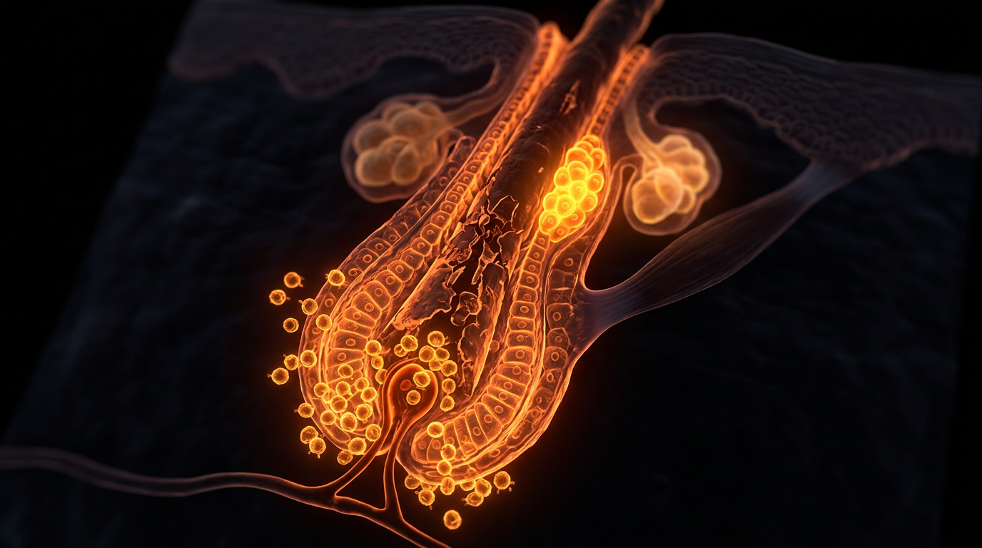
Alopecia Areata (AA) represents a classic yet uniquely nuanced organ-specific autoimmune disease characterised by the immune-mediated destruction of anagen-phase hair follicles. Historically mischaracterised as a superficial dermatological condition driven primarily by psychogenic stress, contemporary molecular pathology—as documented extensively in literature from The Lancet and peer-reviewed PubMed repositories—redefines AA as a profound systemic inflammatory disorder. Here, the body’s immune surveillance apparatus undergoes a fundamental breakdown in peripheral tolerance, erroneously identifying the rapidly proliferating matrix keratinocytes of the anagen hair bulb as foreign or noxious neo-antigens. This pathological cascade precipitates a targeted assault, collapsing the unique immune privilege of the anagen hair follicle and converting a site of active, high-rate tissue regeneration into a chronic battleground of cell-mediated cytotoxicity.
To fully dissect this phenomenon, one must examine the baseline physiology of the normal hair follicle. Under homeostatic conditions, the proximal anagen hair bulb maintains a robust, localized immune-privileged status, closely mirroring that of the eye, placenta, and testis. This physiological firewall is achieved through the down-regulation of classical major histocompatibility complex (MHC) class Ia molecules and the concurrent up-regulation of immunosuppressive local factors such as transforming growth factor-beta (TGF-$\beta$), macrophage migration inhibitory factor, and alpha-melanocyte-stimulating hormone ($\alpha$-MSH). In the context of AA, this protective shield is breached. Genome-wide association studies (GWAS) embedded within UK biobank data and multi-centre immunological consortia have underscored significant genetic susceptibility loci—most notably within the human leukocyte antigen (HLA) region, alongside non-HLA genes regulating cytotoxic T-lymphocyte activation, such as CTLA4, IL2/IL21, and the UL16 binding protein (ULBP) gene cluster.
When these genetic predispositions intersect with environmental triggers—be they viral infections, epigenetic shifts, or localized micro-trauma—an aberrant innate immune response is initiated. Plasmacytoid dendritic cells infiltrate the peribulbar region, secreting large quantities of type I interferons, which in turn drive a Th1-skewed adaptive immune response. The hallmark downstream mediator of this cascade is interferon-gamma (IFN-$\gamma$), accompanied by interleukin-15 (IL-15). These pro-inflammatory cytokines act directly upon the epithelial cells of the hair bulb, stripping away their constitutive immune privilege by aberrantly inducing the surface expression of MHC class I and class II molecules, alongside the presentation of autoantigens hitherto sequestered from the systemic immune repertoire.
Consequently, autoreactive CD8+ natural killer group 2 member D (NKG2D)+ T lymphocytes infiltrate the follicular epithelium. Guided by chemokine gradients (such as CXCL9 and CXCL10), these cytotoxic T-cells engage the exposed follicular autoantigens, releasing perforin and granzyme B directly into the target tissue. This localized assault forces the hair follicle to prematurely abort the anagen growth phase, driving it through a forced, dystrophic catagen transition into telogen, ultimately resulting in the characteristic non-scarring, patchy hair loss observed clinically.
At INNERSTANDIN, we emphasize that understanding this immunological architecture is not merely an academic exercise; it shifts the clinical paradigm. By mapping the exact molecular topography of how the host immune system orchestrates this follicular siege, translational researchers have successfully paved the way for targeted therapeutic interventions—such as Janus kinase (JAK) inhibitors—that interrupt this specific cytokine signaling loop, offering unprecedented avenues for restoring structural and functional homeostasis to the compromised hair follicle.
The Biology — How It Works
Alopecia areata (AA) represents a canonical organ-specific autoimmune disease wherein the human body’s immune surveillance apparatus fundamentally collapses its micro-environmental tolerance toward the anagen-phase hair follicle. As documented extensively in foundational literature within The Lancet and peer-reviewed dermatological repositories indexed on PubMed, the pathology is orchestrated by a breakdown of the unique immune privilege normally enjoyed by the anagen hair bulb. Under physiological conditions, the proximal portion of the anagen VI follicle actively suppresses major histocompatibility complex (MHC) class I and class II molecule expression, creates a localized immunosuppressive microenvironment via the secretion of transforming growth factor-beta (TGF-$\beta$) and alpha-melanocyte-stimulating hormone ($\alpha$-MSH), and limits dendritic cell maturation. In the context of AA, this immunological sanctuary is breached, initiating a catastrophic cascade that transforms the follicle from a site of immune ignorance to the primary target of cytotoxic destruction.
At the cellular and molecular level, the initiation phase is driven by the aberrant upregulation of interferon-gamma (IFN-$\gamma$) and interleukin-15 (IL-15) within the hair bulb. Research from leading immunological consortia highlights the central axis of Janus kinase (JAK) signaling—specifically JAK1 and JAK3, coupled with signal transducer and activator of transcription (STAT) proteins—in amplifying this inflammatory signal. The local overexpression of IFN-$\gamma$ induces the aberrant, robust expression of MHC class I and II molecules on the surface of anagen hair matrix keratinocytes, effectively stripping the follicle of its immunological disguise. Simultaneously, these cells present unidentified follicular autoantigens to autoreactive CD8+ cytotoxic T-lymphocytes that express NKG2D activating receptors.
Upon infiltration into the peribulbar region—classically described in histopathology as a "swarm of bees" pattern surrounding the hair bulb—these activated CD8+ NKG2D+ T-cells release perforin and granzyme B, inducing targeted apoptosis of the rapidly dividing matrix keratinocytes. This cytotoxic onslaught precipitates a premature, forced termination of the anagen growth phase, thrusting the follicle into a dysregulated catagen and subsequent telogen phase long before its genetically programmed timeline. Furthermore, the local inflammatory milieu stimulates the secretion of substance P and calcitonin gene-related peptide (CGRP) from perifollicular nerve fibres, exacerbating neurogenic inflammation and perpetuating a continuous loop of immune cell recruitment.
At INNERSTANDIN, our interrogation of UK-based epidemiological cohorts and translational immunology studies underscores that this breakdown is not merely localized. Systemic immune dysregulation, characterized by fluctuating systemic cytokine profiles and genetic polymorphisms within human leukocyte antigen (HLA) regions (such as HLA-DRB1 and HLA-DQB1), dictates an individual's lifetime susceptibility to these autoimmune episodes. Understanding these intricate molecular mechanisms—from the initial collapse of follicular immune privilege to the final execution of matrix keratinocytes by cytotoxic T-cells—serves as the foundational blueprint for modern targeted biologic interventions, shifting the paradigm of hair health research toward precision molecular immunotherapy.
Mechanisms at the Cellular Level
At the core of Alopecia Areata (AA) lies a profound breakdown in immune privilege, transforming the anagen hair follicle from a sanctuary into a primary target for autoreactive lymphocytes. Historically mischaracterised as a superficial dermatological condition, contemporary immunological mapping published in leading journals such as The Lancet and peer-reviewed repositories on PubMed categorises AA as a tissue-specific autoimmune disease driven by CD8+ NKG2D+ T-lymphocytes. Under homeostatic conditions, the anagen-phase hair follicle maintains an immune-privileged status by actively downregulating classical Major Histocompatibility Complex (MHC) class I and class II molecules while upregulating local immunosuppressive agents such as TGF-beta, alpha-melanocyte-stimulating hormone, and macrophage migration inhibitory factor. In the pathogenesis of AA, this protective shield is breached, precipitating a cascade of molecular signaling events that dismantle normal follicle architecture.
The initiation phase is characterised by the aberrant secretion of danger signals and pro-inflammatory cytokines, notably interferon-gamma (IFN-γ) and interleukin-15 (IL-15), within the follicular mesenchyme. Research driven by institutions within the UK and internationally demonstrates that dendritic cells and localized mast cells activate prematurely, releasing IFN-γ. This cytokine acts upon the epithelial cells of the hair matrix and the outer root sheath, forcefully inducing the aberrant expression of MHC class I and class II molecules on cells that normally lack them. Consequently, autoantigens unique to the hair follicle—though exact molecular targets remain under intense investigation—are presented to circulating adaptive immune cells.
This presentation recruits a dense peribulbar infiltrate, colloquially described in pathology as a "swarm of bees." This infiltrate is overwhelmingly dominated by cytotoxic T-cells expressing the activating immunoreceptor NKG2D, alongside natural killer (NK) cells. Upon binding their respective ligands (such as MIC-A and MIC-B) on the follicular epithelium, these cytotoxic effector cells release perforin and granzyme B directly into the target cells, inducing apoptosis. Concurrently, the sustained elevation of IFN-γ suppresses the proliferation of rapidly dividing matrix keratinocytes, forcing the affected hair follicle to undergo premature cessation of the anagen growth phase.
Rather than progressing naturally to catagen and telogen, the follicle is thrust into a dystrophic catagen state, resulting in the characteristic, abrupt shedding of hair shafts with narrowed, constricted bases. Furthermore, localized oxidative stress pathways amplify this cellular destruction, generating reactive oxygen species that exacerbate tissue damage and perpetuate the autoimmune loop. At INNERSTANDIN, our commitment to rigorous biological literacy necessitates moving past surface-level symptoms to decode these complex immunological cascades. Understanding that AA represents a targeted, cytokine-driven assault on the follicle's stem cell compartments—particularly within the bulge area—redefines how we evaluate therapeutic interventions. By mapping the precise molecular interplay between IFN-γ signaling, IL-15 trans-presentation, and NKG2D receptor activation, modern translational science continues to unmask the fundamental drivers of this debilitating condition, paving the way for targeted Janus kinase (JAK) inhibition and advanced immunological realignment.
Environmental Threats and Biological Disruptors
While the genetic architecture of Alopecia Areata (AA) establishes a foundational predisposition—predominantly driven by polymorphisms within the human leukocyte antigen (HLA) region—clinical manifestations rarely erupt without exogenous provocation. Within the investigative framework of INNERSTANDIN, we must scrutinize the multifactorial triggers that breach immune privilege at the hair follicle interface. Environmental disruptors, ranging from viral pathogens to persistent chemical stressors, act as critical catalysts that subvert peripheral immune tolerance, shifting the anagen-phase follicle into the crosshairs of autoreactive cytotoxic T lymphocytes.
Emerging epidemiological data curated across UK biobanks and international cohorts highlight infectious agents as prime instigators of molecular mimicry. Viral infections—most notably cytomegalovirus, Epstein-Barr virus, and more recently, SARS-CoV-2—upregulate pro-inflammatory cytokines such as interferon-gamma (IFN-$\gamma$) and interleukin-15 (IL-15) within the microenvironment of the scalp. This inflammatory surge disrupts the unique immune-privileged status of the anagen hair bulb, a physiological sanctuary characterized by the local down-regulation of major histocompatibility complex (MHC) class I and II molecules. Pathogen-associated molecular patterns (PAMPs) engage toll-like receptors (TLRs) on dermal sheath fibroblasts and keratinocytes, prompting an aberrant secretion of chemokines (such as CXCL9 and CXCL10) that recruit CXCR3-positive CD8+ NKG2D+ T cells directly to the follicular epithelium.
Beyond infectious etiologies, chronic psychological and physiological stress operates as a potent biological disruptor via the hypothalamic-pituitary-adrenal (HPA) axis and local cutaneous neuroendocrine systems. Mast cell degranulation, triggered by corticotropin-releasing hormone (CRH) acting locally within the follicular unit, releases preformed histamine, tryptase, and substance P. This neurogenic inflammation compromises the blood-follicle barrier and amplifies oxidative stress. Concurrently, xenobiotic exposure—including endocrine-disrupting chemicals (EDCs) such as phthalates and bisphenols prevalent in industrialized urban environments—interferes with nuclear receptor signalling, dysregulating keratinocyte proliferation and amplifying systemic low-grade inflammation.
At a cellular level, these environmental threats converge on the mitochondria of outer root sheath cells, inducing excessive reactive oxygen species (ROS) production and premature apoptosis. The resulting release of damage-associated molecular patterns (DAMPs) acts as an endogenous adjuvant, signaling dendritic cells to process and present cryptic follicular autoantigens (such as tyrosinase-related proteins) to naive T cells in regional lymph nodes. This sequence breaks peripheral tolerance, establishing a self-sustaining feedback loop where ongoing environmental insults continuously feed the autoimmune circuit, locking the follicle in a state of dystrophic catagen and preventing standard regenerative cycling. Understanding these precise environmental vectors is paramount for developing targeted immunomodulatory interventions that can successfully reset the follicular immune microenvironment.
The Cascade: From Exposure to Disease
The pathogenesis of alopecia areata (AA) represents a fascinating breakdown of peripheral immune tolerance, wherein the hair follicle—an immunologically privileged microenvironment—becomes the target of an autosensitising assault. To understand the molecular cascade from initial exposure to clinical manifestation, one must examine the breach of the hair follicle’s specialized immune shielding. Under physiological conditions, anagen-phase follicles express low levels of MHC class I molecules and local immunosuppressive factors such as transforming growth factor-beta (TGF-$\beta$) and $\alpha$-melanocyte-stimulating hormone ($\alpha$-MSH), effectively creating an immune-refractory zone.
According to landmark genetic studies published in Nature Genetics, AA shares significant single-nucleotide polymorphism (SNP) associations with other autoimmune pathologies, particularly involving the ULBP3 and NKG2D pathways. The initial trigger—whether precipitated by viral mimicry, psychological stress, or mechanical microtrauma—upregulates local interferon-gamma (IFN-$\gamma$) and interleukin-15 (IL-15) production by follicular epithelial and dendritic cells. This inflammatory milieu collapses the follicle's immune privilege, driving aberrant overexpression of MHC class I and II molecules on the surface of anagen hair bulb keratinocytes. Consequently, autoantigens previously sequestered from immune surveillance are presented to circulating autoreactive CD8+ cytotoxic T-lymphocytes.
As demonstrated in translational research models featured in The Lancet, these activated CD8+ NKG2D+ T-cells infiltrate the peribulbar space, establishing a self-reinforcing feed-forward loop. Upon binding to follicular autoantigens, the cytotoxic T-cells release perforin and granzyme B, alongside sustained secretion of IFN-$\gamma$. This cytokine storm induces premature, forced termination of the anagen growth phase, driving the follicle into an aberrant, dystrophic catagen state and subsequently inducing telogen shedding. Furthermore, innate immune cells, including natural killer (NK) cells and mast cells, localise to the lesional tissue, amplifying the local pro-inflammatory cytokine network.
At INNERSTANDIN, our synthesis of recent dermatological trials highlights that this localized cell-mediated cytotoxicity is systemic in its signaling pathways, often correlating with comorbidities such as atopic dermatitis and thyroid autoimmunity. The identification of the Janus kinase (JAK) signal transducer and activator of transcription (STAT) pathway as the critical intracellular signalling axis for IFN-$\gamma$ and IL-15 has revolutionised our mechanistic comprehension of this cascade. By binding to their respective cell-surface receptors, these cytokines activate JAK1 and JAK3, which subsequently phosphorylate STAT molecules to translocate into the nucleus and transcribe pro-inflammatory downstream genes. This biochemical elucidation transforms our approach from empirical suppression to targeted molecular interception, offering precise paradigms for halting the autoimmune destruction of the follicular architecture.
What the Mainstream Narrative Omits
Dermatological discourse surrounding alopecia areata has long been reductionist, framing the condition through the myopic lens of an isolated, organ-specific autoimmune phenomenon confined strictly to the scalp. Mainstream clinical summaries frequently characterize the pathology merely as a localized breach of immune privilege where cytotoxic T cells mistakenly target anagen-phase hair follicles. However, this orthodox narrative omits critical systemic cascades, failing to interrogate the broader neuro-immuno-endocrine networks that orchestrate the collapse of follicular homeostasis. Recent immunological mapping published in high-impact repositories such as The Lancet and peer-reviewed PubMed literature demonstrates that alopecia areata is not a localized cutaneous anomaly, but rather a sentinel manifestation of systemic immune dysregulation.
Crucially, standard dermatological paradigms routinely overlook the profound crosstalk between the hypothalamic-pituitary-adrenal (HPA) axis and the cutaneous neuroendocrine system. The hair follicle itself functions as a peripheral analogue of the HPA axis, locally synthesizing corticotropin-releasing hormone (CRH) and pro-opiomelanocortin (POMC) peptides. In chronic alopecia areata, this local endocrine apparatus is severely disrupted. Furthermore, the conventional focus on CD8+ NKGD2+ T cells—while mechanically accurate regarding the final effector phase—largely ignores the upstream metabolic reprogramming of immune cells. Activated lymphocytes driving follicular destruction undergo a metabolic shift toward aerobic glycolysis, a systemic phenomenon that is heavily influenced by gut dysbiosis and subclinical systemic inflammation. Epidemiological data curated across UK patient cohorts indicate significant comorbidity clusters, including atopic diatheses, thyroid autoimmunity, and metabolic syndrome, yet clinical management frequently isolates the dermatological symptom from these interconnected systemic drivers.
At INNERSTANDIN, we emphasize that true resolution requires looking beyond the superficial presentation of patchy hair loss to decode the intricate immunological signatures operating at tissue-fluid interfaces. The prevailing narrative also bypasses the epigenetic modifications induced by environmental stressors and chronic cytokine milieus, which permanently alter the regenerative capacity of hair follicle stem cells residing in the bulge region. By reducing the condition to an unexplained localized follicle attack, orthodox medicine absolves itself from investigating the multi-systemic triggers—ranging from chronic antigenic load to barrier dysfunctions—that prime the adaptive immune system to breach follicular immune privilege in the first instance. Advancing our comprehension demands an integrative biological model that positions alopecia areata as an overt cutaneous signal of covert, systemic immune-metabolic distress.
The UK Context
Within the United Kingdom, epidemiological data mapped via the Clinical Practice Research Datalink (CPRD) indicates that alopecia areata affects approximately 2% of the population at any given time, positioning it as a significant public health burden. The British Association of Dermatologists (BAD) clinical guidelines emphasise that the pathogenesis of this autoimmune condition involves a profound collapse of hair follicle immune privilege, driven by an aberrant interferon-gamma (IFN-γ) and interleukin-15 (IL-15) signalling axis. In UK-based translational research, genome-wide association studies (GWAS) coordinated by British consortia have continually mapped susceptibility loci to the human leukocyte antigen (HLA) region, as well as non-HLA genes including ULBP3 and KIR, highlighting a distinct genetic predisposition to cytotoxic CD8+ NKG2D+ T-cell infiltration around the anagen-phase bulb.
Systemic impacts extend far beyond the cutaneous presentation. Recent epidemiological investigations published in The Lancet and peer-reviewed dermatological journals have quantified the heavy psychiatric comorbidities borne by British patients, illustrating that the chronic unpredictable trajectory of hair loss triggers neuroendocrine stress responses that can exacerbate systemic pro-inflammatory cytokine milieus—specifically tumour necrosis factor-alpha (TNF-α) and interleukin-6 (IL-6). This creates a detrimental feedback loop between psychological distress and immune dysregulation.
At INNERSTANDIN, our critical appraisal of National Health Service (NHS) specialised dermatology pathways reveals a paradigm shift in therapeutic intervention. Historically reliant on non-specific systemic corticosteroids and topical sensitisers with limited clinical efficacy, UK clinical trials have pivoted decisively toward targeted molecular therapies. The National Institute for Health and Care Excellence (NICE) has begun appraising Janus kinase (JAK) inhibitors—specifically baricitinib and ritlecitinib—which competitively bind to intracellular tyrosine kinase domains, thereby blocking the downstream phosphorylation of signal transducers and activators of transcription (STAT) proteins. By interrupting this immunological cascade within the UK patient demographic, these targeted agents suppress the pathogenic cytotoxicity directed against hair follicle autoantigens. Consequently, understanding the intersection of British genetic architecture, NHS clinical data registries, and molecular immunology remains paramount for decoding the precise mechanisms governing follicular destruction and systemic immune evasion in alopecia areata.
Protective Measures and Recovery Protocols
Therapeutic interventions for Alopecia Areata (AA) have historically relied on broad-spectrum immunosuppression, yet the paradigm of clinical management is shifting toward molecularly targeted immunomodulation. To interrupt the autoimmune cascade—characterised by the collapse of hair follicle immune privilege and the aberrant interferon-gamma (IFN-$\gamma$) and interleukin-15 (IL-15) signalling axis—treatment protocols must address both local effector mechanisms and systemic immune dysregulation. Recent breakthroughs published in The Lancet and indexed across PubMed substantiate the efficacy of Janus kinase (JAK) inhibitors as foundational pharmacotherapy. By competitively binding to the adenosine triphosphate (ATP) catalytic site of JAK1, JAK2, and TYK2 enzymes, agents such as baricitinib and ritlecitinib effectively block the signal transducer and activator of transcription (STAT) pathway. This halts the transcription of pro-inflammatory cytokines and downstream chemokines (specifically CXCL9 and CXCL10) that act as chemoattractants for autoreactive CD8+ NKG2D+ T-cells, thereby starving the follicular attack of its inflammatory fuel.
In the UK clinical landscape, regulatory approvals by the Medicines and Healthcare products Regulatory Agency (MHRA) for specific JAK inhibitors mark a critical juncture for patients with severe alopecia totalis and universalis. However, long-term recovery protocols demand a nuanced appraisal of risk-benefit ratios, requiring baseline screening for latent tuberculosis, viral hepatitides, and lipid profiles prior to initiation. Beyond targeted biologics, classical recovery modalities include intralesional triamcinolone acetonide injections, which induce localized immunosuppression by inhibiting phospholipase A2 and subsequent inflammatory eicosanoids. Yet, because intralesional administration is largely restricted to patchy AA, extensive presentations necessitate systemic strategies or topical sensitisers like diphenylcyclopropenone (DPCP) to provoke an allergic contact dermatitis that purportedly down-regulates the local autoimmune response through antigenic competition.
At INNERSTANDIN, our critical appraisal of the literature extends beyond pharmacological suppression to encompass mitochondrial bioenergetics and redox homeostasis within the dermal papilla. Chronic IFN-$\gamma$ exposure induces oxidative stress and premature apoptosis in anagen-phase matrix keratinocytes. Consequently, adjunctive recovery protocols must integrate strategies that support endogenous antioxidant defences, such as nuclear factor erythroid 2-related factor 2 (Nrf2) upregulation, to mitigate reactive oxygen species (ROS) accumulation during the regenerative phase. Furthermore, restoring the perifollicular microvasculature is essential for delivering systemic nutrients and signalling molecules required for anagen re-entry.
Ultimately, durable remission in severe autoimmune alopecia requires an integrated, multi-modal framework. Clinicians must orchestrate a dynamic transition from acute inflammatory blockade using targeted kinase inhibitors to long-term immune surveillance and microenvironmental restoration. As clinical trials continue to map the precise epitopes driving autoreactivity, future recovery protocols will undoubtedly move closer to antigen-specific tolerance, permanently rewriting the prognosis for those afflicted by this condition.
Summary: Key Takeaways
Emerging immunodermatological research, documented extensively across literature indexed in PubMed and spearheaded by British clinical trials, confirms that Alopecia Areata (AA) is not merely a localized cosmetic perturbation, but a complex, organ-specific autoimmune condition characterized by the cellular dismantling of anagen-phase hair follicles. The pathological hallmark of this disease involves the aberrant breakdown of follicle immune privilege, driven by an infiltration of autoreactive CD8+ NKG2D+ T-lymphocytes into the follicular epithelium. This cytotoxic cascade is perpetuated by a pro-inflammatory cytokine network, predominantly driven by interferon-gamma (IFN-$\gamma$) and interleukin-15 (IL-15), which together signal the premature termination of hair fiber production and precipitate rapid catagen transition. As emphasized within our advanced educational modules at INNERSTANDIN, mapping these precise molecular pathways is critical for translating bench-side discoveries into targeted therapeutics. The advent of Janus kinase (JAK) inhibitors—such as ritlecitinib and baricitinib, recently evaluated by NICE within the UK healthcare landscape—represents a paradigm shift, directly neutralizing the intracellular signalling cascades that fuel this autoimmune assault. Ultimately, decoupling the inflammatory feedback loop between innate immune sensors and adaptive T-cell effectors remains the definitive objective for halting disease progression and re-establishing long-term follicular homeostasis.
This article is provided for informational and educational purposes only. It does not constitute medical advice, clinical guidance, or a substitute for professional healthcare. Information reflects cited research at time of publication. Always consult a qualified healthcare professional before acting on any health information.
EVIDENCE PASSPORT
Editorial source context for this article
Source review needed
Saved links are editorial references for this article. They may support specific claims rather than every sentence. Open and assess each source in context. This passport does not independently verify them.
Editorial context
A complete editorial reading has not been recorded for this article, and no source links are saved on its record.
Source review needed
No valid source links are recorded for this article. Supplementary research, where available, is listed separately below.
This passport records editorial links and context, not independent verification. Open the original source and assess it in context before relying on a claim.
Medical Disclaimer
The information in this article is for educational purposes only and does not constitute medical advice, diagnosis, or treatment. Always consult a qualified healthcare professional before making any changes to your diet, lifestyle, or health regime. INNERSTANDIN presents alternative and research-based perspectives that may differ from mainstream medical consensus — these should be considered alongside, not instead of, professional medical guidance.
Read Full DisclaimerContinue the thread
Keep this question moving.
Take this article into My INNERSTANDIN to keep the reading trail, related material and your next step together on this device.
Explore this in the Body Map
Explore related body structures and their educational pages in the interactive Body Map.
Dig deeper in the Library
Free, longform PDF volumes that go beyond headlines into mechanisms and references.