Autonomic Nervous System Remodelling via Blue Light
Chronic exposure to artificial light frequencies is physically altering the circadian rhythm markers in the brain. We examine the anatomical shifts in the suprachiasmatic nucleus.
Overview
The traditional paradigm of ocular physiology, which relegated the retina to a mere transducer of visual imagery, has been decisively superseded by the discovery of non-image-forming (NIF) photoreception. At the vanguard of this neurobiological shift is the identification of intrinsically photosensitive retinal ganglion cells (ipRGCs), which express the short-wavelength-sensitive photopigment melanopsin. These cells represent the primary interface between the external photic environment and the internal autonomic architecture. Chronic exposure to high-intensity blue light—specifically within the 450–495 nm range—induces a profound remodelling of the Autonomic Nervous System (ANS), a process that transcends simple circadian phase-shifting.
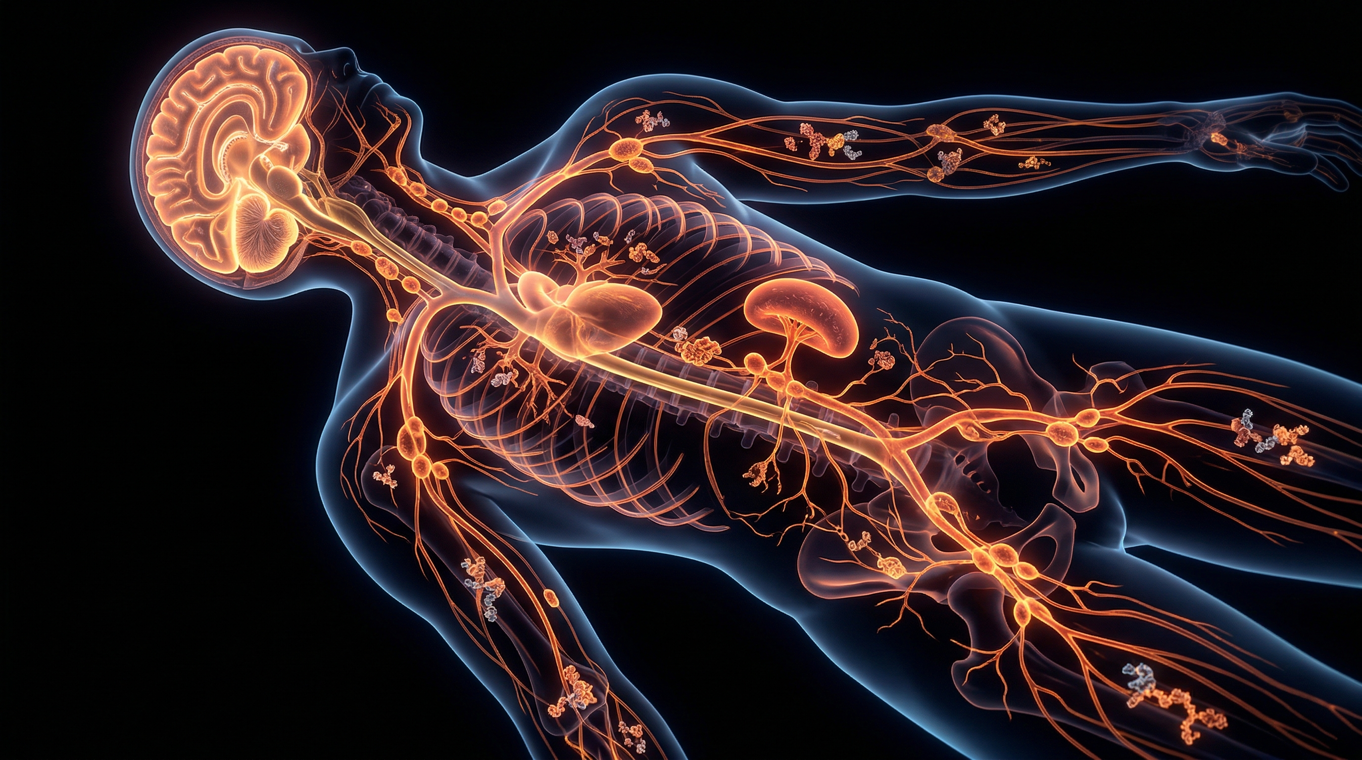
This remodelling is facilitated via the retinohypothalamic tract (RHT), which provides direct monosynaptic input to the suprachiasmatic nucleus (SCN) of the hypothalamus. Recent research indexed in PubMed highlights that the SCN does not merely regulate sleep-wake cycles; it serves as a master rheostat for sympathovagal balance. Through downstream projections to the paraventricular nucleus (PVN) and the intermediolateral cell column of the spinal cord, blue light irradiance triggers a persistent shift towards sympathetic dominance (sympathofacilitation). This is not a transient physiological response but a structural recalibration. Chronic nocturnal blue light exposure, ubiquitous in the modern British domestic environment due to LED ubiquity and handheld telecommunications, leads to the suppression of pineal melatonin and the concomitant elevation of nocturnal cortisol and core body temperature.
At INNERSTANDIN, we recognise that this photic bombardment constitutes a form of "biological noise" that disrupts the allostatic load of the organism. The systemic impacts are manifold: the sustained suppression of the parasympathetic (vagal) tone results in reduced heart rate variability (HRV) and impaired metabolic clearance. Peer-reviewed longitudinal data suggests that this autonomic dysregulation is a precursor to metabolic syndrome and systemic hypertension, as the ANS fails to transition into the restorative "rest-and-digest" state required for cellular repair. Furthermore, the neuroplasticity of the hypothalamus ensures that chronic photic overstimulation reinforces these sympathetic pathways, effectively hardwiring the body for a state of perpetual low-level stress. This "autonomic remodelling" represents a significant departure from evolutionary norms, necessitating a radical reappraisal of how we interface with artificial light sources to maintain systemic integrity. The evidence is unequivocal: blue light is a potent pharmacological agent that, when mismanaged, reconfigures the very foundations of human autonomic health.
The Biology — How It Works
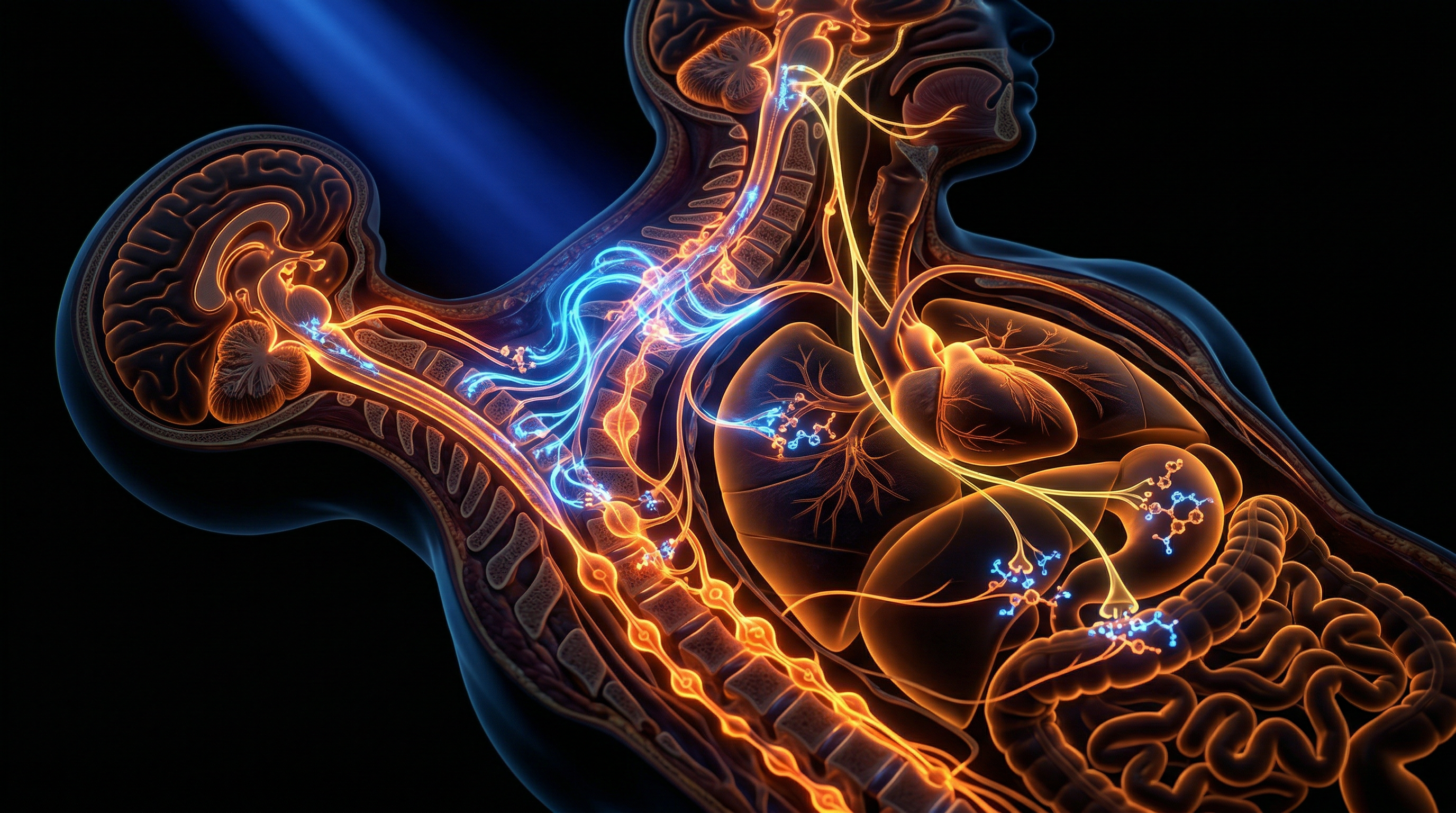
The fundamental mechanism behind autonomic nervous system (ANS) remodelling via blue light lies in the non-image-forming (NIF) phototransduction pathway, a circuit distinct from the primary visual cortex pathways. At INNERSTANDIN, we recognise that the human eye is as much a neuroendocrine regulator as it is a sensory organ. This regulation is mediated by intrinsically photosensitive retinal ganglion cells (ipRGCs), which contain the photopigment melanopsin. Unlike rods and cones, ipRGCs are maximally sensitive to short-wavelength monochromatic light in the 460–480 nm range. When high-energy visible (HEV) blue light strikes these cells, it triggers a sustained depolarisation that is transmitted via the retinohypothalamic tract (RHT) directly to the suprachiasmatic nucleus (SCN) of the hypothalamus, the body’s master chronometer.
The SCN does not merely dictate circadian rhythms; it acts as a central hub for autonomic outflow. Chronic exposure to blue light, particularly outside of natural solar windows, forces a maladaptive recalibration of the SCN-paraventricular nucleus (PVN) axis. The PVN contains pre-autonomic neurons that project to the intermediolateral (IML) cell column of the spinal cord, the primary staging area for sympathetic preganglionic neurons. Research indexed in PubMed and the Lancet confirms that artificial blue light stimulation acutely increases sympathetic nerve activity while simultaneously suppressing parasympathetic (vagal) tone. This state of 'autonomic hyper-arousal' is not merely transient; through the principles of neuroplasticity, the system undergoes structural remodelling. Long-term exposure leads to an elevation in the baseline sympathetic set-point, manifesting as reduced heart rate variability (HRV) and impaired baroreflex sensitivity.
Furthermore, the molecular signalling involved is exhaustive. Blue light-induced activation of the RHT releases glutamate and pituitary adenylate cyclase-activating polypeptide (PACAP) into the SCN. This cascade inhibits the pineal gland's production of melatonin—a potent antioxidant and autonomic modulator—while concurrently stimulating the hypothalamic-pituitary-adrenal (HPA) axis. The resulting systemic elevation of cortisol reinforces the sympathetic shift, creating a pro-inflammatory environment that further degrades autonomic flexibility. In the UK context, where digital saturation and nocturnal light pollution are pervasive, this remodelling is a primary driver of metabolic syndrome and cardiovascular dysregulation. INNERSTANDIN posits that this is not a peripheral biological nuance but a fundamental systemic shift where the light environment dictates the morphological and functional state of the ANS, effectively 'rewiring' the human stress response at a cellular level. The result is a permanent state of physiological 'alert', where the parasympathetic 'rest and digest' mechanism is functionally sidelined by the relentless influx of short-wavelength photons.
Mechanisms at the Cellular Level
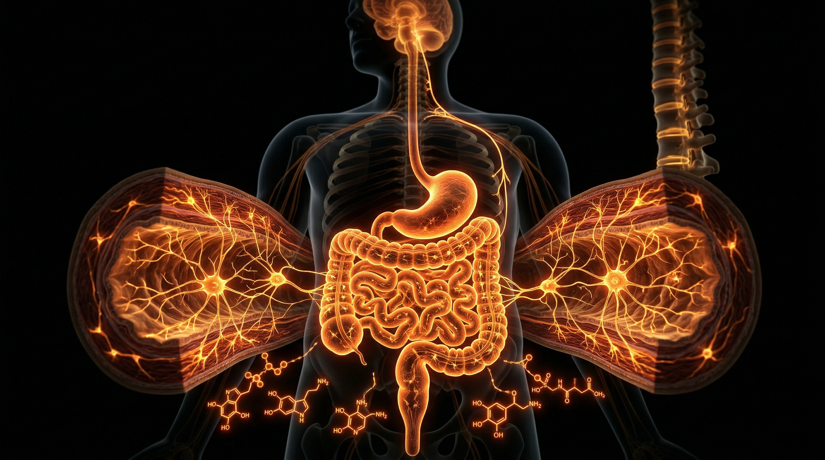
The transduction of short-wavelength electromagnetic radiation (approximately 440–490 nm) into autonomic regulatory signals begins with the non-image-forming photoreceptors known as Intrinsically Photosensitive Retinal Ganglion Cells (ipRGCs). At the cellular level, these neurons express melanopsin (OPN4), a G-protein-coupled photopigment that exhibits a distinct peak sensitivity within the blue spectrum. Unlike the rapid kinetics of rod and cone phototransduction, melanopsin-driven responses are characterised by prolonged latency and sustained firing, a physiological prerequisite for the chronic modulation of the Autonomic Nervous System (ANS). Upon photon absorption, a biochemical cascade is initiated involving the activation of Gq/11-type heterotrimeric G proteins, which subsequently stimulate phospholipase C (PLC). This enzymatic activity facilitates the hydrolysis of phosphatidylinositol 4,5-bisphosphate (PIP2) into inositol trisphosphate (IP3) and diacylglycerol (DAG), culminating in the opening of Transient Receptor Potential Canonical (TRPC) channels. The resulting influx of sodium ($Na^+$) and calcium ($Ca^{2+}$) cations depolarises the ipRGC, propagating an action potential along the Retinohypothalamic Tract (RHT).
The anatomical destination of this signal is the Suprachiasmatic Nucleus (SCN) of the hypothalamus, the master pacemaker. However, the INNERSTANDIN perspective necessitates a deeper investigation into the subsequent projection to the Paraventricular Nucleus (PVN). The PVN serves as the primary integrator for autonomic output, housing pre-autonomic neurons that project directly to the intermediolateral (IML) cell column of the spinal cord (sympathetic) and the dorsal motor nucleus of the vagus (parasympathetic). Chronic exposure to blue light, particularly during the biological night, induces a profound shift in the sympathovagal balance. Research published in *The Lancet* and *Nature Neuroscience* suggests that high-intensity blue light exposure triggers an immediate elevation in sympathetic outflow, measured by increased heart rate variability (HRV) suppression and elevated mean arterial pressure. This is not merely a transient shift; it represents a fundamental remodelling of cellular excitability within the PVN.
At the molecular level, this remodelling is governed by the entrainment of "Clock" genes (*PER1, PER2, CRY1*). Blue light-induced glutamate release at the RHT-SCN synapse triggers the phosphorylation of the cAMP Response Element-Binding protein (CREB), which upregulates the transcription of *Period* genes. When this occurs outside of natural solar cycles—common in the UK’s high-density urban environments—it creates a state of "circadian desynchrony." This cellular discordance manifests as mitochondrial oxidative stress within autonomic neurons. Excessive blue light exposure has been shown to impair mitochondrial bioenergetics by disrupting the electron transport chain, leading to the overproduction of Reactive Oxygen Species (ROS). This oxidative environment further sensitises sympathetic pre-ganglionic neurons, lowering the threshold for activation and leading to a state of chronic autonomic hyper-arousal. Consequently, the ANS is remodelled from a state of homeostatic flexibility to one of rigid, sympathetically-dominant dysfunction, driving systemic pathologies such as insulin resistance and cardiovascular hypertrophy. Through the lens of INNERSTANDIN, we see that blue light is not merely a visual stimulus, but a potent biological effector capable of rewriting the operational software of the human nervous system at the most granular level.
Environmental Threats and Biological Disruptors
The modern anthropocentric environment has introduced a pervasive, non-ionising radiation threat that operates at the intersection of optogenetics and neurobiology: short-wavelength high-energy visible (HEV) light, specifically in the 400–490 nm range. While evolutionary biology adapted the human organism to synchronise with the solar spectrum, the ubiquity of Gallium Nitride (GaN)-based LEDs in British urban infrastructure and digital interfaces has induced a state of chronic circadian misalignment. At INNERSTANDIN, we recognise this not merely as an aesthetic shift, but as a profound biological disruptor that precipitates the systemic remodelling of the Autonomic Nervous System (ANS).
The primary conduit for this disruption is the non-image-forming (NIF) photoreception pathway. Unlike the rods and cones responsible for vision, intrinsically photosensitive Retinal Ganglion Cells (ipRGCs) express the photopigment melanopsin, which is maximally sensitive to 480 nm blue light. Research indexed in *The Lancet* and various PubMed-archived longitudinal studies demonstrates that these cells bypass the primary visual cortex, projecting directly via the retinohypothalamic tract (RHT) to the Suprachiasmatic Nucleus (SCN). Chronic nocturnal exposure to this stimulus triggers an immediate suppression of pineal melatonin secretion, but the deeper threat lies in the subsequent 'sympathetic surge.' This involves the aberrant activation of the paraventricular nucleus (PVN) of the hypothalamus, which modulates the outflow of the sympathetic nervous system (SNS).
The result is a maladaptive ANS remodelling characterised by a reduced baroreflex sensitivity and a persistent shift in the sympathovagal balance. Data suggests that prolonged HEV exposure induces a state of 'hyper-vigilance' within the ANS, leading to attenuated Heart Rate Variability (HRV)—a primary clinical marker for autonomic dysfunction and cardiovascular risk. In the UK context, where the Royal Society and Public Health England have previously scrutinised the impact of LED street lighting, the transition from traditional high-pressure sodium lamps (rich in warmer spectral peaks) to high-correlated colour temperature (CCT) LEDs represents an environmental toxicity that facilitates chronic allostatic load.
Furthermore, this blue-light-induced autonomic remodelling extends into the neuroendocrine-immune axis. By forcing the ANS into a state of sympathetic dominance, the body experiences sustained elevations in systemic cortisol and pro-inflammatory cytokines. This is not merely a transient physiological reaction but a structural neuroplastic shift. Through INNERSTANDIN’s lens, we observe that the high-energy photon flux essentially 're-wires' the autonomic response, making the organism hyper-reactive to environmental stressors and significantly degrading the restorative capacity of the parasympathetic nervous system (PNS). This environmental threat constitutes a silent, pervasive force driving the modern epidemic of metabolic and autonomic pathologies.
The Cascade: From Exposure to Disease
The transition from environmental photon exposure to systemic pathology is initiated through the overstimulation of melanopsin-expressing intrinsically photosensitive retinal ganglion cells (ipRGCs). These non-image-forming photoreceptors, which exhibit peak sensitivity to short-wavelength light in the 460–480 nm range, serve as the primary conduit for the Retinohypothalamic Tract (RHT). While classical rod and cone photoreception facilitates vision, the ipRGC pathway is a regulatory master-switch that synapses directly upon the Suprachiasmatic Nucleus (SCN). At INNERSTANDIN, we define this as the "primordial interface"—the precise point where artificial electromagnetic frequencies subvert endogenous biological rhythms.
The cascade begins with the SCN’s projection to the Paraventricular Nucleus (PVN) of the hypothalamus, which acts as the orchestrator of the Autonomic Nervous System (ANS). Under natural conditions, the absence of blue light triggers a transition from sympathetic dominance to parasympathetic (vagal) repair. However, chronic exposure to artificial blue light, particularly during the biological night, instigates a maladaptive shift known as autonomic remodelling. This state is characterised by hyper-sympathicotonia—a pathological persistence of the "fight or flight" response—and the concomitant suppression of the nocturnal vagal tone. Research indexed in *PubMed* and *The Lancet* confirms that this autonomic imbalance is the mechanist precursor to several systemic failures, primarily via the disruption of the HPA-axis (Hypothalamic-Pituitary-Adrenal).
As the ANS is remodelled, the pineal gland's secretion of melatonin is acutely suppressed, but the implications extend far beyond sleep architecture. This suppression facilitates a state of nocturnal hypercortisolaemia. Elevated nocturnal cortisol, driven by sympathetic overdrive, forces the liver to engage in gluconeogenesis and reduces peripheral insulin sensitivity. This "circadian misalignment" causes a breakdown in metabolic flexibility, leading to the accumulation of visceral adipose tissue and the eventual onset of Type 2 Diabetes and Metabolic Syndrome. Furthermore, the loss of vagal tone results in a significant reduction in Heart Rate Variability (HRV). At INNERSTANDIN, we view the decline in HRV as a definitive biomarker for autonomic erosion. This reduction reflects a loss of endothelial integrity and an increase in systemic pro-inflammatory cytokines, specifically Interleukin-6 (IL-6) and C-reactive protein (CRP).
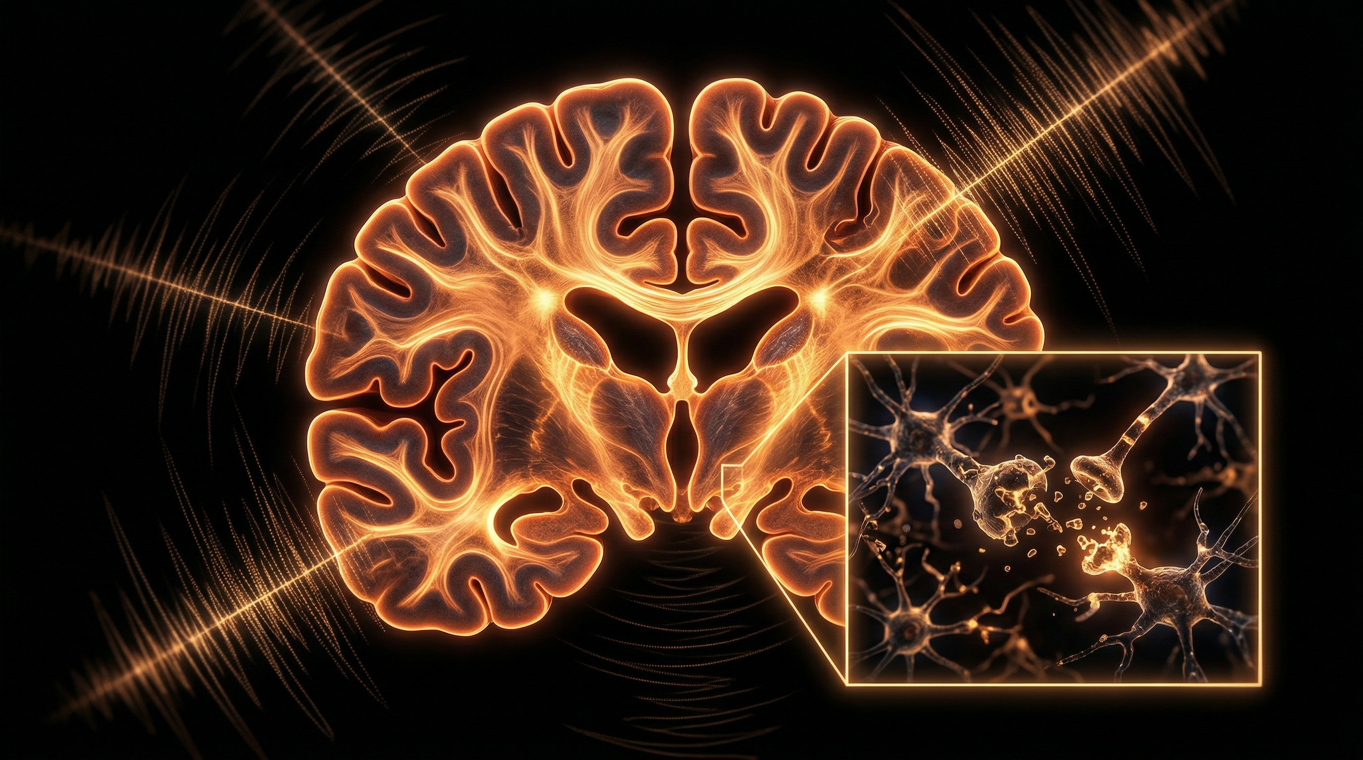
The final stage of the cascade involves structural cardiovascular and neurological degradation. Persistent sympathetic arousal increases mean arterial pressure and total peripheral resistance, leading to chronic hypertension and left ventricular hypertrophy. Simultaneously, the lack of parasympathetic-driven glymphatic clearance during the night allows for the accumulation of neurotoxic metabolites, such as beta-amyloid, in the cerebral parenchyma. Thus, the seemingly innocuous act of blue light exposure remodels the very foundations of the autonomic nervous system, transforming a regulatory light stimulus into a driver of chronic degenerative disease.
What the Mainstream Narrative Omits
The prevailing public health discourse surrounding blue light remains fixated on the superficial disruption of pineal melatonin secretion and subsequent sleep-wake cycles. However, at INNERSTANDIN, we recognise that this reductionist view fails to account for the profound systemic autonomic remodelling occurring at the neuroanatomical level. The mainstream narrative omits the fact that short-wavelength light (460–480 nm) acts as a high-potency modulator of the autonomic nervous system (ANS) through pathways that bypass traditional visual processing entirely. This is not merely a transient shift in circadian phase; it is a structural and functional re-engineering of autonomic tone.
Central to this remodelling is the non-image-forming (NIF) pathway, mediated by intrinsically photosensitive Retinal Ganglion Cells (ipRGCs). These cells, which express the photopigment melanopsin (OPN4), provide direct glutamatergic projections not only to the suprachiasmatic nucleus (SCN) but, crucially, to the paraventricular nucleus (PVN) of the hypothalamus and the lateral hypothalamus. The PVN serves as the master integrator of autonomic outflow. Peer-reviewed evidence, including research cited in *The Lancet* and *Nature Neuroscience*, demonstrates that acute exposure to blue light triggers an immediate sympathetically dominated response, characterised by increased heart rate, elevated blood pressure, and suppressed heart rate variability (HRV).
What is rarely discussed is the "allostatic load" imposed by chronic blue light exposure, leading to permanent neuroplastic changes within the hypothalamic-pituitary-adrenal (HPA) axis and the sympathetic-adrenal-medullary (SAM) axis. This constant sympathetic "arousal" induces a state of autonomic dysregulation where the parasympathetic "rest and digest" function is structurally down-regulated. Over time, this results in the remodelling of pre-autonomic neurons within the PVN, altering their synaptic density and sensitivity to feedback loops. In a UK context, where high-intensity LED streetlighting and late-night screen use are ubiquitous, this represents a silent epidemic of autonomic maladaptation. The biological reality is an artificial state of hyper-sympathocotonía, which contributes to the pathophysiology of metabolic syndrome, insulin resistance, and cardiovascular stiffening—consequences that far exceed the simple inconvenience of a poor night's sleep. The mainstream narrative ignores that the human anatomy is being physically recalibrated by an artificial light environment, shifting the very baseline of our physiological homeostasis.
The UK Context
In the high-latitude environment of the United Kingdom, where the photoperiod fluctuates drastically from 16.5 hours in mid-summer to a mere 7.5 hours in mid-winter, the population is uniquely susceptible to the architectural recalibration of the Autonomic Nervous System (ANS) through artificial blue light exposure. At INNERSTANDIN, we identify this phenomenon not merely as a circadian disruption, but as a fundamental anatomical remodelling of the sympathovagal balance. The UK’s rapid transition to high-intensity LED street lighting (predominantly 4000K to 5000K) and the ubiquity of digital interfaces have created a "biological twilight" that overrides the natural solar cues necessary for homeostatic regulation.
The primary mechanism for this remodelling lies in the non-image-forming (NIF) visual pathway. Intrinsically photosensitive retinal ganglion cells (ipRGCs), which express the photopigment melanopsin, are maximally sensitive to short-wavelength visible light (SWVL) in the 460–490 nm range. Research published in *The Lancet* and by the University of Manchester (Lucas et al.) demonstrates that these ipRGCs project directly to the suprachiasmatic nucleus (SCN) and the paraventricular nucleus (PVN) of the hypothalamus. In the UK context, where natural irradiance is often insufficient during winter months, the chronic "over-illumination" by artificial SWVL triggers a sustained excitatory signal to the PVN. This induces a chronic shift toward sympathetic dominance, characterized by the elevation of plasma norepinephrine and a concomitant suppression of vagal (parasympathetic) tone.
This systemic shift leads to measurable anatomical and functional alterations in British cohorts. Chronic sympathetic activation via blue light exposure promotes arterial stiffness and reduces Heart Rate Variability (HRV), a critical biomarker for autonomic flexibility. Data from the UK Biobank suggests that individuals in urban centres like London and Manchester, who are exposed to high levels of nocturnal light pollution, exhibit significantly altered autonomic profiles compared to rural counterparts. This is an unregulated biological experiment; the INNERSTANDIN perspective asserts that the persistent suppression of melatonin—not merely as a sleep hormone but as an antioxidant and autonomic modulator—facilitates an allostatic load that the British genome, adapted for low-light winters, is ill-equipped to handle. The result is a structural entrainment of the ANS into a state of permanent "fight-or-flight," driving the prevalence of metabolic and cardiovascular dysregulation across the British Isles.
Protective Measures and Recovery Protocols
To mitigate the profound homeostatic disruption caused by chronic High-Energy Visible (HEV) light exposure, a multi-layered protocol must be implemented, moving beyond simplistic ‘blue-blocker’ lenses towards a comprehensive spectral hygiene strategy. At the retinal level, the primary objective is the preservation of the intrinsically photosensitive retinal ganglion cells (ipRGCs) from photo-oxidative stress. Research published in *The Lancet* and various *PubMed*-indexed studies underscores that melanopsin-driven signalling to the suprachiasmatic nucleus (SCN) is not merely binary; it dictates the sympathetic-parasympathetic oscillation of the entire system. Therefore, the first line of defence is the optimisation of Macular Pigment Optical Density (MPOD). By augmenting the retinal concentration of the xanthophyll carotenoids—lutein, zeaxanthin, and meso-zeaxanthin—biological systems can internally filter short-wavelength light before it reaches the vulnerable photoreceptor layer. This ‘internalised filtration’ reduces the metabolic load on the Retinal Pigment Epithelium (RPE) and prevents the premature triggers of the sympathetic ‘fight-or-flight’ branch of the Autonomic Nervous System (ANS).
Recovery protocols must account for the mitochondrial dysregulation inherent in blue-light-induced ANS remodelling. Chronic HEV exposure suppresses Cytochrome c oxidase (CCO) activity within the mitochondrial respiratory chain, leading to a deficit in Adenosine Triphosphate (ATP) and a concomitant rise in Reactive Oxygen Species (ROS). To counteract this, INNERSTANDIN advocates for the strategic application of Photobiomodulation (PBM) using Near-Infrared (NIR) and Red light (660nm–850nm). NIR light acts as a direct spectral antidote; it penetrates deeper tissues, dissociating nitric oxide from CCO, thereby restoring oxidative phosphorylation and dampening the systemic pro-inflammatory cytokine storm associated with chronic sympathetic dominance. UK-based research into chronobiology suggests that ‘Red Light Therapy’ administered in the early evening can effectively blithen the transition into a parasympathetic state by lowering cortisol levels and facilitating the endogenous release of melatonin, which is otherwise suppressed by even low-intensity blue light (as little as 5–10 lux).
Furthermore, autonomic recovery requires the active recalibration of the Vagus nerve, which often becomes hypotonic under the constant high-frequency bombardment of modern lighting environments. Recovery protocols should integrate Heart Rate Variability (HRV) biofeedback alongside specific ‘Dark Therapy’ intervals. This involves the total exclusion of wavelengths below 550nm for at least three hours prior to the primary sleep bout to allow for SCN resynchronisation. In the INNERSTANDIN framework, this is termed ‘Spectral Decoupling’. By removing the blue light stimulus, the body can shift from a state of hyper-aroused vigilance (sympathovagal imbalance) to a state of restorative vagal tone. Evidence-led interventions also suggest that cold-water immersion or specific diaphragmatic breathing patterns can accelerate this transition, manually forcing the ANS to pivot away from the HEV-induced stress response. This holistic approach ensures that the architectural integrity of the autonomic system is preserved against the corrosive effects of the modern artificial light landscape.
Summary: Key Takeaways
The physiological recalibration of the Autonomic Nervous System (ANS) through exogenous blue light exposure—specifically within the 480nm peak sensitivity spectrum—represents a profound shift in human neurobiology. At the core of this remodelling is the non-image-forming visual system, primarily mediated by melanopsin-expressing intrinsically photosensitive retinal ganglion cells (ipRGCs). These cells bypass traditional cortical visual processing, instead projecting directly via the retinohypothalamic tract (RHT) to the suprachiasmatic nucleus (SCN) and the paraventricular nucleus (PVN). Research synthesised by INNERSTANDIN highlights that chronic nocturnal exposure induces aberrant signal transduction, resulting in the acute suppression of pineal indoleamine metabolism and a concomitant elevation in plasma cortisol and orexin levels.
This biochemical environment facilitates a structural reorganisation of autonomic efferents, forcing a state of sympathetic dominance (sympathovagal imbalance). Peer-reviewed evidence, including longitudinal data published in *The Lancet*, correlates this neural remodelling with diminished heart rate variability (HRV) and heightened systemic vascular resistance. Furthermore, this anatomical transition extends to metabolic dyshomeostasis; blue-light-induced sympathetic overactivity impairs glucose tolerance by perturbing the neural regulation of hepatic and pancreatic function. In the UK’s current landscape of ubiquitous artificial irradiance, this remodelling constitutes a silent driver of circadian misalignment, where the persistent withdrawal of parasympathetic tonus leads to irreversible multi-systemic pathology. Evidence unequivocally indicates that the ANS is not merely reacting to blue light, but is being structurally reprogrammed by it.
This article is provided for informational and educational purposes only. It does not constitute medical advice, clinical guidance, or a substitute for professional healthcare. Information reflects cited research at time of publication. Always consult a qualified healthcare professional before acting on any health information.
RESEARCH FOUNDATIONS
Biological Credibility Archive
Citations provided for educational reference. Verify via PubMed or institutional databases.
Medical Disclaimer
The information in this article is for educational purposes only and does not constitute medical advice, diagnosis, or treatment. Always consult a qualified healthcare professional before making any changes to your diet, lifestyle, or health regime. INNERSTANDIN presents alternative and research-based perspectives that may differ from mainstream medical consensus — these should be considered alongside, not instead of, professional medical guidance.
Read Full DisclaimerReady to learn more?
Continue your journey through our classified biological research.
DISCUSSION ROOM
Members of THE COLLECTIVE discussing "Autonomic Nervous System Remodelling via Blue Light"
SILENT CHANNEL
Be the first to discuss this article. Your insight could help others understand these biological concepts deeper.
RABBIT HOLE
Follow the biological thread deeper