Episiotomy Biology: The Myth of Clean Incisions
Episiotomies are often performed under the false premise that they prevent larger tears. Biologically, these surgical incisions are harder to heal and cause more long-term nerve damage than natural tears.
Overview
The episiotomy—a surgical incision of the perineum and the posterior vaginal wall during the second stage of labour—remains one of the most common yet biologically misunderstood procedures in modern obstetrics. For decades, the mainstream medical narrative has presented the episiotomy as a protective measure: a "clean, controlled incision" that is allegedly easier to repair and faster to heal than a "jagged" natural tear. However, as we delve into the histological, neurological, and biomechanical realities of the female pelvic floor, this narrative dissolves into a dangerous myth.
Biologically, the human body is not a piece of fabric that "rips" unpredictably. It is a complex, multi-layered architecture of muscle, fascia, nerves, and vasculature designed to distend under specific hormonal and mechanical conditions. When a surgeon introduces a cold blade into this hyper-distended tissue, they are not preventing trauma; they are inducing a specific, deep-tissue injury that bypasses the body’s innate protective mechanisms.
In this comprehensive analysis, we will expose the biological fallacy of the "clean incision." We will explore how episiotomies disrupt the extracellular matrix (ECM), cause permanent axonal transection of the pudendal nerve branches, and initiate a cascade of chronic pelvic dysfunction that natural tearing—even of the same degree—rarely triggers. From the cellular response to the structural integrity of the pelvic floor, the evidence is clear: the episiotomy is a relic of "industrialised birth" that prioritises the practitioner’s convenience over the biological longevity of the mother.
Fact: Despite the prevalence of the procedure, Cochrane reviews and systematic meta-analyses have repeatedly found no evidence that routine episiotomy reduces the risk of severe perineal trauma (3rd and 4th-degree tears). In fact, an episiotomy is often the primary risk factor for such injuries.
---
The Biology — How It Works
To understand why the "clean incision" is a biological disaster, one must first understand the anatomy of the perineal body. The perineal body is a pyramidal fibromuscular structure located between the vaginal vestibule and the anal canal. It serves as the "anchor point" for the pelvic floor, where several key muscles converge:
- —Bulbospongiosus
- —Superficial transverse perineal muscle
- —External anal sphincter
- —Levator ani (specifically the puborectalis fibre)
The Biomechanics of Distension
During the second stage of labour, the perineum undergoes a process of remodelling. Under the influence of relaxin and oestrogen, the collagen fibres in the pelvic fascia become more pliable. As the fetal head descends, the tissue does not merely stretch; it thins and fan-folds. This is a progressive, physiological adaptation.
In a natural birth, if a tear occurs, it typically follows the "path of least resistance." Biologically, this means the tissue separates along the natural cleavage lines (Langer’s lines) of the skin and the interstices of the muscle fibres. A natural tear is often superficial, affecting only the skin and vaginal mucosa. Even when it involves muscle, the "jagged" edges that surgeons complain about are actually a biological advantage: they provide a larger surface area for fibrin attachment and allow the edges to "knit" back together in their original anatomical orientation.
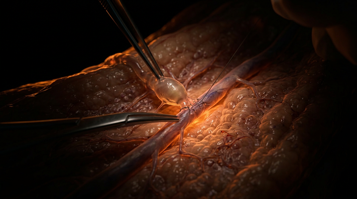
The Surgical Insult
An episiotomy, conversely, is a transverse or mediolateral cut that ignores these natural cleavage lines. Because the tissue is under extreme tension when the cut is made, the wound edges immediately "spring" apart, often receding deep into the pelvic cavity.
- —Midline Episiotomy: Cuts directly into the central tendon of the perineal body toward the anus. This structurally weakens the very anchor of the pelvic floor.
- —Mediolateral Episiotomy: Cuts at a 45-degree angle. While intended to avoid the anal sphincter, it slices through the thickest part of the bulbocavernosus and transverse perineal muscles, often severing the muscle bellies rather than separating the fibres.
By cutting through these muscles while they are at maximum stretch, the surgeon induces a "snap-back" effect. The muscle fibres, once severed, retract. This makes anatomical realignment during suturing nearly impossible to achieve perfectly, leading to the formation of "dead space" and subsequent fibrosis.
---
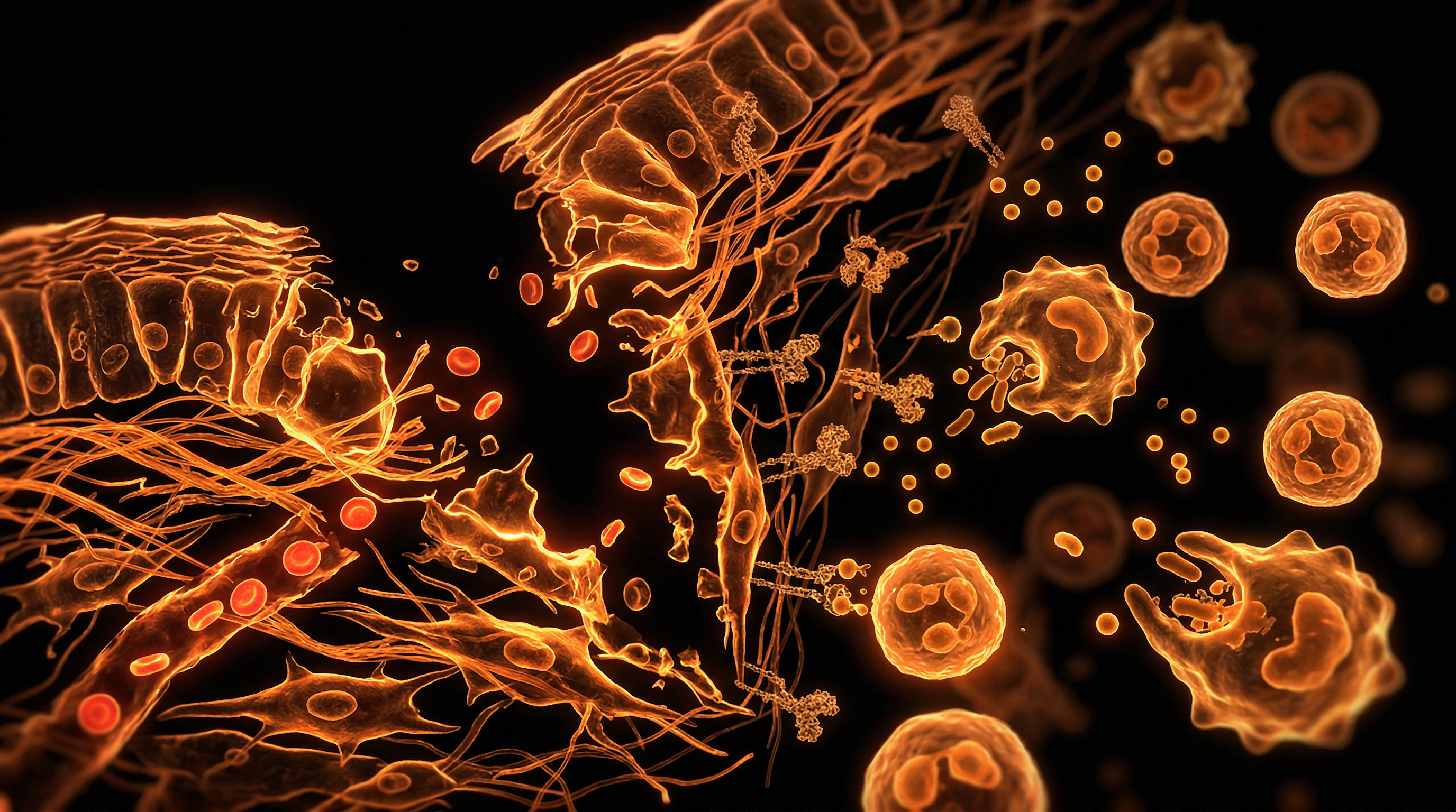
Mechanisms at the Cellular Level
At the microscopic level, the difference between a natural tear and a surgical incision is the difference between a controlled biological adaptation and an acute traumatic event.
Disruption of the Extracellular Matrix (ECM)
The ECM of the perineum is a complex web of collagen type I and III, elastin, and glycosaminoglycans. During a natural birth, the ECM undergoes mechanical "uncoiling." When a tear occurs, the body’s fibroblasts are already in an activated state due to the hormonal environment.
A surgical incision, however, causes an immediate and massive release of damage-associated molecular patterns (DAMPs). Because the incision is deep and uniform, it severs the micro-vasculature (capillaries) in a way that creates a localized "hypoxic zone." While a jagged tear preserves many of these micro-vessels, a straight cut cauterises or collapses them.
The Problem of Primary Intention
Surgeons argue that "clean" wounds heal by primary intention (direct closure), which is theoretically faster. However, in the perineum, this is a fallacy. Because the area is subject to constant movement (walking, sitting, bowel movements) and is naturally colonized by bacteria, these "clean" edges are rarely as stable as they appear.
Furthermore, the act of suturing an episiotomy introduces foreign body reactions. The sutures themselves create "islands" of inflammation. In a natural tear that is left to heal or sutured loosely, the inflammatory response is diffuse and manageable. In an episiotomy, the tension required to pull the retracted muscle edges back together causes pressure necrosis of the tissue trapped within the suture loops.
Neurological Severing: The Pudendal Nerve Damage
This is perhaps the most suppressed biological truth of the episiotomy. The pudendal nerve and its branches (the inferior rectal nerve, the perineal nerve, and the dorsal nerve of the clitoris) provide sensory and motor innervation to this region.
- —Axonal Transection: A surgical blade does not discriminate between muscle and nerve. An episiotomy frequently severs the terminal branches of the perineal nerve.
- —Neuroma Formation: When a nerve is cut cleanly, the regenerating axons often fail to find their target on the other side of the incision. They instead form a "tangled ball" of nerve tissue known as a traumatic neuroma. This is a primary cause of chronic dyspareunia (painful intercourse) and localized vulvodynia following birth.
- —Denervation Atrophy: When the motor nerves to the transverse perineal muscles are cut, those muscles lose their "tone." Over time, this leads to atrophy, even if the "skin" looks healed. This is why women with episiotomies have significantly higher rates of pelvic organ prolapse in later life.
---
Environmental Threats and Biological Disruptors
While we often view the episiotomy as a purely mechanical intervention, its failure is exacerbated by the obstetric environment—the modern hospital setting which acts as a biological disruptor.
The Lithotomy Position: A Mechanical Disruptor
The most significant "environmental" factor contributing to the perceived "need" for episiotomy is the lithotomy position (lying on the back with legs in stirrups). Biologically, this is the worst possible position for the perineum. It:
- —Reduces the pelvic outlet diameter by up to 30%.
- —Places the entire weight of the foetus on the perineum rather than the vaginal opening.
- —Forces the mother to push "uphill" against gravity.
In this position, the perineum becomes unnaturally taut and pale (ischaemic). A practitioner seeing this "white" tissue often panics and performs an episiotomy. Had the mother been in an upright, squatting, or all-fours position, the biological "stretch" would be distributed evenly, likely avoiding the need for any cut.
Synthetic Oxytocin (Syntocinon/Pitocin)
The use of synthetic oxytocin to "speed up" labour is a major biological disruptor. Natural oxytocin is released in pulses, allowing the perineal tissues time to "rest" and reperfuse with blood between contractions. Syntocinon creates hyper-tonic contractions—longer, stronger, and closer together.
This gives the perineal tissues no time to adapt. The collagen doesn't have time to "uncoil." Under the relentless pressure of a Syntocinon-driven second stage, the tissue is more likely to appear as if it will tear, leading to the "preventative" episiotomy.
The Sterile Field vs. The Microbiome
The hospital environment’s obsession with sterility also disrupts healing. The vagina and perineum have a specific microbiome (dominated by *Lactobacillus*) that protects against pathogenic colonization. The use of harsh antiseptic washes (like chlorhexidine) before an episiotomy wipes out this protective flora. This leaves the surgical wound vulnerable to hospital-acquired infections (MRSA or *E. coli*), which are far more difficult to treat than the natural flora exposure a tear would face at home.
---
The Cascade: From Exposure to Disease
The episiotomy is not a "one-and-done" event. It initiates a biological cascade that can lead to lifelong morbidity.
Stage 1: The Acute Inflammatory Overload
Within hours of the procedure, the body is flooded with pro-inflammatory cytokines (IL-1, IL-6, TNF-alpha). In a natural tear, this response is proportional. In an episiotomy, because muscle bellies were severed, the "haematoma" (internal bruising) is often significant. This trapped blood serves as a medium for bacteria, leading to the high rates of post-episiotomy infections.
Stage 2: Fibrotic Replacement (Scarring)
Because the severed muscle fibres retract, the body fills the gap with Type III collagen—inelastic scar tissue. This is the "Myth of the Clean Incision" laid bare. While the skin looks like a straight line, the sub-dermal layers are a disorganized mess of fibrosis.
- —This scar tissue does not stretch.
- —During subsequent births, this "scar" acts as a weak point. Because scar tissue lacks the elasticity of healthy muscle, it often "zips" open, leading to even more severe 3rd or 4th-degree tears in future deliveries.
Stage 3: Pelvic Floor Dysfunction (PFD)
The structural integrity of the pelvic diaphragm is compromised. This leads to a cascade of issues:
- —Urinary Incontinence: Damage to the nerve supply of the urethral sphincter.
- —Anal Incontinence: If the episiotomy extends (the "zipper effect") or if the repair of the perineal body is inadequate, the internal and external anal sphincters lose their structural support.
- —Organ Prolapse: Without the "anchor" of the perineal body, the uterus, bladder, and rectum begin to descend into the vaginal vault.
Stage 4: Psychosomatic Trauma and PTSD
The biological trauma to the nerves also has a psychological component. The pudendal nerve is intimately linked to the autonomic nervous system. Chronic pain from a neuroma or an incorrectly healed episiotomy keeps the mother in a state of "sympathetic dominance" (fight or flight). This disrupts the bonding process, breastfeeding (due to suppressed oxytocin from pain), and can lead to postpartum PTSD.
Important Fact: Women who receive episiotomies report significantly lower sexual satisfaction scores at 6 and 12 months postpartum compared to women who experienced spontaneous 1st or 2nd-degree tears. The "clean cut" is a direct thief of sexual health.
---
What the Mainstream Narrative Omits
If the biological evidence against episiotomy is so overwhelming, why is it still performed? The mainstream narrative omits three key factors:
1. The Convenience of the Practitioner
A natural birth requires "patience of the perineum." It may take 30 to 60 minutes of "crowning" for the tissue to distend safely. An episiotomy takes seconds. In a busy obstetric ward, the "cut" is a tool for time management. It accelerates the second stage of labour, allowing the doctor to move to the next "patient."
2. The Training Gap
Many modern obstetricians are experts at surgery but have lost the art of "supporting the perineum." They are trained to "manage" birth rather than facilitate it. Consequently, they lack the skills to guide a mother through a slow crowning process that prevents tearing. They view the knife as a "control" mechanism in an otherwise "unpredictable" biological event.
3. The Fallacy of "Easier to Repair"
The claim that a straight line is easier to sew is a half-truth. It is easier for the surgeon to *place* the stitches, but it is much harder for the *body* to heal the result. Surgeons prefer symmetry, but biology prefers biocompatibility. A "jagged" natural tear allows for an "interlocking" of tissue edges, which provides superior structural strength once healed.
---
The UK Context
In the United Kingdom, the National Health Service (NHS) has made strides in reducing routine episiotomies, yet the rates remain alarmingly high in "instrumental" deliveries (forceps or ventouse).
NICE Guidelines and the OASI Care Bundle
The National Institute for Health and Care Excellence (NICE) explicitly recommends against routine episiotomy. However, the UK has recently seen the introduction of the OASI (Obstetric Anal Sphincter Injury) Care Bundle. While intended to reduce severe tears, some critics argue it has inadvertently increased the episiotomy rate by encouraging a "mediolateral" cut at exactly 60 degrees during instrumental births.
The Consent Crisis
A significant issue within the UK context is the "implied consent" or "emergency consent" during the heat of labour. Many British women report that the episiotomy was performed without their prior knowledge or against their birth plan, often with the justification that it was "necessary to save the baby." Biologically, however, the "emergency" is often a result of the very interventions (like the lithotomy position or Syntocinon) that the hospital imposed.
The "Montgomery" Standard
Following the Montgomery v Lanarkshire Health Board (2015) ruling, UK law is clear: doctors must disclose the risks of a procedure and the alternatives. The failure to mention that an episiotomy increases the risk of a 3rd-degree tear—the very thing it claims to prevent—is a failure of the legal and biological duty of care.
---
Protective Measures and Recovery Protocols
For those navigating the birth process, understanding how to protect the biological integrity of the perineum is vital.
Antenatal Perineal Massage
Beginning at 34 weeks, perineal massage can help "pre-stretch" the tissues. This is not just about physical stretching; it is about "desensitising" the nerves and increasing blood flow to the area, encouraging the collagen to become more pliable.
The "Hands-Off" Approach
The "Ritgen manoeuvre" (where the practitioner applies pressure to the perineum) has been shown in some studies to *increase* the risk of trauma. A "hands-poised" or "hands-off" approach, where the midwife only intervenes if necessary, allows the tissue to distend at its own biological pace.
Warm Compresses
The application of warm, moist cloths to the perineum during crowning is one of the most effective biological supports. The heat causes vasodilation, bringing oxygen-rich blood to the stretching tissues and allowing the muscles to relax more effectively than any chemical intervention.
Recovery: Pelvic Floor Physiotherapy
If an episiotomy has been performed, the standard "wait and see" approach is biologically inadequate.
- —Scar Massage: Once the wound is closed (around 6 weeks), manual therapy to break up the fibrotic adhesions is essential to restore blood flow and nerve function.
- —Low-Level Laser Therapy (LLLT): Can be used to stimulate mitochondrial activity in the injured cells, accelerating ATP production and collagen realignment.
- —Pelvic Floor Rehab: Specifically focusing on the "release" of the pelvic floor rather than just "Kegels." Many women with episiotomies develop a "hypertonic" (overly tight) pelvic floor due to pain, which requires specialized physiotherapy to correct.
---
Summary: Key Takeaways
- —The "Clean Cut" Myth: Surgical incisions are more traumatic than natural tears. They cut through muscle bellies and nerves, whereas natural tears often follow the path of least resistance between fibres.
- —Neurological Damage: Episiotomies frequently cause axonal transection of the pudendal nerve, leading to chronic pain, neuromas, and loss of sensation.
- —Structural Integrity: By severing the perineal body, an episiotomy weakens the entire pelvic floor, increasing the long-term risk of prolapse and incontinence.
- —The "Zipper Effect": A surgical incision creates a high-tension weak point that is more likely to extend into a 3rd or 4th-degree tear than a natural, shallow tear.
- —Environmental Factors: Birthing positions (lithotomy) and synthetic hormones (Syntocinon) are the primary drivers of the "need" for episiotomies, not biological inadequacy.
- —The UK Reality: While NICE guidelines discourage routine use, "instrumental" births in the NHS still carry a high risk of surgical intervention, often without true informed consent.
The episiotomy is a triumph of "obstetric efficiency" over "biological wisdom." By framing it as a "protective snip," the medical establishment has obscured the reality of deep-tissue trauma. True perinatal health requires a return to respecting the biomechanical and histological genius of the female body—a body that was designed to stretch, not to be cut.
This article is provided for informational and educational purposes only. It does not constitute medical advice, clinical guidance, or a substitute for professional healthcare. Information reflects cited research at time of publication. Always consult a qualified healthcare professional before acting on any health information.
RESEARCH FOUNDATIONS
Biological Credibility Archive
Citations provided for educational reference. Verify via PubMed or institutional databases.
Medical Disclaimer
The information in this article is for educational purposes only and does not constitute medical advice, diagnosis, or treatment. Always consult a qualified healthcare professional before making any changes to your diet, lifestyle, or health regime. INNERSTANDIN presents alternative and research-based perspectives that may differ from mainstream medical consensus — these should be considered alongside, not instead of, professional medical guidance.
Read Full DisclaimerReady to learn more?
Continue your journey through our classified biological research.
DISCUSSION ROOM
Members of THE COLLECTIVE discussing "Episiotomy Biology: The Myth of Clean Incisions"
SILENT CHANNEL
Be the first to discuss this article. Your insight could help others understand these biological concepts deeper.
THE ARSENAL
Based on Birth Trauma & Perinatal Health — products curated by our research team for educational relevance and biological support.
Magnesium Blend – The Most Important Mineral

Clean Slate – Detoxes thousands of chemicals,heavy metals, pesticides, allergens, mold spores and fungus
Vegan Essential Amino Acids – Plant-Powered Protein Building
INNERSTANDING may earn a commission on purchases made through these links. All products are selected based on rigorous educational relevance to our biological research.
RABBIT HOLE
Follow the biological thread deeper