Iron Infusion Dynamics: Managing Ferritin Without Toxicity
Rapid iron correction requires careful monitoring to prevent oxidative damage to the vasculature. This study outlines the UK protocols for treating iron deficiency anemia via parenteral routes.
# Iron Infusion Dynamics: Managing Ferritin Without Toxicity
Overview
In the modern landscape of haematology and functional medicine, the management of iron deficiency (ID) and iron deficiency anaemia (IDA) has undergone a radical shift. The traditional "wait and see" approach, reliant on oral supplementation that often proves poorly tolerated and biologically inefficient, is being superseded by the rapid-correction paradigm of parenteral iron infusions. However, as we accelerate the delivery of this highly reactive transition metal directly into the vascular compartment, we enter a precarious biological theatre.
Iron is a double-edged sword: it is the fundamental catalyst for life, enabling oxygen transport via haemoglobin and energy production through the mitochondrial electron transport chain. Yet, it is also a potent pro-oxidant. When introduced intravenously, bypassing the tightly regulated intestinal barrier (the mucosal block), it presents a significant challenge to the body’s homeostatic mechanisms.
At INNERSTANDING, we recognise that "normal" ferritin levels do not always equate to optimal health. This article explores the intricate dynamics of iron infusions, the biochemical fallout of rapid iron loading, and the sophisticated UK protocols designed to mitigate the risk of oxidative damage. We must look beyond the simple numbers on a pathology report to understand the cellular consequences of modern iron management.
According to the World Health Organisation, iron deficiency remains the most common nutritional disorder globally, affecting over 2 billion people. However, the move toward intravenous correction in the UK has seen a 300% increase in the last decade, necessitating a deeper understanding of infusion toxicity.
The Biology — How It Works
To understand the dynamics of an infusion, one must first appreciate the systemic regulation of iron. Under normal physiological conditions, iron homeostasis is governed by the Hepcidin-Ferroportin axis. Hepcidin, a hormone produced by the liver, acts as the master regulator. When iron levels are sufficient, hepcidin rises, binding to and degrading ferroportin—the only known cellular iron exporter—effectively locking iron within the enterocytes of the gut and the macrophages of the reticuloendothelial system.
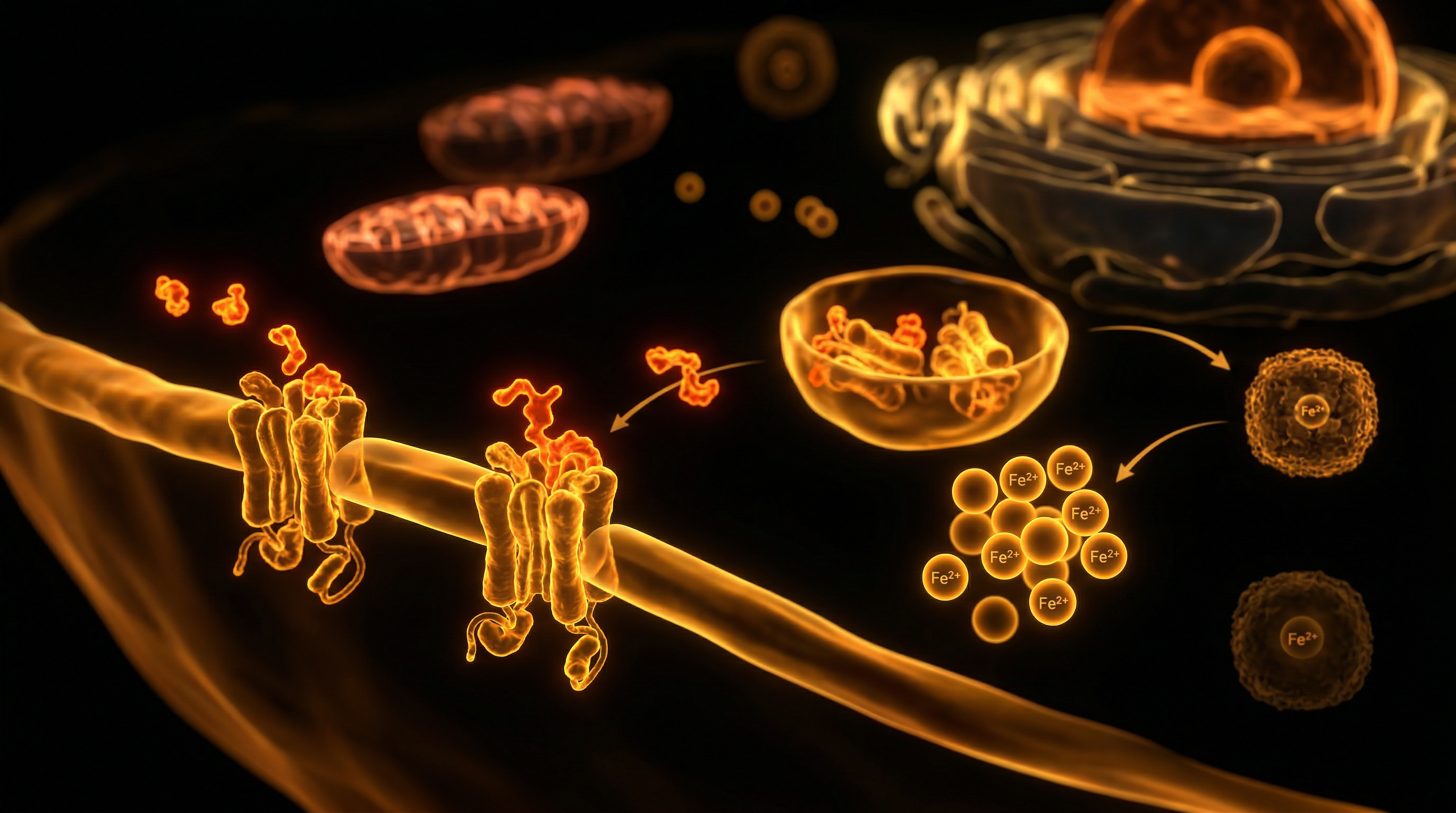
The Parenteral Bypass
When iron is administered intravenously, the intestinal gateway is entirely bypassed. The iron is typically delivered in a complexed form—a core of polynuclear iron(III)-hydroxide surrounded by a carbohydrate shell (such as carboxymaltose, sucrose, or isomaltoside). This shell is designed to ensure a controlled release of iron, preventing the immediate saturation of transferrin, the body’s primary iron transport protein.
Once the infusion enters the bloodstream:
- —The iron-carbohydrate complexes are taken up by the macrophages of the liver, spleen, and bone marrow.
- —Within these cells, the complex is broken down in the lysosomes.
- —The released iron is either stored as ferritin or exported via ferroportin into the plasma to be picked up by transferrin for delivery to the bone marrow for erythropoiesis (red blood cell production).
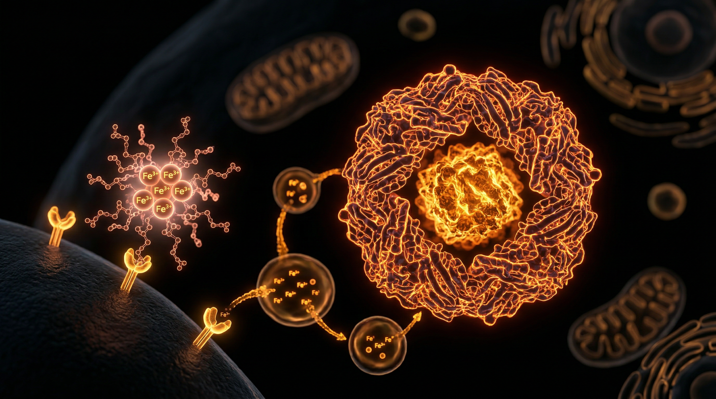
The Danger of NTBI
The primary risk during an infusion is the formation of Non-Transferrin Bound Iron (NTBI). If the rate of iron release from the carbohydrate complex exceeds the binding capacity of transferrin (measured as Total Iron Binding Capacity or TIBC), free iron begins to circulate. NTBI is highly reactive and is rapidly taken up by the liver, heart, and endocrine organs, where it can induce severe cellular damage.
Mechanisms at the Cellular Level
At the cellular level, the toxicity of iron is almost entirely mediated by its ability to participate in single-electron transfer reactions. This is the "dark side" of iron’s catalytic power.
The Fenton Reaction
The most significant mechanism of iron-induced toxicity is the Fenton Reaction. In this process, ferrous iron ($Fe^{2+}$) reacts with hydrogen peroxide ($H_2O_2$), a natural byproduct of mitochondrial respiration, to produce the hydroxyl radical ($OH^\bullet$).
- —$Fe^{2+} + H_2O_2 \rightarrow Fe^{3+} + OH^\bullet + OH^-$
The hydroxyl radical is the most reactive and damaging of all reactive oxygen species (ROS). It possesses an extremely short half-life but can inflict immediate damage on any biological molecule it encounters, including DNA, proteins, and lipids.
Lipid Peroxidation and Ferroptosis
The vascular endothelium—the lining of our blood vessels—is particularly vulnerable during an infusion. The presence of NTBI triggers lipid peroxidation, a chain reaction where ROS attack the polyunsaturated fatty acids in cell membranes. This leads to the formation of lipid peroxyl radicals, which eventually cause membrane rupture and cell death.
A newly identified form of regulated cell death, termed Ferroptosis, is specifically driven by iron-dependent lipid peroxidation. Unlike apoptosis, ferroptosis is characterised by the catastrophic failure of the cell’s antioxidant defences, specifically the glutathione-dependent enzyme GPX4. When an iron infusion overwhelms this system, the result is localized tissue damage and systemic inflammation.
Mitochondrial Dysfunction
Iron is essential for the cytochromes in the electron transport chain. However, an excess of labile (unbound) iron within the mitochondria leads to the production of superoxide radicals. This creates a "mitochondrial bottleneck" where the cell’s energy production is compromised by the very element meant to enhance it.
Statistics show that up to 20% of patients receiving rapid-dose ferric carboxymaltose may experience transient "hypophosphatemia," a condition where phosphate levels drop dangerously low due to iron’s impact on the hormone FGF23, which regulates renal phosphate excretion.
Environmental Threats and Biological Disruptors
The challenge of managing iron is exacerbated by the modern environment. Our biological systems are currently under siege from factors that disrupt iron metabolism, making infusions more volatile than they were in previous generations.
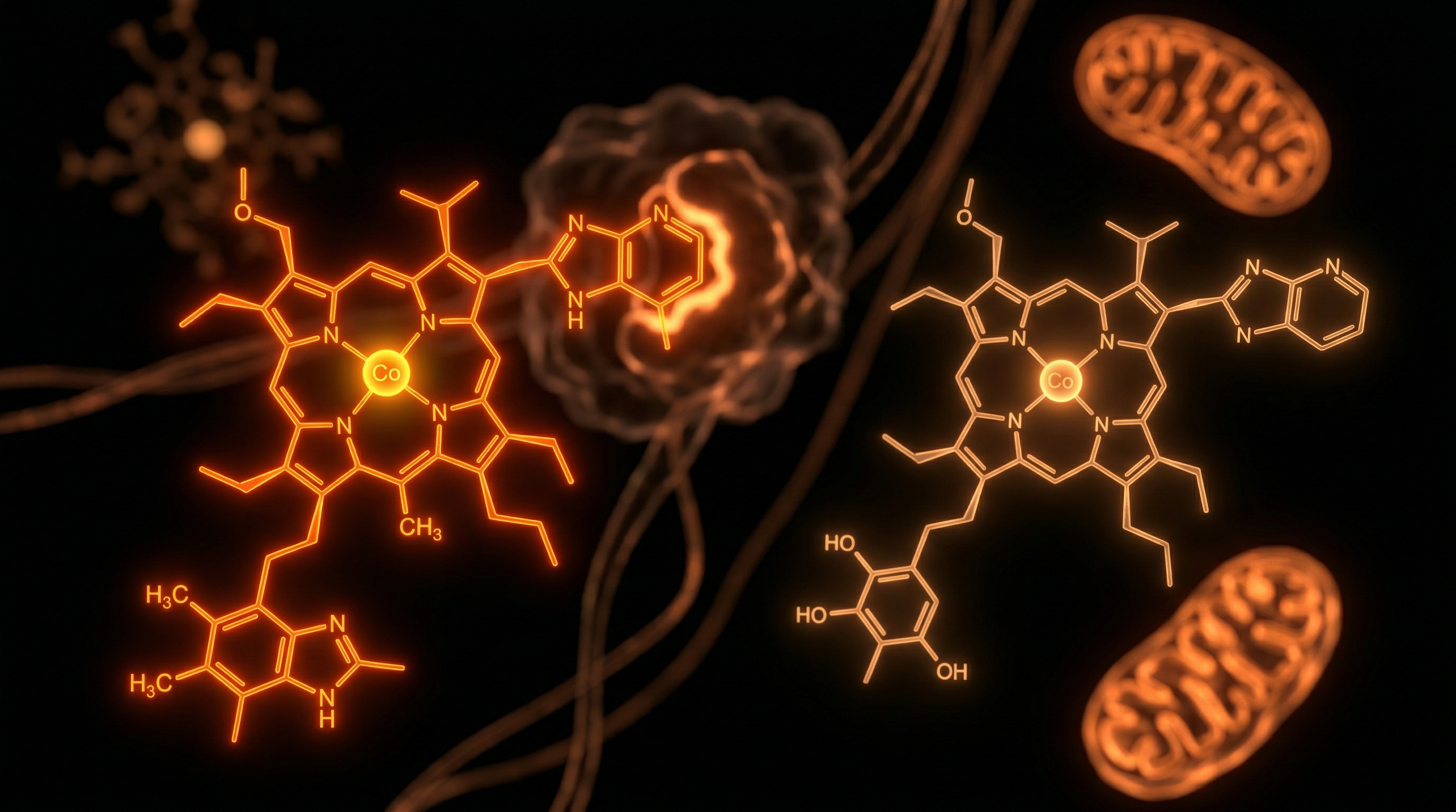
- —Glyphosate Exposure: The most widely used herbicide in the world acts as a powerful mineral chelator. It has been shown to disrupt the body’s ability to utilise manganese and cobalt, while simultaneously interfering with the shikimate pathway in the gut microbiome. This leads to intestinal permeability (leaky gut), which can cause systemic inflammation—triggering hepcidin and "locking" iron in the tissues even when the body is functionally anaemic.
- —Electromagnetic Fields (EMFs): Emerging research suggests that EMF exposure can alter the gating of voltage-gated calcium channels. Because iron and calcium share certain transport pathways, EMF-induced ion dysregulation can increase the intracellular uptake of labile iron, heightening the risk of oxidative stress during an infusion.
- —Heavy Metal Burden: Presence of lead, cadmium, or aluminium competes with iron for binding sites on transport proteins. If a patient has a high body burden of toxic metals, the "safe" storage capacity for iron is reduced, making the infusion more likely to result in NTBI.
- —The Copper Paradox: Modern agricultural practices have depleted soil copper. Copper is a prerequisite for the function of ceruloplasmin (the ferroxidase enzyme). Without adequate bioavailable copper, iron cannot be converted from its toxic ferrous form to its transportable ferric form. This creates a state of "unbound iron" that the mainstream medical narrative frequently ignores.
The Cascade: From Exposure to Disease
When iron infusions are administered without regard for the patient’s underlying oxidative stress levels or mineral co-factors, a pathological cascade can be triggered.
- —Phase I: Immediate Oxidative Burst. Within minutes of infusion, markers of lipid peroxidation (such as malondialdehyde) rise in the plasma. This causes the "infusion reaction"—nausea, metallic taste, and flushing.
- —Phase II: Endothelial Damage. The hydroxyl radicals damage the glycocalyx (the protective lining of the blood vessels). This can lead to increased vascular permeability and, in some cases, the initiation of atherosclerotic plaques.
- —Phase III: The Inflammatory Loop. Damage to the liver by NTBI triggers the release of pro-inflammatory cytokines like IL-6. This, in turn, stimulates further hepcidin production. Paradoxically, a high-dose infusion intended to cure anaemia can "shut down" iron recycling, making the patient dependent on further infusions as their own stores remain "locked" in the reticuloendothelial system.
- —Phase IV: Organ Deposition. If the iron is not successfully integrated into haemoglobin, it deposits in the liver (haemosiderosis) or the heart. Long-term, this contributes to metabolic syndrome and insulin resistance, as iron in the pancreas interferes with insulin secretion.
What the Mainstream Narrative Omits
The current medical consensus treats iron deficiency as a simple "empty tank" problem. If the tank is empty (low ferritin), you fill it up (infusion). This reductionist view omits several critical biological truths:
The Anemia of Chronic Inflammation (ACD)
A low ferritin level is often a sign of true iron deficiency. However, a *high* or *normal* ferritin level in the presence of low serum iron often indicates Anemia of Chronic Disease. In this state, the body is intentionally sequestering iron to keep it away from pathogens (many bacteria thrive on iron). Infusing iron into a body that is actively trying to hide it is a recipe for disaster, essentially "feeding the fire" of inflammation or infection.
The Myth of Ferritin as an Accurate Marker
Ferritin is an acute-phase reactant. It rises in response to stress, infection, or trauma. Relying solely on ferritin to guide infusion therapy is dangerous. One can have high ferritin (indicating systemic inflammation) and still have zero functional iron in the mitochondria. Conversely, a "low" ferritin of 30 ng/mL might be perfectly healthy for an individual with an highly efficient iron-recycling system.
The Role of Bioavailable Copper
Mainstream haematology rarely tests for Ceruloplasmin. Without ceruloplasmin, iron is essentially "stuck." Iron infusions given to copper-deficient individuals cannot be properly mobilised, leading to tissue loading and oxidative damage. The "Innerstanding" view is that we do not have an iron deficiency epidemic; we have a copper bio-availability crisis.
The Phosphate Sink
The medical community has only recently begun to acknowledge the phenomenon of 6-H (FGF23-mediated hypophosphatemic osteomalacia). Certain iron formulations (specifically Ferric Carboxymaltose) trigger an explosion in the hormone FGF23, which tells the kidneys to dump phosphate. Low phosphate leads to profound muscle weakness, bone pain, and heart arrhythmias—symptoms often dismissed by doctors as "post-infusion fatigue."
The UK Context
In the United Kingdom, the administration of parenteral iron is strictly governed by the NICE (National Institute for Health and Care Excellence) guidelines and the MHRA (Medicines and Healthcare products Regulatory Agency).
Current Protocols
The UK approach typically prioritises oral iron first (such as ferrous sulphate or ferrous fumarate), despite their 80% side-effect rate. When oral iron fails or is inappropriate (e.g., in cases of IBD, CKD, or preoperative optimization), IV iron is sanctioned.
The two dominant formulations in the UK are:
- —Ferinject (Ferric Carboxymaltose): Allows for rapid, high-dose administration (up to 1000mg in 15 minutes). While convenient, it is the formulation most associated with hypophosphatemia.
- —Monofer/Monoferric (Iron Isomaltoside 1000): A more stable carbohydrate complex that arguably releases iron more slowly, potentially reducing the risk of NTBI, though still requiring careful monitoring.
The Ganzoni Formula
UK clinicians use the Ganzoni Formula to calculate the iron deficit:
- —*Total Iron Deficit [mg] = Body weight [kg] x (Target Hb - Actual Hb) [g/dL] x 2.4 + Iron stores [mg]*
While mathematically precise, this formula is biologically crude. It assumes that every gram of infused iron will be used for haemoglobin, ignoring the systemic "sequestration" that occurs in inflamed patients.
Safety and Monitoring
UK hospitals are required to have resuscitation equipment on hand during infusions due to the risk of anaphylaxis. However, the "delayed" reactions—the oxidative stress and phosphate depletion—are often not monitored post-discharge, leaving a gap in patient care that "Innerstanding" practitioners must fill.
Protective Measures and Recovery Protocols
If an infusion is deemed necessary, it should never be performed as a standalone procedure. A comprehensive biological shield must be constructed to protect the vasculature and mitochondria from the inevitable oxidative burst.
1. Pre-Infusion Priming
- —The Copper-Retinol Connection: Ensure the patient has adequate levels of Whole Food Vitamin C (which contains the enzyme tyrosinase), bioavailable copper (from organ meats or bee pollen), and Vitamin A (Retinol). Retinol is essential for the loading of iron onto transferrin.
- —Glutathione Support: Supplementing with Liposomal Glutathione or its precursor, N-Acetyl Cysteine (NAC), for five days prior to infusion can bolster the cell’s primary defence against the Fenton Reaction.
2. The "Antioxidant Umbrella"
During the infusion, the body’s antioxidant stores are rapidly depleted.
- —Vitamin E (Alpha and Gamma Tocopherols): Essential for stopping the chain reaction of lipid peroxidation in the cell membranes.
- —Selenium: A critical cofactor for GPX4, the enzyme that prevents ferroptosis.
3. Managing the Phosphate Sink
For those receiving Ferinject, monitoring phosphate levels is non-negotiable.
- —Patients should be advised to increase dietary phosphate and consider supplementation if levels drop.
- —Avoiding high-dose Vitamin D immediately after an infusion can be beneficial, as Vitamin D can interact with the FGF23 pathway in complex ways.
4. Post-Infusion Detoxification
Iron that has been "misplaced" in the tissues must be mobilised.
- —Systemic Enzymes: Proteolytic enzymes like Serrapeptase may help reduce the inflammation caused by NTBI deposition.
- —Sweat Therapy: Using an Infrared Sauna can help move systemic toxins that may be compounding the iron-induced oxidative stress, though this should be delayed until the patient is haemodynamically stable.
5. Monitoring Beyond Ferritin
Post-infusion monitoring should include:
- —Full Blood Count (FBC): To ensure erythropoiesis is occurring.
- —Serum Phosphate: Checked at day 7 and 14.
- —GGT (Gamma-Glutamyl Transferase): A sensitive marker of liver stress and glutathione depletion.
- —Transferrin Saturation (TSAT): To ensure the iron is being transported and not sitting as NTBI.
Summary: Key Takeaways
The management of iron via infusion is a potent medical intervention that requires a shift from "dosage-centric" to "biology-centric" thinking. To navigate the dynamics of iron without succumbing to toxicity, we must respect the following truths:
- —Iron is a Catalyst for Oxidative Stress: Through the Fenton Reaction, unbound iron generates hydroxyl radicals that damage the vascular endothelium and mitochondrial DNA.
- —The Mucosal Block is There for a Reason: Bypassing the gut via IV therapy removes a critical evolutionary safety valve. We must compensate by providing the body with the co-factors it would normally use to manage iron.
- —Ferritin is an Imperfect Metric: A high ferritin post-infusion does not necessarily mean the patient is "cured." It may indicate that the iron is being sequestered due to inflammation.
- —Copper is the Master Key: Without bioavailable copper and ceruloplasmin, iron remains toxic and stagnant. True iron management is, in reality, mineral management.
- —UK Protocols are Just the Baseline: While NICE guidelines provide a safety framework, they do not account for the environmental disruptors (EMFs, Glyphosate) or the subtle nutritional deficiencies (Retinol, Vitamin E) that dictate a patient’s oxidative resilience.
In the pursuit of "Innerstanding," we must view the iron infusion not as a simple refill of a depleted nutrient, but as a high-stakes biochemical event. By supporting the body's natural antioxidant defences and ensuring mineral synergy, we can harness the benefits of iron correction while shielding the delicate machinery of human life from the fires of oxidative damage.
This article is provided for informational and educational purposes only. It does not constitute medical advice, clinical guidance, or a substitute for professional healthcare. Information reflects cited research at time of publication. Always consult a qualified healthcare professional before acting on any health information.
RESEARCH FOUNDATIONS
Biological Credibility Archive
Citations provided for educational reference. Verify via PubMed or institutional databases.
Medical Disclaimer
The information in this article is for educational purposes only and does not constitute medical advice, diagnosis, or treatment. Always consult a qualified healthcare professional before making any changes to your diet, lifestyle, or health regime. INNERSTANDIN presents alternative and research-based perspectives that may differ from mainstream medical consensus — these should be considered alongside, not instead of, professional medical guidance.
Read Full DisclaimerReady to learn more?
Continue your journey through our classified biological research.
DISCUSSION ROOM
Members of THE COLLECTIVE discussing "Iron Infusion Dynamics: Managing Ferritin Without Toxicity"
SILENT CHANNEL
Be the first to discuss this article. Your insight could help others understand these biological concepts deeper.
RABBIT HOLE
Follow the biological thread deeper