Iron: Oxygen Transport, Ferritin Storage and the Dangers of Overload
Iron is central to haemoglobin and myoglobin function, but excess iron is a potent pro-oxidant. This article explores haem vs non-haem iron, ferritin as an acute-phase reactant, iron dysregulation in chronic disease and the overlooked problem of iron overload in the UK.

Overview
Iron is perhaps the most paradoxical element within the human biological landscape. It is the "spark of life," the fundamental metallic core that allows us to harness the power of oxygen, yet it is also a potent catalyst for oxidative destruction. In the UK, the public health narrative has been singularly obsessed with iron deficiency, a tunnel-visioned approach that has blinded both the medical establishment and the citizenry to the silent epidemic of iron dysregulation and pathological overload.
At the heart of our physiology, iron sits within the haem group of haemoglobin, binding oxygen in the lungs and releasing it to gasping tissues. It resides within myoglobin in our muscles and powers the cytochromes within our mitochondria, facilitating the electron transport chain that generates ATP. Without iron, we cease to respire; we fade into the lethargy of anaemia. However, because iron is chemically "promiscuous"—meaning it readily swaps electrons—it is capable of generating the hydroxyl radical, the most reactive and damaging free radical known to science.
The human body has evolved no active excretory pathway for iron. Aside from blood loss or the sloughing of skin and intestinal cells, iron remains trapped within our systems. This biological "hoarding" mechanism was an evolutionary advantage in an era of scarce nutritional resources and frequent parasitic infections. In the modern UK context, characterised by mandatory flour fortification, the ubiquity of red meat, and a lack of regular blood loss in the male and post-menopausal female populations, this hoarding mechanism has become a liability.
This article aims to expose the mechanisms by which iron, when misplaced or excessive, drives chronic inflammatory states, neurodegeneration, and organ failure. We will deconstruct the "ferritin myth," explore the molecular carnage of the Fenton Reaction, and provide a blueprint for regaining iron homeostasis in a world that is increasingly—and dangerously—saturated with this heavy metal.
##
The Biology — How It Works
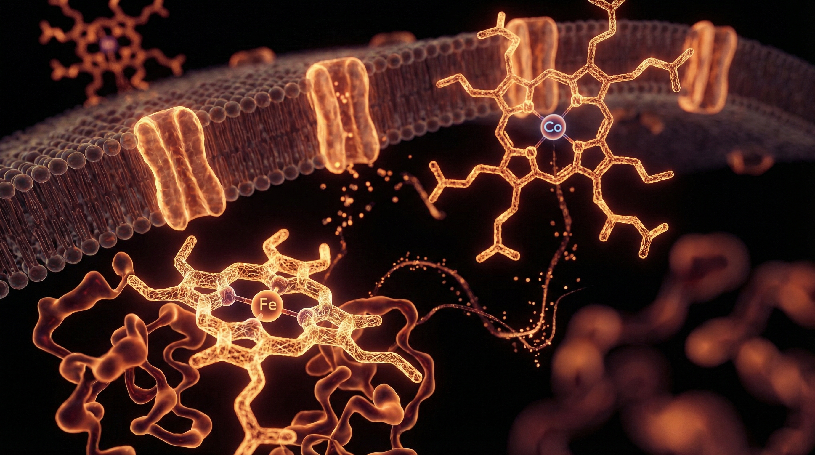
To understand iron’s role, we must first look at its transport and storage architecture. In the healthy body, iron is never "free." Ionic iron is so reactive that it must be chaperoned at all times by specialised proteins.
Haemoglobin and Myoglobin: The Oxygen Carriers
Approximately 70% of the body's iron is found in haemoglobin, the protein in red blood cells that carries oxygen from the lungs to the tissues. Each haemoglobin molecule contains four iron atoms. Myoglobin, found in muscle tissue, stores oxygen for use during physical exertion. The iron in these molecules is in the ferrous (Fe2+) state when binding oxygen. The efficiency of this system is what allows for the high metabolic rate of mammals.
The Absorption Gateway: Haem vs. Non-Haem
Dietary iron exists in two forms, and the body treats them very differently:
- —Haem Iron: Found in animal tissues (meat, fish, poultry). It is absorbed with high efficiency (15-35%) via the HCP1 (Haem Carrier Protein 1). Its absorption is relatively unaffected by other dietary factors.
- —Non-Haem Iron: Found in plants and fortified foods (grains, legumes, spinach). It is poorly absorbed (2-10%). It must be converted from the ferric (Fe3+) to the ferrous (Fe2+) state by the enzyme duodenal cytochrome b (Dcytb) before it can enter the intestinal cells via the DMT1 (Divalent Metal Transporter 1).
Transferrin: The Chaperone
Once iron enters the bloodstream, it is immediately bound by transferrin. This protein acts as a secure transport vehicle, ensuring that iron cannot engage in runaway chemical reactions while in transit. Under normal conditions, transferrin is about 30% saturated with iron. When this saturation exceeds 45-50%, the risk of Non-Transferrin Bound Iron (NTBI) increases. NTBI is highly toxic and is rapidly taken up by the heart, liver, and endocrine organs, leading to tissue damage.
Ferritin: The Biological Safe
Ferritin is the primary storage protein for iron. Shaped like a hollow sphere, a single ferritin molecule can sequester up to 4,500 iron atoms, keeping them in a non-toxic, mineralised state. While traditionally viewed as a simple measure of iron "stores," modern research reveals that ferritin is an acute-phase reactant. This means it rises significantly during infection, inflammation, and chronic stress, regardless of the body's actual iron levels.
CRITICAL FACT: A high ferritin reading on a standard NHS blood test does not always indicate high iron stores; it may indicate that the body is "locking away" iron to starve invading pathogens or as a response to systemic inflammation.
##
Mechanisms at the Cellular Level
At the cellular level, the management of iron is a high-stakes balancing act. The cell must have enough iron for its enzymes and mitochondria but not so much that it triggers programmed cell death.
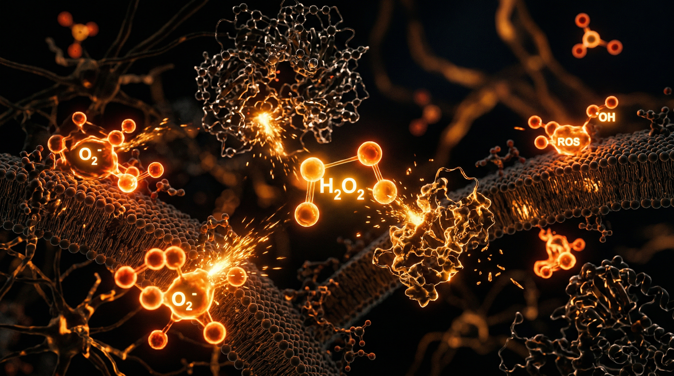
The Fenton Reaction: The Engine of Oxidative Stress
The primary danger of iron lies in its ability to participate in the Fenton Reaction. In this process, ferrous iron (Fe2+) reacts with hydrogen peroxide (H2O2)—a natural byproduct of metabolism—to produce the hydroxyl radical (•OH).
- —Fe2+ + H2O2 → Fe3+ + •OH + OH−
The hydroxyl radical is a biological "wrecking ball." It has no enzymatic neutraliser; once formed, it instantly attacks lipids in cell membranes, proteins, and DNA. This leads to lipid peroxidation, particularly of the polyunsaturated fatty acids (PUFAs) that make up our cell membranes, creating a chain reaction of destruction.
Hepcidin: The Master Regulator
Iron levels are not regulated by excretion, but by the hormone hepcidin, produced by the liver. Hepcidin acts as the body's iron "gatekeeper." When iron levels are high or inflammation is present, the liver secretes hepcidin, which binds to and degrades ferroportin—the only known protein that exports iron out of cells.
- —When ferroportin is destroyed, iron remains trapped inside the intestinal cells (which are eventually sloughed off) and inside the macrophages (the body's "recycling" cells).
- —This results in low serum iron but high cellular iron, a condition known as iron sequestration.
Ferroptosis: A New Form of Cell Death
In 2012, scientists identified a unique form of programmed cell death called ferroptosis. Unlike apoptosis (orderly cell death), ferroptosis is driven by iron-dependent lipid peroxidation. When cellular iron levels are too high, or the antioxidant system (specifically glutathione and the enzyme GPX4) fails, the cell literally "rusts" from the inside out and implodes. This pathway is now being linked to the massive loss of neurons in Alzheimer’s and Parkinson’s disease.
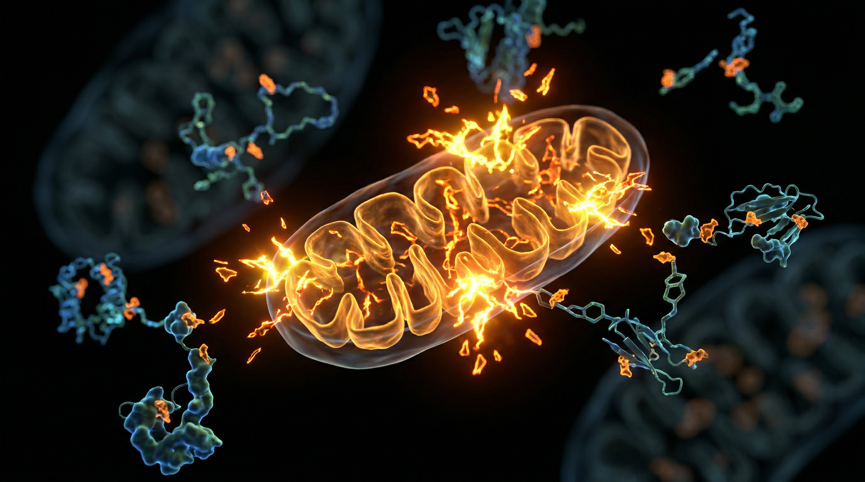
Mitochondrial Iron-Sulphur Clusters
Iron is essential for the Iron-Sulphur (Fe-S) clusters found in the proteins of the electron transport chain. These clusters are responsible for the actual transfer of electrons that produces energy. However, the mitochondria are also the primary site of ROS (Reactive Oxygen Species) production. If iron is not tightly regulated within the mitochondria, it creates a "perfect storm" for mitochondrial DNA damage and metabolic collapse.
##
Environmental Threats and Biological Disruptors
The modern environment, particularly in the UK, is saturated with factors that disrupt iron metabolism and promote overload.
Mandatory Flour Fortification
Since the Bread and Flour Regulations 1998, all white wheat flour in the UK must be fortified with iron (along with calcium, thiamine, and niacin). This iron is usually in the form of powdered elemental iron or ferrous sulphate.
- —Elemental iron is essentially fine iron filings.
- —Ferrous sulphate is highly oxidative and irritating to the gut lining.
This mandatory intervention applies to almost all commercially produced bread, biscuits, and pastries. While designed to prevent anaemia in a small subset of the population, it subjects the entire population—including those at risk of overload—to a lifelong, unavoidable intake of highly reactive iron.
Industrial Contaminants
Heavy metals like lead, cadmium, and aluminium interfere with iron metabolism. Lead, for instance, inhibits the enzyme ferrochelatase, which is responsible for inserting iron into the haem molecule. This can result in "sideroblastic anaemia," where the body has plenty of iron, but cannot use it, leading to iron accumulating in the mitochondria of developing red blood cells (ringed sideroblasts).
Endocrine Disruptors and Hepcidin
Xenooestrogens, such as BPA (Bisphenol A) and certain phthalates ubiquitous in plastic packaging, can interfere with liver function and the signalling pathways that control hepcidin. This can lead to inappropriate hepcidin suppression, causing the body to absorb more iron than it requires, even when stores are already saturated.
The "Iron-Enriched" Cereal Industry
Many breakfast cereals marketed to children in the UK contain up to 100% of the daily "recommended" intake of iron in a single serving. This iron is often added as a spray-on coating. For a growing child, some of this is utilised; however, for an adult male or a post-menopausal woman consuming these products daily, it represents a significant and unnatural iron burden that the body has no way to eliminate.
ALARMING STATISTIC: Research published in the *British Journal of Nutrition* indicates that mandatory fortification in the UK may contribute to a significant percentage of the population exceeding the 'Tolerable Upper Intake Level' for iron, particularly among men and the elderly.
##
The Cascade: From Exposure to Disease
Unchecked iron accumulation and dysregulation do not cause immediate illness but rather a slow, decades-long cascade of systemic decay.
Stage 1: The Asymptomatic Accumulation
In the early stages, iron begins to accumulate in the liver's Kupffer cells and hepatocytes. Serum ferritin begins to creep upward, often dismissed by GPs as being "within the normal range" (which in the UK can extend up to 300-400 ng/mL—levels many experts consider far too high).
Stage 2: Oxidative Stress and Insulin Resistance
As iron accumulates in the pancreas, it specifically damages the beta cells, which are highly sensitive to oxidative stress. This impairs insulin secretion. Simultaneously, iron in the liver promotes gluconeogenesis (the production of sugar) and interferes with insulin signalling, leading to Type 2 Diabetes. This is often referred to as "Bronze Diabetes" when associated with hereditary haemochromatosis.
Stage 3: Cardiovascular and Organ Damage
Iron catalyzes the oxidation of LDL cholesterol. Oxidised LDL is what actually gets trapped in the arterial walls, forming the basis of atherosclerosis. In the heart, iron deposits in the myocardium lead to cardiomyopathy and arrhythmias. The liver, under the constant strain of iron-induced lipid peroxidation, begins to develop fibrosis, which can progress to cirrhosis and hepatocellular carcinoma.
Stage 4: Neurodegeneration
The brain is highly susceptible to iron overload. Because the brain is rich in lipids and has a high oxygen demand, the presence of excess "labile" (unbound) iron is catastrophic.
- —In Alzheimer’s disease, iron is found concentrated within amyloid plaques.
- —In Parkinson’s disease, iron accumulates in the substantia nigra, the area of the brain responsible for dopamine production, driving the ferroptotic death of neurons.
Pathogenic Exploitation
Almost all pathogens—bacteria, fungi, and even certain viruses—require iron to replicate. The body’s innate immune response is to hide iron during an infection (via hepcidin). However, if the body is systemically overloaded, this "nutritional immunity" fails. Chronic iron overload is associated with increased susceptibility to infections and may even play a role in the persistence of "stealth" pathogens and biofilms.
##
What the Mainstream Narrative Omits
The mainstream medical approach to iron is outdated, often relying on "reference ranges" that are based on an increasingly sick population rather than optimal health.
The Ferritin Fallacy
The NHS typically considers a ferritin level above 15-20 ng/mL to be "normal." However, lower levels (around 25-50 ng/mL) are often associated with better longevity and lower cancer risk. Conversely, "high-normal" ferritin is often a marker of Metabolic Syndrome and silent inflammation. The medical establishment frequently fails to distinguish between iron *deficiency* and iron *anemia of chronic disease* (where iron is present but locked away). Giving iron supplements to someone with inflammation-induced low serum iron is like throwing petrol on a fire.
The Missing "Full Iron Panel"
Most UK patients only receive a Ferritin test. To truly understand iron status, one must see the whole picture:
- —Serum Iron: The amount of iron in the blood.
- —TIBC (Total Iron Binding Capacity): A measure of how much transferrin is available.
- —Transferrin Saturation (%): The most critical marker for overload. Anything over 45% is a red flag.
- —GGT (Gamma-Glutamyl Transferase): A liver enzyme that, when elevated alongside ferritin, strongly suggests iron-driven oxidative stress.
The Role of Copper and Retinol
Mainstream nutrition ignores the "Iron-Copper-Retinol" triad. Iron cannot be properly utilised or moved without the copper-dependent enzyme ceruloplasmin (the ferroxidase). Ceruloplasmin requires bioavailable copper and Vitamin A (retinol) to function.
SUPPRESSED TRUTH: Many cases of "iron-deficiency anaemia" are actually copper or Vitamin A deficiencies. Without these cofactors, iron gets stuck in the tissues and cannot be loaded onto transferrin, leading to a false appearance of deficiency on blood tests.
The Gender Bias
While the focus is often on women of childbearing age being "deficient," the medical narrative ignores the fact that men and post-menopausal women are at the highest risk for overload. By the age of 60, the average man has 3-4 times more iron than the average woman. This correlates perfectly with the higher rates of heart disease and early mortality in men.
##
The UK Context
The United Kingdom presents a unique landscape for iron issues due to historical policy and genetic prevalence.
Hereditary Haemochromatosis: The "Celtic Curse"
The UK has some of the highest rates of Hereditary Haemochromatosis (HH) in the world, particularly the C282Y mutation of the HFE gene. It is often called the "Celtic Curse" because of its prevalence in people of Irish, Scottish, and Welsh descent.
- —Approximately 1 in 10 people in the UK are carriers of the gene.
- —1 in 200 have two copies of the gene, putting them at extreme risk of massive iron overload.
Despite this, many people with HH go undiagnosed until they suffer permanent organ damage in their 40s or 50s. The NHS does not currently screen for this condition routinely.
The Bread and Flour Regulations: A Failed Policy?
As mentioned, the mandatory fortification of flour is a "blunt instrument" policy. While it was intended to help women of childbearing age, data suggests it has had a negligible impact on anaemia rates while contributing to the cumulative iron burden of the rest of the population. The Food Standards Agency (FSA) and the Department of Health and Social Care have resisted calls to make fortification voluntary or to use more bioavailable, less oxidative forms of iron.
The NHS Reference Range Problem
In many UK labs, the upper limit for ferritin is set as high as 400 ng/mL for men. Functional medicine practitioners and researchers in the field of longevity suggest that any ferritin level over 100 ng/mL is associated with increased all-cause mortality. The "normal" range is a statistical average of the population, but in a country where obesity and chronic inflammation are rampant, the "normal" range is not the "healthy" range.
##
Protective Measures and Recovery Protocols
If you suspect iron dysregulation or overload, the path to recovery involves both reducing intake and actively "unloading" the excess.
1. Phlebotomy: The Gold Standard
The most effective way to reduce iron is through blood donation (phlebotomy). Each pint of blood donated removes approximately 250mg of iron. For those with clinical haemochromatosis, this is the primary treatment. Even for those without the genetic condition, donating blood 2-3 times a year can be a powerful anti-ageing strategy.
- —Action: Check your transferrin saturation. If it’s above 45%, or ferritin is consistently above 150 ng/mL, consider becoming a regular donor via the NHS Blood and Transplant service.
2. Dietary Modifications
- —Avoid Fortified Foods: Read labels on bread, cereals, and flour. Opt for organic or artisan flours that do not contain added "iron" or "reduced iron."
- —Tea and Coffee with Meals: The tannins and polyphenols in tea and coffee strongly inhibit the absorption of non-haem iron. Drinking a cup of tea with a meal can reduce iron absorption by up to 60-70%.
- —Calcium Interference: Calcium competes with iron for absorption. Consuming dairy or calcium-rich foods with iron-rich meals can mitigate uptake.
3. Natural Iron Chelators
Certain natural compounds can bind to iron and help usher it out of the body or neutralise its oxidative potential:
- —IP6 (Inositol Hexaphosphate): Found in the hulls of seeds and grains, IP6 is a potent iron chelator. It has been studied for its ability to remove excess iron from cells and even inhibit the growth of iron-dependent cancer cells.
- —Quercetin and Curcumin: These polyphenols are not only anti-inflammatory but also act as mild iron chelators.
- —Lactoferrin: This protein, found in colostrum and whey, is an iron-regulator. It has the unique ability to "grab" free iron and deliver it to where it is needed (like red blood cells) while keeping it away from pathogens.
4. Addressing Co-Factors
Ensure you are not "functionally" deficient. To move iron, your body needs:
- —Copper: From organic liver (the most bioavailable source), oysters, or high-quality supplements (e.g., copper glycinate).
- —Vitamin A (Retinol): Real Vitamin A from animal sources (cod liver oil, eggs, butter). Beta-carotene from carrots is often poorly converted and does not provide the same support for ceruloplasmin.
- —Magnesium: Essential for over 300 enzymatic reactions, including those involved in the antioxidant defence system that protects against iron-induced damage.
5. Proper Testing
Demand a full iron panel. Do not settle for just a ferritin test.
- —Optimal Ferritin: 30–80 ng/mL.
- —Optimal Transferrin Saturation: 25–35%.
- —Optimal GGT: Below 20 U/L (an indicator that iron isn't causing liver stress).
IMPORTANT CALLOUT: If you are taking a multi-vitamin, ensure it is "Iron-Free." Most men and post-menopausal women should never supplement with iron unless a clear, diagnosed deficiency (via low haemoglobin AND low ferritin) is present.
##
Summary: Key Takeaways
The reality of iron is far removed from the simplistic "more is better" narrative found in high-street supplement shops and government fortification policies.
- —Iron is a Double-Edged Sword: Vital for oxygen transport but a primary driver of oxidative stress and lipid peroxidation via the Fenton Reaction.
- —The UK Overload Problem: Mandatory flour fortification and a high prevalence of the "Celtic Curse" (haemochromatosis) make the UK population particularly vulnerable to iron overload.
- —Ferritin is Not Just Storage: It is a marker of inflammation. High ferritin often reflects systemic "rusting" and metabolic distress rather than healthy iron levels.
- —The "Anaemia" Deception: Many people diagnosed with iron deficiency actually have "iron recycling" issues caused by a lack of bioavailable copper and Vitamin A, or they have "anaemia of chronic disease" where iron is trapped in tissues.
- —Ferroptosis: This iron-dependent cell death is a major mechanism in neurodegenerative diseases like Alzheimer’s and Parkinson’s.
- —Take Control: Regular blood donation, avoiding fortified grains, and focusing on copper/retinol co-factors are essential steps for anyone looking to escape the "iron trap."
In the pursuit of health, we must recognise that iron is not a passive nutrient but a powerful, reactive metal that requires meticulous management. To ignore the dangers of iron overload is to invite the slow, oxidative decay of the human frame. It is time to move beyond the "iron-poor blood" marketing of the 20th century and embrace a more sophisticated, biologically accurate understanding of mineral homeostasis.
This article is provided for informational and educational purposes only. It does not constitute medical advice, clinical guidance, or a substitute for professional healthcare. Information reflects cited research at time of publication. Always consult a qualified healthcare professional before acting on any health information.
RESEARCH FOUNDATIONS
Biological Credibility Archive
Citations provided for educational reference. Verify via PubMed or institutional databases.
Medical Disclaimer
The information in this article is for educational purposes only and does not constitute medical advice, diagnosis, or treatment. Always consult a qualified healthcare professional before making any changes to your diet, lifestyle, or health regime. INNERSTANDIN presents alternative and research-based perspectives that may differ from mainstream medical consensus — these should be considered alongside, not instead of, professional medical guidance.
Read Full DisclaimerReady to learn more?
Continue your journey through our classified biological research.
DISCUSSION ROOM
Members of THE COLLECTIVE discussing "Iron: Oxygen Transport, Ferritin Storage and the Dangers of Overload"
SILENT CHANNEL
Be the first to discuss this article. Your insight could help others understand these biological concepts deeper.
THE ARSENAL
Based on Vitamins, Minerals & Botanicals — products curated by our research team for educational relevance and biological support.
Magnesium L-Threonate
Clean Slate – Detoxes thousands of chemicals,heavy metals, pesticides, allergens, mold spores and fungus
Fulvic Minerals – Natural Rare Earth Minerals. The essential trace elements missing from modern processed foods.
INNERSTANDING may earn a commission on purchases made through these links. All products are selected based on rigorous educational relevance to our biological research.
RABBIT HOLE
Follow the biological thread deeper