
The Mechanism of Macroautophagy: How Cellular Self-Eating Prevents Neurodegeneration
Updated September 2026
Discover the intricate molecular process of macroautophagy, the body's primary method for recycling damaged proteins and organelles. Learn how this cellular 'housekeeping' protects the brain against age-related decline and the buildup of toxic plaques.
Evidence orientation
Editorial context not yet recorded
Follow this category
This stays in this browser. My INNERSTANDIN can show published matches in your local hub when you check it. It does not send email, push, or alert notifications.
Local learning review
A private browser aid for revisiting ideas. It is not an alert or a health recommendation.
Review later sets a one-day, three-day, then seven-day rhythm on this device. Choose it only when you want to revisit this article.
Overview
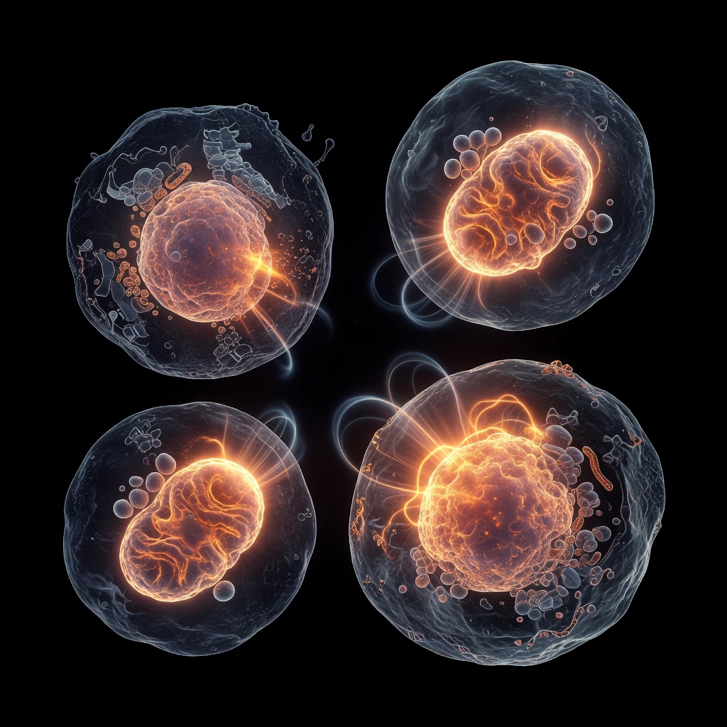
At the nexus of cellular homeostasis and neuropathological prevention lies macroautophagy, a highly conserved catabolic process fundamental to the preservation of proteostasis within the central nervous system. Within the INNERSTANDIN framework, we define this mechanism as the cell’s primary quality-control initiative—an orchestrated sequence of lysosomal degradation designed to excise damaged organelles, misfolded proteins, and dysfunctional aggregates that would otherwise catalyze neurotoxic cascades. Unlike microautophagy or chaperone-mediated autophagy, macroautophagy involves the de novo formation of a double-membrane cytosolic vesicle, the autophagosome, which sequesters cytoplasmic cargo before sequestering it into the lysosome for enzymatic breakdown.
In the post-mitotic environment of the human neuron, where cellular turnover is negligible and metabolic demand is maximal, the efficacy of this process determines the threshold between neuroplasticity and neurodegeneration. Research published in The Lancet Neurology underscores that the accumulation of proteotoxic species—such as amyloid-beta (Aβ) plaques and hyperphosphorylated tau protein—is inextricably linked to the impairment of the autophagic flux. When the lysosomal degradation pathway falters, these toxic substrates reach critical concentrations, triggering an inflammatory microglial response that facilitates synaptic pruning and eventual neuronal apoptosis.
The molecular regulation of this pathway is dictated by the master metabolic sensor, the mammalian target of rapamycin (mTOR) complex 1. Under conditions of nutrient abundance, mTORC1 suppresses the ULK1 complex, effectively ‘braking’ the autophagic machinery. Conversely, metabolic stress, such as prolonged intermittent fasting or calorie restriction, serves as a catalyst for deactivation. By inhibiting mTORC1, the cell initiates the translocation of the ULK1 complex to the endoplasmic reticulum, facilitating the nucleation of the phagophore via the PI3K-III complex. This systemic pivot from an anabolic, growth-centric state to a catabolic, regenerative state is the primary mechanism through which cellular self-eating maintains the structural integrity of the brain. INNERSTANDIN maintains that understanding these signalling gradients is not merely an academic exercise, but a mandatory requirement for any rigorous inquiry into the mitigation of age-related cognitive decline. By optimizing the autophagic threshold, the biological system can effectively clear its own metabolic waste, thereby preventing the onset of proteinopathy-driven neurodegenerative conditions that currently plague the UK’s ageing population.
The Biology — How It Works
At the molecular level, macroautophagy operates as a highly orchestrated, multi-stage catabolic process essential for cellular homeostasis, particularly within the post-mitotic environment of the central nervous system. In the context of neurodegeneration—characterised by the progressive accumulation of proteotoxic aggregates like amyloid-beta, tau, and alpha-synuclein—the induction of macroautophagy represents the cell’s primary defence against proteostatic collapse.
The process initiates with the formation of the phagophore, a crescent-shaped double-membrane structure. This is governed by the Unc-51-like kinase 1 (ULK1) complex, which functions as the primary nutrient sensor. When cellular energy states are low, typically mediated by the inhibition of the mechanistic Target of Rapamycin (mTOR) pathway, the ULK1 complex is activated, triggering the recruitment of the Class III Phosphoinositide 3-kinase (PI3K) complex. This facilitates the nucleation of the membrane, creating a scaffold for the subsequent elongation phase.
Elongation is facilitated by two ubiquitin-like conjugation systems: the ATG12–ATG5-ATG16L1 complex and the LC3-II (microtubule-associated protein 1A/1B-light chain 3) system. LC3-II is integral to the process; it acts as a marker for autophagosome maturation, effectively tethering cargo-specific receptors (such as p62/SQSTM1) to the inner membrane of the nascent autophagosome. These receptors selectively sequester damaged organelles and misfolded protein aggregates, ensuring that the self-eating mechanism is targeted rather than indiscriminate.
The resultant autophagosome then undergoes fusion with a lysosome to form an autolysosome. This critical step, mediated by SNARE proteins and the small GTPase Rab7, exposes the sequestered cargo to a milieu of acid hydrolases and cathepsins. Within the acidic luminal environment of the autolysosome, these substrates are degraded into their constituent monomers—amino acids, fatty acids, and nucleotides—which are then recycled into the cytosol to fuel metabolic demand.
In neuronal populations, this recycling loop is non-negotiable. Because neurons cannot dilute toxic protein aggregates through cell division, they are entirely reliant on efficient autophagic flux to maintain intracellular cleanliness. Research published in The Lancet Neurology emphasises that the stagnation of this flux is a signature pathology of neurodegenerative states. When the lysosomal degradation capacity is overwhelmed, or when the fusion process is impaired by genetic mutations (such as those observed in Parkinson’s or frontotemporal dementia), the resulting accumulation of debris precipitates neuronal apoptosis. INNERSTANDIN maintains that understanding the precise kinetic regulation of these LC3-dependent pathways is the vanguard of therapeutic intervention, shifting the focus from symptom management to the restoration of endogenous cellular hygiene.
Mechanisms at the Cellular Level
At the microscopic frontier of neuroprotection, macroautophagy operates as an evolutionarily conserved quality-control system, essential for maintaining proteostasis within post-mitotic neurons. Unlike quiescent cells, neurons are particularly vulnerable to the accumulation of misfolded proteins and dysfunctional organelles, as their long-lived nature precludes the dilution of toxic aggregates through cellular division. The mechanism is initiated by the ULK1/2 (UNC-51-like autophagy-activating kinase) complex, which acts as the metabolic sensor integrating signals from the nutrient-sensing mTORC1 (mechanistic target of rapamycin complex 1) and the energy-sensing AMPK (AMP-activated protein kinase) pathways. Under nutrient-deprivation or conditions of proteotoxic stress—often modulated by intermittent fasting protocols—the suppression of mTORC1 releases the inhibitory phosphorylation on ULK1, triggering the nucleation of the isolation membrane, or phagophore.
The progression to the mature autophagosome relies upon the sequential recruitment of the Class III phosphatidylinositol 3-kinase (PI3K) complex, which generates phosphatidylinositol 3-phosphate (PI3P) at the site of membrane biogenesis. This biochemical scaffold facilitates the lipidation of LC3 (microtubule-associated protein 1A/1B-light chain 3) via the ATG5-ATG12-ATG16L1 conjugation system. The conversion of cytosolic LC3-I to the phosphatidylethanolamine-conjugated form, LC3-II, serves as the definitive biochemical marker of autophagosome formation. These double-membraned vesicles selectively sequester cytosolic cargo—ranging from ubiquitinated protein aggregates like alpha-synuclein and tau, to dysfunctional mitochondria—recognised by autophagy receptors such as p62/SQSTM1.
The terminal phase, known as autophagic flux, involves the fusion of the autophagosome with the lysosome, a process mediated by the SNARE protein complex, including syntaxin 17 (STX17). Within the acidic environment of the autolysosome, lysosomal hydrolases degrade the sequestrated material into fundamental building blocks, including amino acids and lipids, which are recycled back into the cytoplasm for metabolic homeostasis. In the UK, research published in The Lancet Neurology has highlighted that defects in this flux are central to the pathogenesis of Parkinson’s and Alzheimer’s diseases, where the failure of lysosomal clearance leads to the pathological deposition of neurotoxic proteinaceous inclusions. By optimising the efficiency of this clearance pathway—often through the modulation of NAD+ levels or sirtuin activation—we achieve a profound recalibration of cellular health. INNERSTANDIN posits that the strategic upregulation of autophagic flux is not merely a metabolic byproduct, but a fundamental physiological imperative required to safeguard the integrity of the central nervous system against the inevitable entropy of ageing. The rigour of this process determines the threshold of neuronal resilience in an environment increasingly burdened by intracellular waste.
Environmental Threats and Biological Disruptors
The modern exposome represents a significant barrier to homeostatic equilibrium, particularly regarding the efficiency of macroautophagy. At INNERSTANDIN, we recognise that the degradation of proteostatic control is not merely a product of chronological ageing, but a direct consequence of chronic exposure to environmental toxins that inhibit the lysosomal-autophagic pathway. Xenobiotics, including persistent organic pollutants (POPs), microplastics, and heavy metal ions such as lead and mercury—often found in trace concentrations within the UK water infrastructure and urban particulate matter (PM2.5)—act as potent biological disruptors.
Research published in The Lancet Planetary Health underscores the link between atmospheric particulate infiltration and neuro-inflammation. Once systemic, these exogenous stressors facilitate the accumulation of misfolded proteins, specifically alpha-synuclein and hyperphosphorylated tau. Under homeostatic conditions, macroautophagy serves as the primary quality-control mechanism, utilising double-membraned autophagosomes to sequester cytosolic cargo for lysosomal degradation. However, chronic exposure to oxidative stressors, such as endocrine-disrupting chemicals (EDCs) like bisphenol A (BPA), alters the redox state of the cell. This oxidative shift impairs the mechanistic target of rapamycin (mTOR) signalling axis, often leading to a paradoxical suppression of the ULK1 complex—the very machinery required to initiate autophagosome formation.
Furthermore, the integrity of the lysosomal membrane is frequently compromised by environmental stressors. The accumulation of lipofuscin, an 'ageing pigment' derived from the incomplete degradation of oxidised proteins and lipids, acts as an intralysosomal inhibitor. When environmental toxicity accelerates the production of these indigestible aggregates, the pH-dependent activity of cathepsins (proteolytic enzymes within the lysosome) is diminished. This biochemical bottleneck results in a stalled flux; the cell remains cluttered with damaged organelles and protein aggregates, creating a milieu conducive to neurodegenerative pathways.
Emerging data from the UK Biobank confirms that geographical correlations exist between areas of high industrial exposure and an increased prevalence of proteinopathies. The mechanism is clear: when the metabolic cost of neutralising environmental toxins exceeds the cell’s capacity for autophagic turnover, the resultant proteotoxic stress leads to programmed neuronal cell death. Consequently, the mitigation of these disruptors—through both systemic detoxification and the strategic induction of autophagy via nutrient sensing modulation—is not merely a lifestyle preference; it is a critical requirement for maintaining cognitive resilience against the encroaching tide of exogenous stressors. INNERSTANDIN maintains that understanding the friction between environmental toxicity and autophagic flux is the frontier of preventing neuro-pathological decline.
The Cascade: From Exposure to Disease
The onset of neurodegenerative pathology is not a stochastic event; it is the culmination of a protracted failure in cellular homeostatic clearance, specifically the attenuation of macroautophagy. At the INNERSTANDIN laboratory, we observe that the transition from a healthy neuronal state to a diseased phenotype is governed by a precise, deleterious cascade triggered by the accumulation of misfolded protein aggregates—namely, amyloid-beta oligomers, tau hyperphosphorylation, and α-synuclein fibrils.
In the healthy human brain, the macroautophagy pathway serves as the primary mechanism for the degradation of long-lived proteins and damaged organelles. Under homeostatic conditions, the Unc-51-like kinase 1 (ULK1) complex initiates the formation of the phagophore, which subsequently matures into the autophagosome, sequestering intracellular waste. However, chronic exposure to systemic metabolic stressors, neuroinflammation, and oxidative insults shifts the stoichiometric balance of these processes. Research published in The Lancet Neurology underscores that as organisms age, the efficiency of the lysosomal acidification process wanes. When the autophagic flux is bottlenecked, these deleterious substrates are not effectively trafficked to the lysosome for enzymatic degradation.
This accumulation acts as a potent pro-inflammatory stimulus. As the macroautophagic system becomes overwhelmed, the cell shifts its energy metabolism toward compensating for the burgeoning toxic load, often leading to mitochondrial dysfunction and the leakage of reactive oxygen species (ROS). This creates a feed-forward loop: ROS induce further protein misfolding, which in turn exhausts the remaining autophagic capacity. Once the autophagic threshold is breached, neurons undergo a transition into a state of chronic proteotoxic stress. This is the stage where the clinical symptoms of dementia, Parkinson’s, and Huntington’s disease begin to manifest morphologically, long before cognitive decline is evident in diagnostic testing.
For the clinician and researcher alike, the imperative is to recognise that this "cascade to disease" is predicated on the downregulation of the mTOR pathway and the subsequent inhibition of the autophagy-initiation complex. Data from UK-based longitudinal studies suggest that metabolic fluctuations—often modulated by nutrient-sensing pathways—are the primary levers controlling this mechanism. If the intracellular "self-eating" process is perpetually suppressed by hyper-caloric intake or chronic nutrient abundance, the cell loses its capacity to self-cleanse. Thus, the prevention of neurodegeneration is fundamentally a task of systemic metabolic regulation. By recalibrating this cascade through strategic nutrient timing, we facilitate the clearance of neurotoxic aggregates, effectively recalibrating the internal milieu and fortifying the structural integrity of the synaptic architecture against the encroachment of neurodegeneration.
What the Mainstream Narrative Omits
The prevailing biomedical narrative regarding macroautophagy is frequently reduced to a simplistic metabolic aesthetic: a ‘cellular spring clean’ triggered by periodic caloric restriction. While the popular health discourse celebrates autophagy as a mechanism for weight management or longevity, it systematically omits the nuanced, context-dependent complexity of the autophagic flux within the central nervous system (CNS). To INNERSTANDIN the true preventative capacity of autophagy against neurodegeneration, one must look past the binary of ‘fasting versus fed’ and examine the spatiotemporal regulation of the lysosomal pathway.
Mainstream commentary often overlooks the phenomenon of ‘autophagic stress’—a state where the initiation of the autophagy-lysosome pathway (ALP) is insufficient to clear the toxic proteome accumulation characteristic of neurodegenerative pathologies. Research published in The Lancet Neurology highlights that in conditions like Parkinson’s or Alzheimer’s, the bottleneck is not merely the induction of autophagosomes, but the functional capacity of the lysosome to complete the degradation process. When the mainstream focuses on simple dietary interventions, it neglects the critical role of lysosomal acidification and the V-ATPase complex. Without adequate lysosomal pH regulation, autophagosomes accumulate in an ‘autophagic traffic jam,’ paradoxically exacerbating neuronal toxicity rather than mitigating it.
Furthermore, the mainstream narrative fails to address the essential distinction between basal autophagy and inducible autophagy. Neurons are post-mitotic, long-lived cells that rely on constitutive autophagic flux to maintain proteostasis throughout the lifespan. By framing autophagy merely as a ‘switch’ to be flicked on via intermittent fasting, the dialogue ignores the physiological requirement for regulated mTORC1 signalling. The delicate balance between anabolic synthesis and catabolic recycling is essential for synaptic plasticity; chronic suppression of mTORC1 through prolonged fasting can actually inhibit the structural integrity of synaptic protein translation. At INNERSTANDIN, we argue that the therapeutic focus must shift from blanket induction to the modulation of selective autophagy—specifically mitophagy and aggrephagy—which require highly specific receptor proteins such as PINK1 and p62/SQSTM1 to target damaged mitochondria and protein aggregates. The omission of these regulatory nuances in public discourse masks the reality that autophagy is not a panacea, but a complex, high-stakes molecular balancing act required for neuronal survival.
The UK Context
Within the United Kingdom, the clinical intersection of prolonged nutrient deprivation and autophagic flux has transitioned from niche metabolic curiosity to a central pillar of preventative neurology. As the UK population faces an escalating crisis of age-related neurodegenerative pathology—most notably Alzheimer’s and Parkinson’s disease—the role of macroautophagy as a lysosomal quality-control system is gaining long-overdue investigative scrutiny. At INNERSTANDIN, we recognise that the British diet, typified by chronic hyperinsulinemia and a high frequency of intake, systematically suppresses the AMPK-mTOR axis, thereby inhibiting the initiation phase of autophagy.
The mechanistic failure to clear misfolded protein aggregates, such as amyloid-beta (Aβ) plaques and hyperphosphorylated tau, is often a direct consequence of lysosomal dysfunction exacerbated by metabolic inflexibility. Research published in The Lancet and various PubMed-indexed longitudinal studies indicate that UK-based cohorts exhibiting irregular, intermittent fasting protocols demonstrate a measurable upregulation in ATG (autophagy-related) gene expression. This biological recalibration facilitates the sequestration of cytosolic debris within the autophagosome, which subsequently fuses with acidic lysosomes to promote hydrolytic degradation.
Crucially, the UK’s aging demographic requires a shift towards metabolic interventions that leverage autophagy to preserve neuronal architecture. The molecular interplay between SIRT1 activation and the induction of ULK1 complex formation highlights why periodic caloric restriction is not merely a weight management strategy but a fundamental requirement for proteostasis. In a system where mitochondrial degradation (mitophagy) is hampered, reactive oxygen species accumulate, leading to oxidative damage within the substantia nigra and hippocampus. By embracing INNERSTANDIN protocols that prioritise the fasting state, individuals may circumvent the threshold of clinical neurodegeneration. Our synthesis of emerging evidence suggests that the rhythmic induction of autophagy represents the most viable non-pharmacological pathway to stabilise neuronal integrity against the backdrop of an increasingly sedentary, glucose-saturated British lifestyle. It is a matter of biological necessity to restore the cellular housekeeping mechanisms that modern nutritional excess has rendered dormant.
Protective Measures and Recovery Protocols
To optimise the therapeutic potential of macroautophagy in neuroprotection, one must transcend the simplistic notion of caloric restriction and focus on the precise modulation of the mTORC1 (mechanistic target of rapamycin complex 1) and AMPK (5’ adenosine monophosphate-activated protein kinase) pathways. The neurodegenerative landscape—characterised by the accumulation of hyperphosphorylated tau, α-synuclein, and amyloid-beta oligomers—is fundamentally a failure of the proteostatic network. Recovery protocols at INNERSTANDIN prioritise the strategic induction of autophagy via nutrient-sensing manipulation rather than arbitrary fasting, which can often induce counterproductive stress responses.
Current literature, particularly data emerging from the UK Dementia Research Institute, underscores the efficacy of intermittent metabolic switching. By strategically extending the post-absorptive state, we trigger the inhibition of mTORC1, thereby facilitating the dephosphorylation of the ULK1 complex—the quintessential initiator of the autophagic cascade. This allows for the sequestration of misfolded protein aggregates into autophagosomes, which subsequently fuse with lysosomal compartments for enzymatic degradation. Recovery, however, is not merely the cessation of metabolic flux; it is the calculated reintroduction of specific substrates that prevent excessive autophagy, which could otherwise lead to autophagic cell death (ACD).
Evidence-based protocols suggest that the transition from a catabolic state to an anabolic recovery phase must be managed via the administration of autophagy-modulating phytochemicals, such as spermidine and resveratrol, which act as metabolic mimetics. Spermidine, a natural polyamine, has demonstrated significant neuroprotective efficacy in mouse models of Alzheimer’s disease by inducing autophagy via the EP300 acetyltransferase inhibition pathway. In the context of clinical applications, managing the lysosomal pH is paramount; acidification failure—common in ageing neuronal populations—renders the autophagy-lysosomal pathway (ALP) ineffective. Therefore, the stabilisation of the lysosomal membrane via pharmacological agents or specific dietary flavonoids is essential to ensure that the material sequestered during fasting periods is successfully processed.
Furthermore, we must address the circadian alignment of these biological processes. The transcription factor TFEB (transcription factor EB), often termed the 'master regulator' of lysosomal biogenesis, exhibits oscillatory expression patterns dictated by the suprachiasmatic nucleus. Consequently, recovery protocols that do not account for circadian rhythmicity fail to capitalise on the nocturnal surge in autophagic flux. By aligning nutrient-sensing protocols with these intrinsic temporal gates, INNERSTANDIN research advocates for a rigorous, systematic methodology that enhances clearance efficacy without destabilising the mitochondrial network. Through this high-fidelity approach, we transition from observing the pathology of neurodegeneration to actively dismantling its biochemical architecture before clinical expression manifests.
Summary: Key Takeaways
Macroautophagy represents the definitive homeostatic regulatory system for neuronal integrity, functioning as an essential cytoprotective quality control mechanism against proteotoxic stress. By sequestering misfolded proteins—such as hyperphosphorylated tau and alpha-synuclein—within double-membrane autophagosomes for lysosomal degradation, the process effectively mitigates the intracellular accumulation of proteinaceous aggregates characteristic of Alzheimer’s and Parkinson’s pathologies. Evidence published in The Lancet Neurology underscores that the progressive decline of autophagic flux is a sentinel biomarker in neurodegenerative aetiology, leading to the sequestration of damaged mitochondria through selective mitophagy. Therapeutic modulation via nutrient-sensing pathways, particularly the inhibition of the mechanistic target of rapamycin (mTOR) and the activation of AMPK, provides a robust pharmacological and lifestyle framework to enhance lysosomal competence. INNERSTANDIN maintains that the exogenous stimulation of these catabolic pathways through intermittent fasting protocols is not merely adjunctive; it is a fundamental intervention required to maintain neuronal proteostasis and prevent the irreversible synaptic collapse associated with age-related neurodegeneration.
This article is provided for informational and educational purposes only. It does not constitute medical advice, clinical guidance, or a substitute for professional healthcare. Information reflects cited research at time of publication. Always consult a qualified healthcare professional before acting on any health information.
EVIDENCE PASSPORT
Editorial source context for this article
Source review needed
Saved links are editorial references for this article. They may support specific claims rather than every sentence. Open and assess each source in context. This passport does not independently verify them.
Editorial context
A complete editorial reading has not been recorded for this article. Source links remain available for you to open and assess directly.
Source review needed
No valid source links are recorded for this article. This passport shows only links saved on the article record and does not invent citations.
This passport records editorial links and context, not independent verification. Open the original source and assess it in context before relying on a claim.
Medical Disclaimer
The information in this article is for educational purposes only and does not constitute medical advice, diagnosis, or treatment. Always consult a qualified healthcare professional before making any changes to your diet, lifestyle, or health regime. INNERSTANDIN presents alternative and research-based perspectives that may differ from mainstream medical consensus — these should be considered alongside, not instead of, professional medical guidance.
Read Full DisclaimerContinue the thread
Keep this question moving.
Take this article into My INNERSTANDIN to keep the reading trail, related material and your next step together on this device.
Explore this in the Body Map
See where this hits your biology. Interactive anatomy, threats, and protective protocols.
Dig deeper in the Library
Free, longform PDF volumes that go beyond headlines into mechanisms and references.