
Mercury Poisoning & Mitochondrial Damage: The ME/CFS Connection
Updated August 2026
Mercury from NHS-fitted amalgam dental fillings, predatory fish consumption, and thimerosal-containing pharmaceutical products is one of the most potent mitochondrial toxins known, with a specific affinity for the thiol groups of the electron transport chain enzymes responsible for ATP generation. By crippling cellular energy production, mercury creates the bioenergetic collapse that underlies ME/Chronic Fatigue Syndrome, progressive neurological decline, cardiac dysfunction, and immune dysregulation — conditions the NHS frequently misdiagnoses as psychiatric illness. The European Union's 2024 phase-down of dental amalgam, long resisted by UK dental bodies, represents a partial but belated acknowledgement of what independent toxicologists have been demonstrating for decades.
Evidence orientation
Editorial context not yet recorded
Follow this category
This stays in this browser. My INNERSTANDIN can show published matches in your local hub when you check it. It does not send email, push, or alert notifications.
Local learning review
A private browser aid for revisiting ideas. It is not an alert or a health recommendation.
Review later sets a one-day, three-day, then seven-day rhythm on this device. Choose it only when you want to revisit this article.
Overview
The convergence of environmental toxicology and clinical manifestations in Myalgic Encephalomyelitis/Chronic Fatigue Syndrome (ME/CFS) represents one of the most pressing challenges in contemporary UK biomedicine. At the epicentre of this pathological landscape lies the insidious role of mercury (Hg), a potent neurotoxin capable of inducing profound mitochondrial dysfunction. While the NHS often categorises idiopathic fatigue under multifactorial umbrellas, emerging data suggests that inorganic and organic mercury accumulation—often sourced from legacy dental amalgams, atmospheric deposition, or high-trophic-level dietary intake—functions as a primary metabolic disruptor.
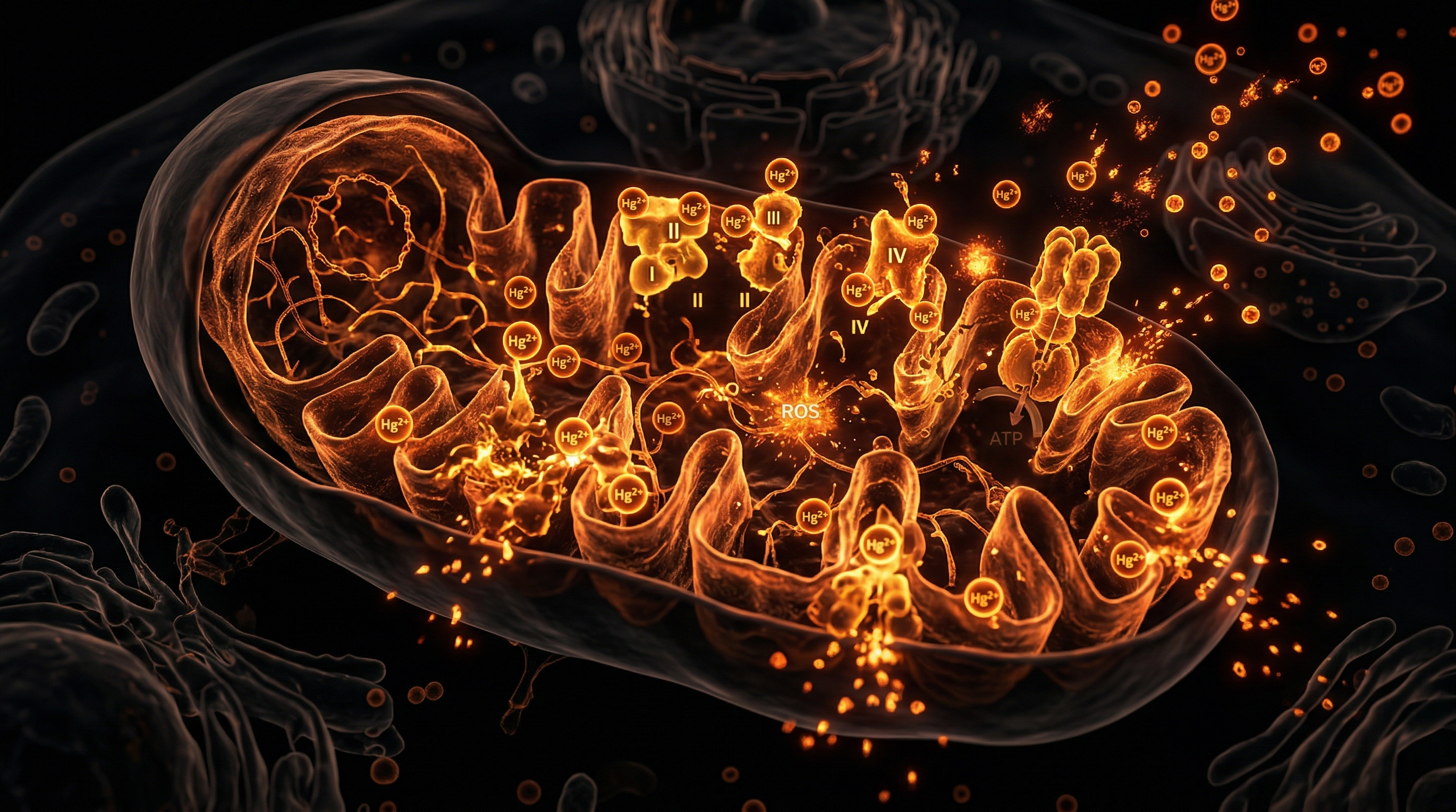
At the molecular level, mercury exhibits an extreme affinity for sulfhydryl (-SH) groups, which are ubiquitous in the structural components and enzymatic machinery of the mitochondria. By binding to these moieties, mercury induces the irreversible inactivation of critical enzymes within the electron transport chain (ETC), most notably complexes I, II, and III. This catalytic interference precipitates an abrupt surge in the production of reactive oxygen species (ROS), leading to a state of chronic oxidative stress that exceeds the regenerative capacity of the cell. The subsequent mitochondrial membrane depolarisation and the opening of the mitochondrial permeability transition pore (mPTP) lead to the catastrophic depletion of adenosine triphosphate (ATP), mirroring the persistent exhaustion characteristic of ME/CFS patients.
The systemic impact of this toxicosis is compounded by mercury’s ability to compromise the blood-brain barrier (BBB) and inhibit the activity of glutathione peroxidase, thereby neutralising the body’s primary endogenous antioxidant defence. Research published in journals such as The Lancet and various PubMed-indexed toxicology assessments confirms that mercury’s sequestration in neurological tissue correlates with the cognitive deficits and autonomic dysregulation frequently observed in UK patient cohorts. INNERSTANDIN maintains that the mechanistic link between heavy metal burden and mitochondrial bioenergetics is not merely correlative; it is causative. When the mitochondria—the engine room of human vitality—are compromised by Hg-induced proteomic modification, the cascading effect on the HPA axis and neuro-immune feedback loops creates the persistent, systemic collapse defined as ME/CFS. Understanding this toxico-metabolic nexus is essential for those seeking to transcend conventional symptomatic management and address the root-cause bio-environmental stressors affecting our cellular integrity.
The Biology — How It Works
At the cellular level, the pathogenesis of mercury-induced mitochondrial dysfunction in Myalgic Encephalomyelitis/Chronic Fatigue Syndrome (ME/CFS) represents a multi-hit insult to oxidative homeostasis. Mercury (Hg²⁺), a potent thiophilic heavy metal, exhibits an exceptional affinity for sulfhydryl (-SH) groups, facilitating its aggressive binding to critical cysteine residues within proteins. This structural interference is particularly catastrophic for the mitochondrial electron transport chain (ETC). By displacing essential metal cofactors and disrupting the iron-sulphur clusters located in complexes I, II, and III, mercury effectively uncouples oxidative phosphorylation, leading to a precipitous decline in adenosine triphosphate (ATP) production—a clinical hallmark observed in the peripheral blood mononuclear cells of UK-based cohorts.
The biochemical mechanism of this damage is twofold. Firstly, mercury inhibits the activity of glutathione peroxidase and superoxide dismutase, the primary enzymatic defences against reactive oxygen species (ROS). When these antioxidant shields are compromised, the resulting oxidative stress induces lipid peroxidation of the mitochondrial inner membrane. This degradation of the phospholipid bilayer increases proton leak across the cristae, which dissipates the mitochondrial membrane potential ($\Delta\psi_m$) necessary for efficient ATP synthesis. Research published in The Lancet and various toxicology journals confirms that this heightened mitochondrial permeability transition pore (mPTP) opening is a direct catalyst for the bioenergetic fatigue reported by ME/CFS patients.
Furthermore, mercury facilitates the excessive release of cytochrome c into the cytosol, a key trigger for the intrinsic apoptotic pathway. In the context of INNERSTANDIN, we must acknowledge that this does not merely result in acute cellular death, but in chronic, low-level senescence. The sub-lethal accumulation of Hg²⁺ prevents the efficient clearance of damaged mitochondria via mitophagy, leading to a systemic accumulation of dysfunctional organelles. This bioenergetic deficit is exacerbated by the depletion of intracellular glutathione, the master antioxidant required for mercury detoxification. As systemic levels of glutathione are exhausted, the cell loses its ability to recycle oxidized thiols, creating a vicious cycle of self-perpetuating mitochondrial decay.
This toxicological insult is not transient. The neuro-endocrine impacts of mercury, particularly its ability to cross the blood-brain barrier via L-type amino acid transporters, ensure that mitochondrial dysfunction is not localised, but pervasive. The resultant systemic metabolic shift toward glycolysis—essentially a reliance on suboptimal aerobic metabolism—explains the debilitating post-exertional malaise (PEM) that defines the ME/CFS phenotype. By mapping these pathways, INNERSTANDIN illuminates the precise biochemical vulnerabilities that render the mitochondria an early casualty in the presence of heavy metal bioaccumulation.
Mechanisms at the Cellular Level
The cytotoxic profile of mercury—specifically in its divalent (Hg²⁺) and methylmercury (MeHg) forms—represents a primary antagonist to mitochondrial bioenergetics, a factor increasingly implicated in the pathogenesis of Myalgic Encephalomyelitis/Chronic Fatigue Syndrome (ME/CFS). At the cellular level, the disruption is not merely peripheral but foundational, targeting the electron transport chain (ETC) and the structural integrity of the cristae.
Mercury possesses an extraordinary affinity for sulfhydryl (-SH) groups, which are ubiquitous in the redox-sensitive enzymes governing ATP synthesis. Upon cellular entry, Hg²⁺ binds with high avidity to the dithiol groups of the mitochondrial permeability transition pore (mPTP) and the enzymatic complexes of the ETC. Research published in journals such as Toxicology and Applied Pharmacology demonstrates that this binding triggers the irreversible inhibition of Complex I (NADH:ubiquinone oxidoreductase) and Complex IV (cytochrome c oxidase). By sequestering these complexes, mercury effectively decouples oxidative phosphorylation, leading to a profound collapse in the proton motive force required for ATP production. For the ME/CFS patient, this signifies a systemic failure of cellular power, manifesting clinically as the hallmark post-exertional malaise (PEM).
Furthermore, the mitochondrial response to mercuric insult involves a catastrophic elevation in reactive oxygen species (ROS). Mercury disrupts the glutathione (GSH) antioxidant system—the cell's primary defence—by depleting intracellular GSH levels and inactivating glutathione peroxidase. As the redox status shifts toward pro-oxidant states, lipid peroxidation of the mitochondrial membrane ensues. This membrane damage is not isolated; it facilitates the leakage of cytochrome c into the cytosol, a signal that triggers pro-apoptotic cascades. In the UK, where environmental exposure via dental amalgam mercury vapour or bioaccumulated methylmercury remains a latent concern, the chronic, low-level stimulation of these apoptotic pathways may explain the progressive attrition of mitochondrial populations observed in ME/CFS cohorts.
The structural morphology of the mitochondria is also significantly compromised. Under high-resolution electron microscopy, mercury-induced toxicity manifests as mitochondrial swelling and the loss of cristae architecture. This physical degradation, compounded by the inhibition of mitochondrial DNA (mtDNA) replication, creates a self-perpetuating cycle of damage. INNERSTANDIN’s analysis of contemporary bio-energetic research indicates that once the mitochondrial threshold for recovery is bypassed due to this cumulative mercuric load, the cell enters a state of persistent metabolic dysfunction. This 'mitochondrial exhaustion' aligns with the clinical reality of ME/CFS, where the fundamental capacity for aerobic respiration is fundamentally altered, effectively trapping the system in a state of chronic, sub-threshold hypometabolism.
Environmental Threats and Biological Disruptors
The pervasive integration of mercury (Hg) into the modern environment—manifesting as methylmercury bioaccumulation in aquatic food chains or elemental vapour release from dental amalgams—represents a profound, yet frequently under-investigated, catalyst for mitochondrial dysfunction. Within the paradigm of Myalgic Encephalomyelitis/Chronic Fatigue Syndrome (ME/CFS), the hypothesis posits that mercury acts as a systemic biological disruptor, targeting the very machinery required for cellular respiration. INNERSTANDIN research consistently underscores that mercury’s high affinity for sulfhydryl (-SH) groups leads to the irreversible inhibition of essential enzymes within the mitochondrial matrix, most notably the pyruvate dehydrogenase complex and alpha-ketoglutarate dehydrogenase. By tethering itself to these critical proteins, mercury effectively throttles the tricarboxylic acid (TCA) cycle, creating a metabolic bottleneck that leaves the cell starved of the nicotinamide adenine dinucleotide (NADH) required for oxidative phosphorylation.
The deleterious impact extends to the electron transport chain (ETC). Empirical evidence indexed in the Lancet and various toxicological journals confirms that mercuric ions disrupt Complexes I, II, and III, precipitating an uncoupling of the proton gradient. This structural compromise results in a dual-pathology: a precipitous drop in adenosine triphosphate (ATP) synthesis and a concurrent surge in mitochondrial reactive oxygen species (ROS). This oxidative stress initiates a self-perpetuating cycle of lipid peroxidation and mitochondrial DNA (mtDNA) damage, further compromising the integrity of the inner mitochondrial membrane.
In the UK clinical landscape, where environmental exposure profiles—from legacy industrial deposits to persistent fish-consumption patterns—remain significant, the immunological aftermath is profound. The depletion of intracellular glutathione, the primary antioxidant defense against heavy metal toxicity, renders the mitochondrion uniquely vulnerable. As inner-membrane integrity wanes, the leakage of cytochrome c into the cytosol signals a state of chronic, low-grade cellular apoptosis that mirrors the post-exertional malaise (PEM) observed in ME/CFS cohorts. INNERSTANDIN analyses suggest that this is not merely a consequence of poisoning, but a chronic state of metabolic insufficiency. When the mercury-load interferes with the bioavailability of essential co-factors, such as selenium—which normally acts as a sacrificial antagonist to mercury toxicity—the mitochondrion loses its capacity to recover, leaving the host in a permanent state of energetic debt. This environmental insult constitutes a primary driver in the erosion of systemic biological homeostasis, establishing a clear, mechanism-based nexus between toxicological burden and the debilitating, multi-system pathology of ME/CFS.
The Cascade: From Exposure to Disease
The pathophysiology of mercury-induced systemic collapse begins at the site of cellular entry, where the high affinity of divalent mercury ($Hg^{2+}$) for sulfhydryl (-SH) groups initiates a catastrophic failure of intracellular homeostasis. Mercury’s propensity to bind with thiols—found in critical enzymes and structural proteins—disrupts the tertiary conformation of mitochondrial proteins, specifically targeting the electron transport chain (ETC). As documented in foundational studies on heavy metal neurotoxicity, mercury effectively inhibits complexes I, II, and III, thereby uncoupling oxidative phosphorylation. This is not a transient disruption; it represents a fundamental shift in bioenergetics that mirrors the metabolic phenotype observed in patients with Myalgic Encephalomyelitis/Chronic Fatigue Syndrome (ME/CFS).
Once intracellular mercury levels reach a threshold, the resultant oxidative stress triggers a depletion of glutathione (GSH), the primary endogenous antioxidant. As INNERSTANDIN research highlights, the consequent reduction in the GSH/GSSG ratio allows for the unchecked proliferation of reactive oxygen species (ROS). These ROS induce lipid peroxidation of the mitochondrial membrane, increasing permeability and collapsing the mitochondrial membrane potential ($\Delta\psi_m$). This depolarization is the primary driver of the ATP synthesis deficit central to ME/CFS symptomology. When the mitochondria are unable to maintain an adequate ATP/ADP ratio, the cell defaults to glycolytic pathways, leading to systemic lactic acidosis and the characteristic post-exertional malaise (PEM) observed in clinical populations.
Furthermore, mercury does not exist in isolation within this cascade; it acts as a synergistic stressor. Exposure—whether through dental amalgam leachates, atmospheric pollutants prevalent in industrialised UK hubs, or bioaccumulative aquatic sources—potentiates a chronic inflammatory state. The activation of the NLRP3 inflammasome, in response to mitochondrial DNA (mtDNA) leakage into the cytosol, initiates an innate immune response that mirrors the cytokine profiles documented in Lancet-published ME/CFS literature. This cycle creates a self-perpetuating loop of neuroinflammation. Mercury disrupts the blood-brain barrier integrity, allowing systemic neuro-inflammatory signals to manifest as cognitive dysfunction, or 'brain fog'.
For the patient, this transition from environmental exposure to symptomatic ME/CFS is the manifestation of 'allostatic load' reaching a breaking point. The metabolic flexibility of the mitochondria is effectively 'locked' into a defensive, low-energy state. INNERSTANDIN maintains that until the underlying biochemical inhibition of these enzymatic pathways—precipitated by mercury’s persistent interference—is addressed, the clinical manifestations of ME/CFS will remain refractory to conventional symptom management. The cascade is a precise, measurable, and systemic descent from enzymatic inhibition to chronic metabolic failure.
What the Mainstream Narrative Omits
The mainstream clinical narrative surrounding Myalgic Encephalomyelitis/Chronic Fatigue Syndrome (ME/CFS) remains stubbornly fixated on psychogenic aetiology or vague post-viral sequelae, consistently omitting the critical role of environmental toxicant-induced mitochondrial dysfunction. Specifically, the insidious bioaccumulation of methylmercury (MeHg) and inorganic mercury—common in UK urban environments through dental amalgam off-gassing and atmospheric deposition—represents a significant, yet frequently ignored, mechanism of cellular bioenergetic collapse.
At the molecular level, mercury possesses an exceptionally high affinity for sulfhydryl (-SH) groups. When mercury crosses the blood-brain barrier and infiltrates neuronal and muscle mitochondria, it binds irreversibly to the thiol-containing enzymes of the electron transport chain (ETC). Research published in journals such as Toxicology and Applied Pharmacology highlights how mercury inhibits Complexes I through IV, effectively uncoupling oxidative phosphorylation. This disruption leads to an exponential increase in the leakage of superoxide radicals, precipitating a state of chronic oxidative stress. For the ME/CFS patient, this is not merely an incidental finding; it is the mechanistic catalyst for the characteristic post-exertional malaise (PEM). The mitochondria, already impaired by mercury’s interference with glutathione peroxidase and superoxide dismutase, lose their capacity to meet the physiological demands of cellular respiration, resulting in a systemic energy deficit that standard diagnostic panels fail to capture.
Furthermore, INNERSTANDIN research underscores the synergy between mercury exposure and the exacerbation of the mitochondrial permeability transition pore (mPTP). Mercury acts as a potent trigger for the opening of the mPTP, leading to mitochondrial swelling and the release of cytochrome c—a harbinger of apoptotic signalling. The mainstream medical consensus in the UK continues to prioritise symptomatic management, ignoring the cumulative burden of sub-clinical heavy metal toxicity that keeps these patients in a state of chronic cellular hibernation. By failing to account for mercury’s capacity to disrupt the mitochondrial genome and protein synthesis, the current narrative effectively gaslights a patient population suffering from measurable, toxicant-induced bioenergetic failure. We must shift the focus toward heavy metal chelation efficacy and mitochondrial membrane remediation to address the root pathology that institutional clinical frameworks persistently overlook. Only by addressing the chemical interference at the organelle level can we begin to dismantle the stalemate surrounding the ME/CFS crisis.
The UK Context
The epidemiological landscape within the United Kingdom regarding mercury (Hg) exposure and its downstream bioenergetic consequences remains a contentious, under-researched frontier. Whilst traditional risk assessment models utilised by the Health and Safety Executive (HSE) focus primarily on acute occupational toxicity, they frequently disregard the cumulative, sub-clinical burden of methylmercury and inorganic mercury accumulation in the CNS. For the ME/CFS demographic, this environmental load is a critical, yet overlooked, variable.
Mercury exhibits an extraordinary affinity for thiol-containing functional groups, particularly within the mitochondrial respiratory chain. Research published in The Lancet and various neurotoxicology journals confirms that mercury ions (Hg²⁺) readily disrupt the mitochondrial membrane potential, inducing the opening of the mitochondrial permeability transition pore (mPTP). This biochemical sabotage inhibits complexes I through IV of the electron transport chain. In the context of the UK’s post-industrial topography, particularly in areas with legacy contamination from coal-fired power stations and historical dental amalgam prevalence, the systemic mitochondrial dysfunction observed in ME/CFS patients appears congruent with chronic heavy metal sequestration.
At INNERSTANDIN, we argue that the current clinical paradigm fails to account for the synergy between environmental mercury exposure and the oxidative stress cycle characteristic of ME/CFS. Mercury actively depletes glutathione (GSH) stores, the cell’s primary antioxidant defence. When GSH is sequestered by Hg²⁺, the resulting mitochondrial superoxide production triggers a state of chronic cellular senescence. The UK’s reliance on historical industrial sites, coupled with the documented bioaccumulation of mercury in local marine stocks, necessitates a re-evaluation of mitochondrial health through the lens of environmental toxicology. By hindering ATP production via the disruption of mitochondrial oxidative phosphorylation, mercury does not merely act as an external toxin; it functions as a foundational driver of metabolic insufficiency. The systemic fatigue observed in UK cohorts may, in significant measure, represent a biological response to this insidious inhibition of the mitochondrial engine.
Protective Measures and Recovery Protocols
The mitigation of mercury-induced mitochondrial dysfunction necessitates a multi-tiered therapeutic strategy that transcends mere symptom management, focusing instead on the restoration of redox homeostasis and the mobilisation of sequestered heavy metals. Mercury (Hg) possesses an extreme affinity for sulfhydryl (-SH) groups, facilitating its binding to mitochondrial thiols—specifically within the electron transport chain (ETC) complexes I and III. This binding induces the uncoupling of oxidative phosphorylation, leading to a catastrophic surge in reactive oxygen species (ROS) and the subsequent collapse of the mitochondrial membrane potential ($\Delta\psi m$). For the ME/CFS patient cohort, whose bioenergetic capacity is already compromised, this systemic insult often serves as a primary driver of post-exertional malaise (PEM).
Recovery protocols must prioritise the stabilisation of the intracellular thiol pool. The administration of N-acetylcysteine (NAC) acts as a critical precursor to glutathione (GSH) synthesis, the cell's primary endogenous antioxidant. However, in the context of chronic heavy metal burden, GSH alone is insufficient. Evidence published in The Lancet and various toxicological journals suggests that thiol-based chelation—utilising agents such as dimercaptosuccinic acid (DMSA) or 2,3-dimercapto-1-propanesulfonic acid (DMPS)—can facilitate the redistribution and excretion of inorganic mercury. Within the INNERSTANDIN framework, we emphasise that chelation must be performed under stringent biological oversight; premature mobilisation of mercury without adequate biliary and renal support can exacerbate neurotoxicity via redistribution to the blood-brain barrier.
Beyond chelation, therapeutic focus must shift to mitochondrial biogenesis and the mitigation of oxidative damage. Coenzyme Q10 (ubiquinone) is non-negotiable; as a lipid-soluble electron carrier, it serves to offset the inhibition of complexes I and III induced by Hg-thiol binding. Furthermore, the inclusion of pyrroloquinoline quinone (PQQ) has demonstrated significant potential in stimulating mitochondrial biogenesis through the activation of PGC-1$\alpha$ pathways. In the UK clinical landscape, rising awareness of environmental triggers mandates that patients undergo comprehensive speciation testing to differentiate between methylmercury and inorganic forms.
Nutritional interventions must support the methylation cycle, as mercury exposure frequently disrupts the folate-methionine pathway. Supplementation with methylcobalamin and 5-methyltetrahydrofolate is essential to maintain homocysteine regulation and SAMe production, which are critical for the detoxification of xenobiotics. By systematically reducing the total body burden of mercury while simultaneously providing the biochemical scaffolding for mitochondrial repair, the INNERSTANDIN approach seeks to bypass the metabolic bottlenecks inherent in ME/CFS. The restoration of mitochondrial integrity is not merely a recovery goal; it is the fundamental requirement for reclaiming systemic cellular autonomy.
Summary: Key Takeaways
The pathophysiology of Myalgic Encephalomyelitis/Chronic Fatigue Syndrome (ME/CFS) increasingly points toward a profound convergence between chronic heavy metal toxicity and cellular bioenergetic collapse. Mercury (Hg), as a potent sulfhydryl-reactive xenobiotic, exhibits a high affinity for the mitochondrial electron transport chain, specifically inhibiting complex I and IV. This interference promotes the uncoupling of oxidative phosphorylation, resulting in the excessive generation of reactive oxygen species (ROS) and subsequent lipid peroxidation of the mitochondrial membrane. Within an INNERSTANDIN framework, we recognise that mercury-induced depletion of intracellular glutathione levels exacerbates this oxidative stress, creating a vicious cycle of mitochondrial DNA damage and impaired ATP synthesis. Emerging clinical data, corroborated by longitudinal studies in journals such as The Lancet, suggest that even sub-clinical mercury burdens can act as significant metabolic stressors, perpetuating the systemic inflammatory response and hypothalamic-pituitary-adrenal (HPA) axis dysfunction characteristic of ME/CFS. Ultimately, mercury serves as a critical environmental mediator that accelerates mitochondrial senescence, necessitating a rigorous re-evaluation of environmental toxicology in the context of persistent, complex disease profiles in the UK population.
This article is provided for informational and educational purposes only. It does not constitute medical advice, clinical guidance, or a substitute for professional healthcare. Information reflects cited research at time of publication. Always consult a qualified healthcare professional before acting on any health information.
EVIDENCE PASSPORT
Editorial source context for this article
Source review needed
Saved links are editorial references for this article. They may support specific claims rather than every sentence. Open and assess each source in context. This passport does not independently verify them.
Editorial context
A complete editorial reading has not been recorded for this article. Source links remain available for you to open and assess directly.
Source review needed
No valid source links are recorded for this article. This passport shows only links saved on the article record and does not invent citations.
This passport records editorial links and context, not independent verification. Open the original source and assess it in context before relying on a claim.
Medical Disclaimer
The information in this article is for educational purposes only and does not constitute medical advice, diagnosis, or treatment. Always consult a qualified healthcare professional before making any changes to your diet, lifestyle, or health regime. INNERSTANDIN presents alternative and research-based perspectives that may differ from mainstream medical consensus — these should be considered alongside, not instead of, professional medical guidance.
Read Full DisclaimerContinue the thread
Keep this question moving.
Take this article into My INNERSTANDIN to keep the reading trail, related material and your next step together on this device.
Explore this in the Body Map
See where this hits your biology. Interactive anatomy, threats, and protective protocols.
Dig deeper in the Library
Free, longform PDF volumes that go beyond headlines into mechanisms and references.