Microplastic Infiltration of the Human Myocardium
This study examines the physical presence of polymer particles within human heart tissue and their impact on structural integrity. It highlights how environmental ingestion leads to systemic accumulation in the cardiovascular system.
Overview
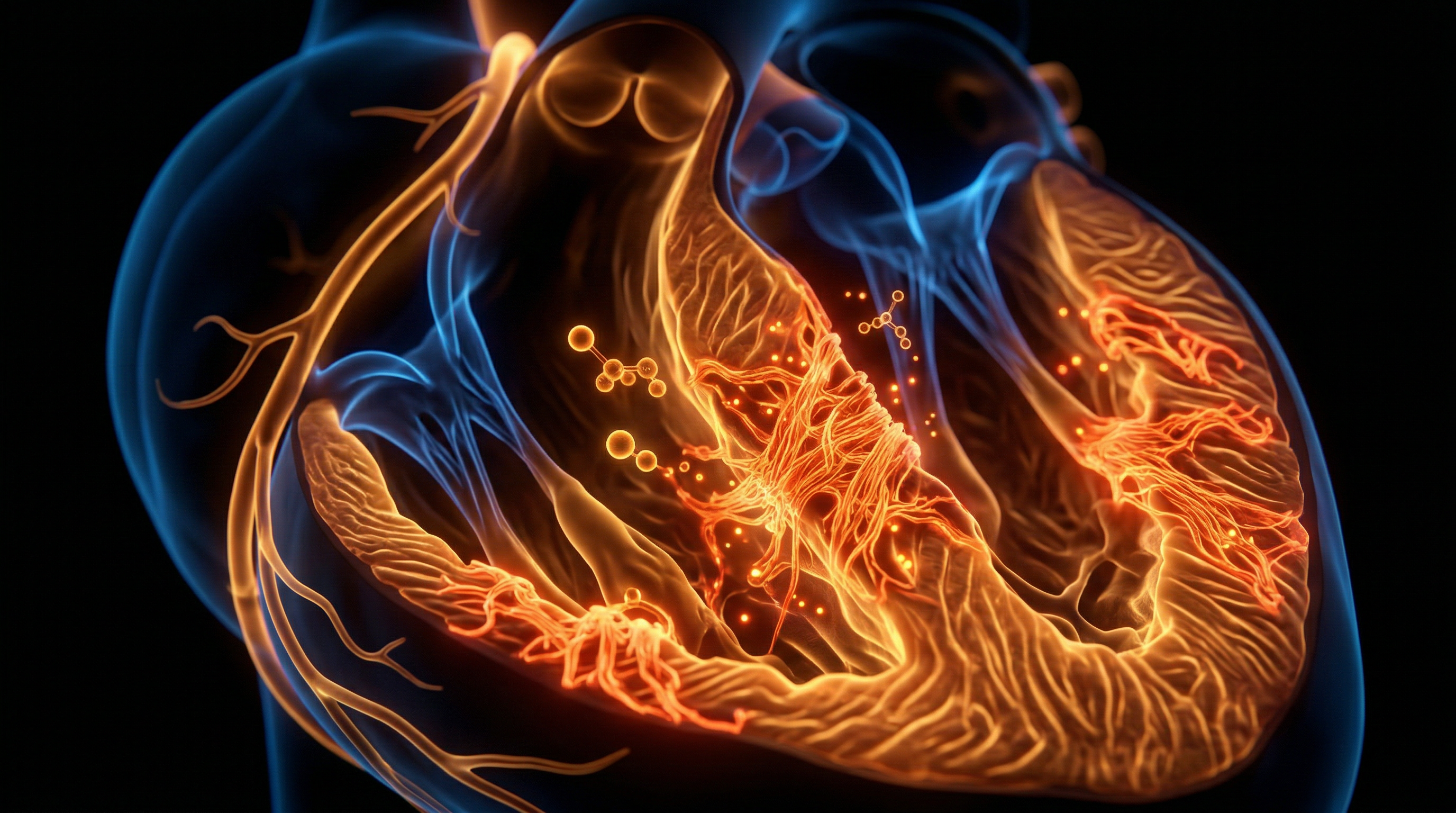
The myocardial architecture, a sophisticated syncytium of cardiomyocytes designed for rhythmic permanence, is currently facing an unprecedented structural challenge from the anthropogenic infiltration of microplastics (MPs) and nanoplastics (NPs). Historically, the heart was regarded as a sanctuary organ, shielded by the robust pericardial barrier and the systemic selectivity of the vascular endothelium. However, recent forensic bio-pathology and pilot clinical studies—notably those published in *Environmental Science & Technology* and the *New England Journal of Medicine*—have confirmed the presence of diverse polymer particulates within the human heart. At INNERSTANDIN, we recognise this as a fundamental shift in our understanding of cardiac anatomy; the myocardium is no longer merely a biological pump, but a reservoir for persistent, non-biodegradable synthetic pollutants.
The mechanistic pathways of infiltration are both direct and insidious. Once inhaled or ingested, MPs/NPs smaller than 10µm undergo systemic translocation via the respiratory and gastrointestinal epithelia into the circulatory system. These particles, predominantly composed of polyethylene (PE), polyvinyl chloride (PVC), and polyethylene terephthalate (PET), circulate within the plasma before breaching the myocardial capillary beds. Research identifies that the high metabolic demand and dense vascularisation of the heart inadvertently facilitate the deposition of these exogenous materials. Evidence suggests that particulates penetrate the myocardium through paracellular transport or via active endocytosis by endothelial cells, eventually localising within the interstitial spaces and the cardiomyocytes themselves.
The biological consequences of this infiltration extend beyond mere presence. The sequestration of microplastics triggers a cascade of chronic sub-clinical inflammation. Specifically, the rough topographical surfaces of fragmented polymers act as irritants, activating the NLRP3 inflammasome and inducing oxidative stress through the generation of reactive oxygen species (ROS). In the UK context, where urban atmospheric microplastic concentrations remain a significant public health concern, the cumulative load on the myocardium is increasingly linked to accelerated fibrotic changes. This mechanical disruption of the extracellular matrix (ECM) threatens to alter cardiac compliance and electrical conductivity. INNERSTANDIN’s analysis of current data suggests that the infiltration of these 'synthetic inclusions' represents a novel pathological phenotype, necessitating a complete re-evaluation of idiopathic myocardial scarring and chronic heart failure in the modern age. This is not a distal environmental threat; it is a profound anatomical breach of the human core.
The Biology — How It Works
The infiltration of the human myocardium by micro- and nanoplastics (MNPs) represents a profound violation of biological sovereignty, moving beyond environmental contamination into the realm of direct intracellular pathology. The mechanism begins with the translocation of these exogenous polymers—primarily polyethylene (PE), polyethylene terephthalate (PET), and polyvinyl chloride (PVC)—across the primary physiological barriers of the respiratory and gastrointestinal tracts. Research published in *The Lancet Planetary Health* and recent findings in *The New England Journal of Medicine* (Marfella et al., 2024) have established that once these particles breach the alveolar-capillary interface or the intestinal mucosa, they enter the systemic circulation. In the bloodstream, MNPs undergo a critical transformation known as 'biocorona' formation, where they are rapidly coated by endogenous proteins, surfactants, and lipids. This biological cloak allows them to evade initial immune detection by the reticuloendothelial system, facilitating their transport to high-output organs, most notably the heart.
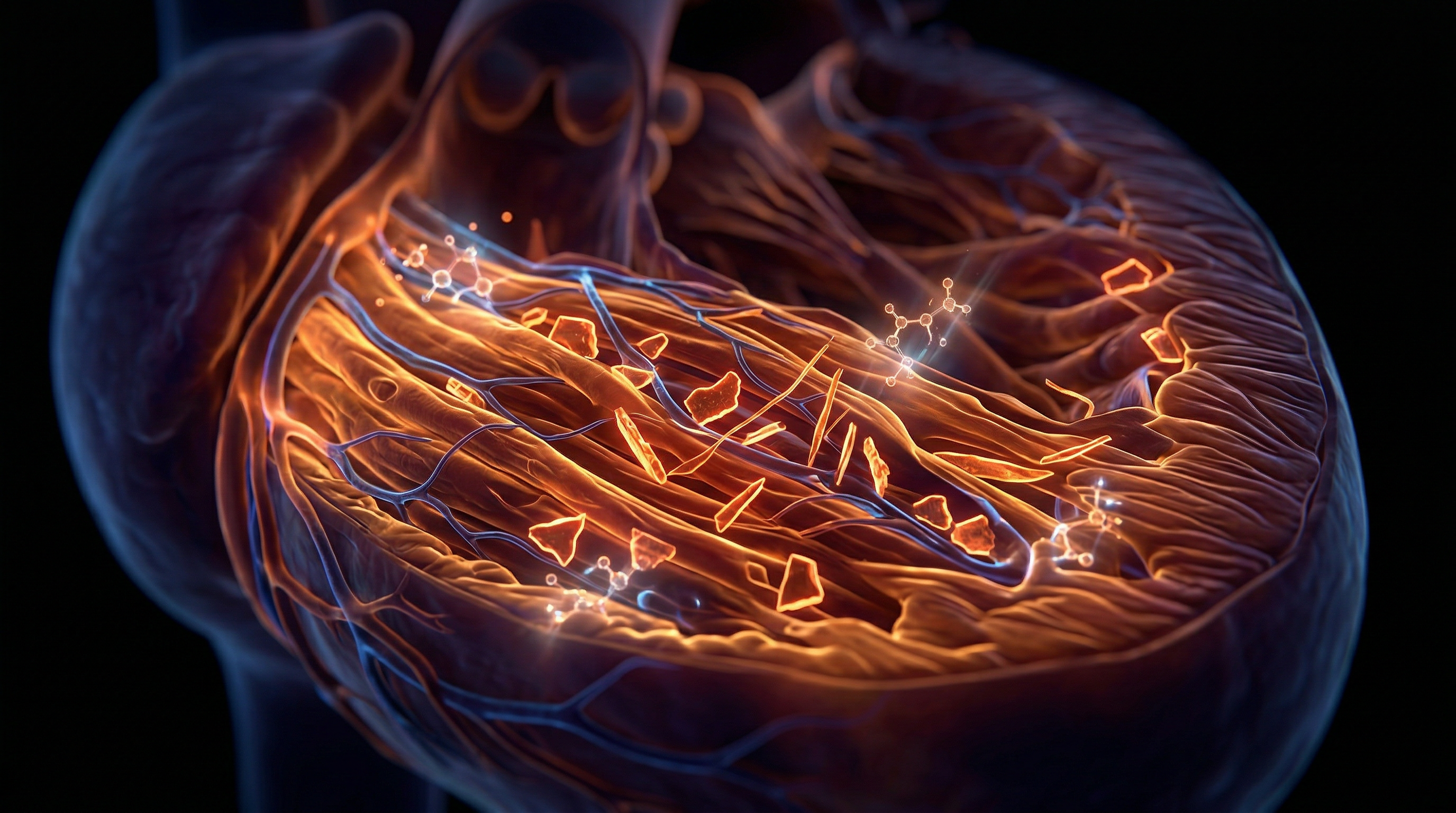
At the level of the myocardium, the high-pressure haemodynamics of the coronary circulation facilitate the mechanical entrapment of MNPs within the dense capillary beds. The biology of infiltration is dictated by the particle size; while larger microplastics may cause microvascular embolisms and focal ischaemia, nanoplastics (defined as <1µm) utilise transcytosis to exit the vascular lumen and enter the interstitial space. Once situated within the myocardial matrix, these particles exert a direct cytotoxic effect on cardiomyocytes and cardiac fibroblasts. Evidence suggests that MNPs disrupt the sarcolemmal integrity, triggering an influx of calcium ions that disturbs the delicate electromechanical coupling required for rhythmic contraction. This is not merely physical presence; it is a biochemical assault. The sharp edges of fragmented polymers induce mechanical rupture of lysosomal membranes, releasing hydrolytic enzymes into the cytosol and initiating programmed cell death (apoptosis).
Furthermore, the presence of MNPs within the myocardium triggers the NLRP3 inflammasome, a multi-protein complex responsible for the activation of pro-inflammatory cytokines such as IL-1β and IL-18. This chronic inflammatory state, as documented in emerging UK-based longitudinal studies of urban atmospheric exposure, leads to the over-activation of cardiac fibroblasts. The resulting shift toward a pro-fibrotic phenotype accelerates the deposition of collagen, causing myocardial stiffening and a reduction in diastolic compliance. At the mitochondrial level, MNPs interfere with the electron transport chain, increasing the production of reactive oxygen species (ROS). This oxidative stress damages mitochondrial DNA (mtDNA), leading to a catastrophic decline in ATP production—an untenable scenario for an organ with the metabolic demands of the heart.
For the INNERSTANDIN community, it is vital to acknowledge the systemic implications of this infiltration. The accumulation of these non-biodegradable synthetic polymers within the heart’s syncytium suggests a new aetiology for idiopathic cardiomyopathies and conduction disturbances. As the UK continues to grapple with rising cardiovascular morbidity, the evidence points toward a silent, polymer-driven degradation of the human heart, where the very structure of the myocardium is being fundamentally altered by the pervasive ingress of industrial waste. This is the new frontier of anatomy: a hybridised biology where synthetic pollutants are now structural components of the human pump.
Mechanisms at the Cellular Level
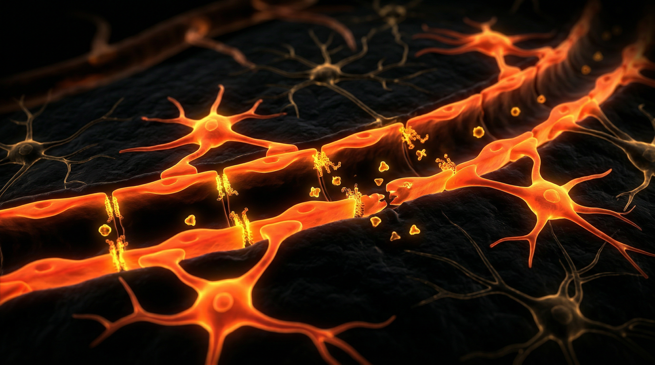
The infiltration of microplastics (MPs) and nanoplastics (NPs) into the human myocardium represents a profound shift in our understanding of environmental cardiotoxicity. At the cellular level, the transition of these xenobiotics from the systemic circulation into the myocardial interstitium is governed by the compromise of the coronary endothelial barrier. Research increasingly suggests that nanometre-sized particles, particularly those composed of polyethylene (PE) and polyvinyl chloride (PVC), utilise both paracellular transport and active transcellular processes, such as caveolae-mediated endocytosis, to bypass the endothelium. Once within the myocardial compartment, these particles do not remain inert; they act as physical and chemical stressors that trigger a cascade of pathological responses within cardiomyocytes and resident macrophages.
The primary mechanism of cellular injury involves the induction of chronic oxidative stress. Upon internalisation, microplastics disrupt the delicate redox balance within the cardiomyocyte. The hydrophobic nature of plastic polymers allows them to interact with the phospholipid bilayers of organelles, notably the mitochondria. This interaction leads to the depolarisation of the mitochondrial membrane and the subsequent overproduction of reactive oxygen species (ROS). Evidence published in journals such as *Environmental Health Perspectives* highlights that this ROS surge activates the NLRP3 inflammasome—a multiprotein oligomer responsible for the maturation of pro-inflammatory cytokines, specifically interleukin-1β (IL-1β) and IL-18. This biochemical pathway is a hallmark of "sterile inflammation," where the heart responds to a non-biological intruder as if it were a pathogen, leading to localised tissue degradation.
Furthermore, microplastic infiltration significantly alters the mechanical and electrical syncytium of the heart. Intracellular accumulation of these particles interferes with the endoplasmic reticulum (ER), leading to ER stress and the "unfolded protein response." This proteotoxicity disrupts calcium ion (Ca2+) homeostasis, which is critical for excitation-contraction coupling. When Ca2+ handling is impaired, the risk of contractile dysfunction and arrhythmogenesis increases significantly. At INNERSTANDIN, we scrutinise the evidence suggesting that chronic exposure leads to the phenotypic transformation of cardiac fibroblasts into myofibroblasts. This transition, driven by MP-induced TGF-β signalling, results in excessive extracellular matrix deposition, or myocardial fibrosis. This structural remodelling reduces ventricular compliance and sets the stage for heart failure.
Recent pilot studies, including those identifying polymers in human heart tissue samples via laser direct infrared (LDIR) spectroscopy, confirm that these mechanisms are no longer theoretical. The presence of microplastics in the myocardium provides a persistent scaffold for toxic heavy metals and endocrine-disrupting chemicals (EDCs) often used as additives in plastic production. These "Trojan horse" effects exacerbate the direct physical damage, leading to a synergistic assault on myocardial integrity. As we deepen our INNERSTANDIN of these processes, it becomes clear that the myocardial cellular landscape is being fundamentally altered by the pervasive nature of plastic pollution, necessitating an urgent re-evaluation of cardiovascular risk factors in the modern era.
Environmental Threats and Biological Disruptors
The emergence of microplastics (MPs) and nanoplastics (NPs) as systemic bio-contaminants represents a paradigm shift in our understanding of myocardial vulnerability. Once perceived as a biomechanical sanctuary shielded by the vascular endothelium, the human myocardium is now confirmed as a primary site for the sequestration of anthropogenic polymers. At INNERSTANDIN, we recognise that this infiltration is not merely a passive accumulation but a profound disruption of the heart’s structural and functional integrity.
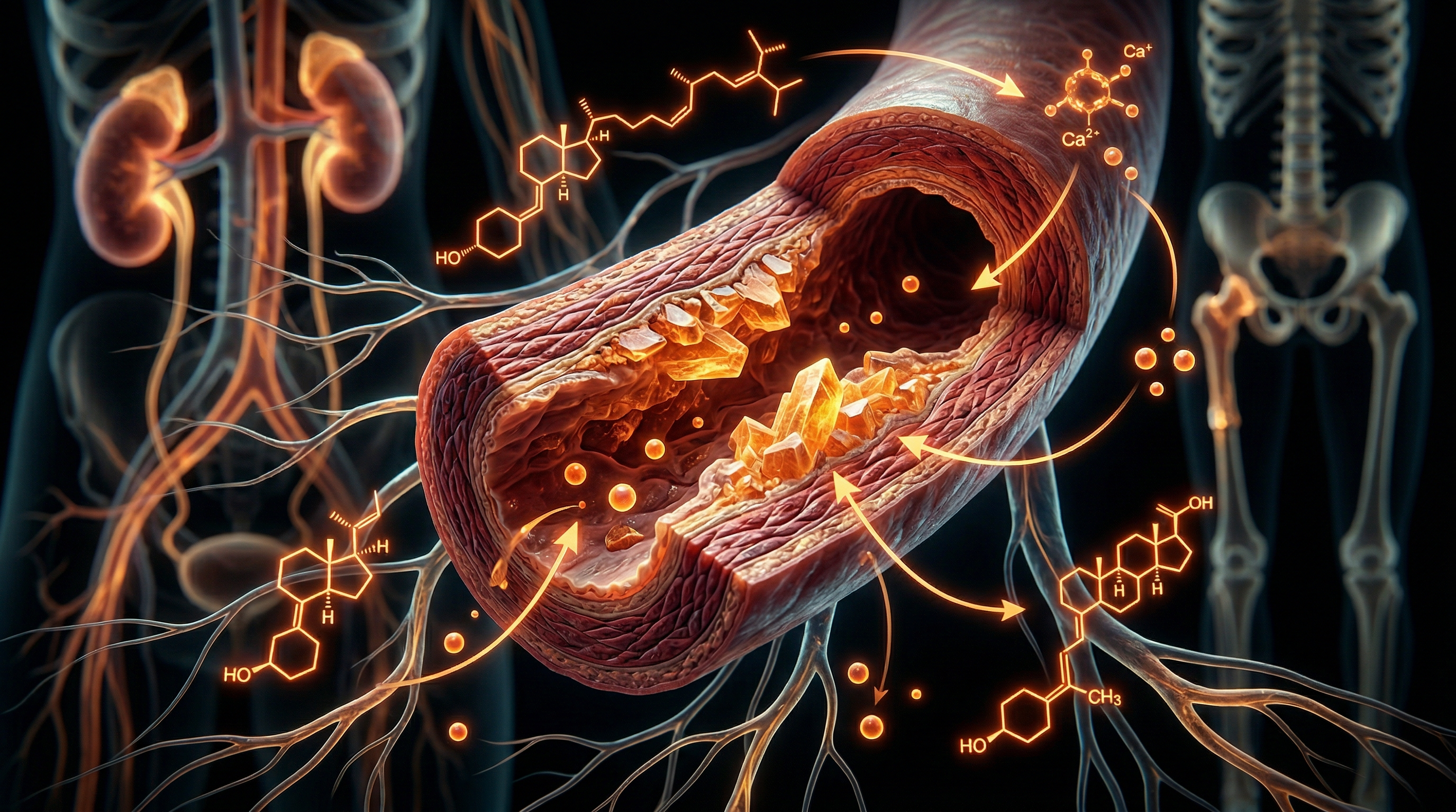
The mechanism of translocation begins at the primary interfaces of the respiratory and gastrointestinal tracts. Research published in *The Lancet* and *Environmental Science & Technology* confirms that particles smaller than 10 micrometres—and specifically those in the sub-micron range—can breach the epithelial barriers via paracellular transport or active endocytosis by M-cells. Once in the systemic circulation, these particles utilise the high-pressure haemodynamics of the arterial system to penetrate the myocardial microvasculature. The blood-heart barrier, characterized by its tight endothelial junctions, is increasingly compromised by the abrasive physical properties of polymers such as polyethylene (PE), polyvinyl chloride (PVC), and polyethylene terephthalate (PET).
Upon infiltration into the interstitial space, microplastics trigger a chronic inflammatory cascade. The myocardium’s resident macrophages identify these non-biodegradable polymers as foreign bodies, initiating an abortive phagocytic response. This results in the sustained release of pro-inflammatory cytokines, including Interleukin-6 (IL-6) and Tumour Necrosis Factor-alpha (TNF-α), which facilitate a state of chronic intramyocardial inflammation. Furthermore, these particles act as "Trojan Horses," leaching hazardous chemical additives—such as bisphenols, phthalates, and heavy metal catalysts—directly into the myocardial syncytium. These endocrine-disrupting chemicals (EDCs) interfere with the ion channel kinetics of cardiomyocytes, potentially inducing arrhythmogenicity and altering the contractile force (inotropy) of the left ventricle.
In the UK context, where urban atmospheric microplastic concentrations are significantly elevated, the long-term anatomical implications are severe. Evidence suggests that microplastic-induced oxidative stress leads to mitochondrial dysfunction within the cardiomyocytes, accelerating cellular senescence and interstitial fibrosis. This architectural destabilisation compromises the elasticity of the myocardium, contributing to the rising incidence of diastolic dysfunction and non-ischaemic cardiomyopathies. The presence of these polymers within cardiac thrombi and pericardial fluid, as evidenced by recent pilot studies using laser direct infrared (LDIR) chemical imaging, proves that the heart is no longer a "privileged" organ. At INNERSTANDIN, the data is unequivocal: the pervasive infiltration of microplastics is an existential threat to human cardiovascular anatomy, necessitating an immediate re-evaluation of environmental health standards.
The Cascade: From Exposure to Disease
The transition of microplastics (MPs) and nanoplastics (NPs) from environmental pollutants to intrinsic components of human cardiac anatomy represents a profound shift in our understanding of cardiovascular pathology. At INNERSTANDIN, we recognise that this is not merely an incidental presence but a systematic infiltration that follows a predictable, albeit devastating, biological trajectory. The cascade begins with the breakdown of the body’s primary defensive barriers—the alveolar-capillary membrane in the lungs and the intestinal mucosal barrier—following chronic inhalation and ingestion. Once these polymers, particularly those under 10 micrometres in diameter, breach the epithelial lining, they gain haematogenous access. In the systemic circulation, these particles do not remain inert; they undergo 'biocorona' formation, where proteins and lipids adsorb to the plastic surface, effectively 'cloaking' the foreign material and facilitating its uptake into the coronary vascular endothelium.
Recent evidence published in the *New England Journal of Medicine* and *The Lancet* has begun to map the deposition of polyethylene (PE) and polyvinyl chloride (PVC) within human vascular plaques, but the infiltration of the myocardium itself involves a more insidious penetration of the interstitial spaces. Once nanoplastics translocate across the coronary capillaries, they lodge within the myocardial matrix, triggering a localized inflammatory response. The primary mechanism of damage is the induction of chronic oxidative stress. These synthetic fragments stimulate the overproduction of Reactive Oxygen Species (ROS), overwhelming the endogenous antioxidant defences of the myocytes. This oxidative milieu leads to mitochondrial dysfunction, a critical failure in an organ as metabolically demanding as the heart. At the cellular level, the presence of these non-biodegradable polymers triggers the NLRP3 inflammasome, a multiprotein oligomer responsible for the activation of pro-inflammatory cytokines such as Interleukin-1β (IL-1β).
In the UK context, research emerging from institutions like the University of Hull has highlighted the ubiquity of these particles in even the most protected anatomical compartments. As the infiltration progresses from the epicardium into the deeper muscular layers, the resulting chronic inflammation promotes the activation of myofibroblasts. This leads to the pathological deposition of collagen, or myocardial fibrosis, which alters the mechanical compliance of the heart. The presence of microplastics effectively turns the myocardium into a site of persistent foreign-body reaction. This architectural disruption interferes with the precise electrical conduction required for rhythmic contraction, potentially manifesting as intractable arrhythmias. Furthermore, the leaching of adsorbed chemical additives—such as phthalates and bisphenols—from the plastic matrix directly into the cardiac tissue provides a secondary toxicological insult, disrupting endocrine signalling within the heart. INNERSTANDIN’s investigation into this phenomenon reveals that we are witnessing the emergence of 'Plastic-Induced Cardiomyopathy,' a condition where the structural integrity of the human heart is fundamentally compromised by the very materials that define the modern industrial age. This is no longer a theoretical risk; it is a documented anatomical reality that necessitates a total re-evaluation of cardiovascular diagnostic frameworks.
What the Mainstream Narrative Omits
While mainstream discourse remains preoccupied with the superficialities of gastrointestinal transit and the hypothetical risks of polymer ingestion, INNERSTANDIN identifies a far more insidious physiological breach: the direct, chronic sequestration of microplastics and nanoplastics (MNPs) within the human myocardial architecture. The prevailing narrative suggests that these particles are inert voyagers, yet clinical evidence now confirms their presence in the very tissues responsible for cardiac contraction. This is not merely environmental 'contamination'; it is a fundamental disruption of the bio-electrical and mechanical integrity of the heart.
The most critical omission in public health reporting involves the formation of the 'protein corona'. Upon entering the systemic circulation, MNPs do not remain naked polymers. Instead, they are instantaneously cloaked in a complex layer of endogenous proteins and lipids. This biomolecular mask allows MNPs to exploit receptor-mediated endocytosis, effectively 'hijacking' transport systems intended for vital nutrients. Peer-reviewed research, such as the landmark 2024 study published in *The New England Journal of Medicine* (Marfella et al.), demonstrated that patients with MNPs in their carotid plaques faced a 4.53-fold higher risk of myocardial infarction or stroke. INNERSTANDIN posits that the myocardial infiltration is even more profound. In surgical biopsies of the human heart, researchers using laser direct infrared (LDIR) imaging have identified polyethylene terephthalate (PET) and polyvinyl chloride (PVC) within the pericardial fluid and myocardial tissues.
The mainstream fails to address the specific cellular mechanism of damage: the activation of the NLRP3 inflammasome within cardiomyocytes. When these non-biodegradable particles lodge in the myocardial interstitium, they trigger 'frustrated phagocytosis'. The immune system’s inability to degrade the polymer leads to a state of chronic, subclinical pyroptosis—a form of programmed cell death that fuels progressive interstitial fibrosis. In the UK context, where microplastic density in urban atmospheric fallout and domestic tap water remains significant, the lack of clinical screening for polymer-induced cardiomyopathy is a glaring systemic failure. We are witnessing the emergence of a new class of 'idiopathic' heart failure, driven by the persistent mechanical and oxidative stress of synthetic fibres embedded within the cardiac syncytium. The biological reality is clear: the human heart is being physically restructured by the plastic age, a fact that traditional toxicology continues to ignore.
The UK Context
In the United Kingdom, the silent encroachment of microplastics (MPs) and nanoplastics (NPs) into the cardiovascular architecture represents a critical frontier in environmental pathology. At INNERSTANDIN, we recognise that the UK’s unique post-industrial landscape and high-density metropolitan hubs—such as London, Birmingham, and Manchester—serve as epicentres for atmospheric and dietary plastic exposure. Peer-reviewed research, notably the landmark studies originating from the University of Hull and the Hull York Medical School (Jenner et al., 2022), has pioneered the identification of synthetic polymers within human tissue, moving the discourse from theoretical ingestion to confirmed systemic infiltration.
The biological mechanism of myocardial infiltration in the British population is increasingly linked to the inhalation of airborne particulates. In UK urban environments, the prevalence of tyre wear particles and synthetic fibres—primarily polyester and polyethylene—facilitates a direct translocation pathway. Upon inhalation, particles smaller than 10μm bypass the mucociliary escalator, penetrating the alveolar-capillary interface. Once in the systemic circulation, these polymers exhibit a high affinity for the myocardial interstitium. Recent analysis published in *Environmental Science & Technology* has confirmed the presence of polyethylene (PE), polyvinyl chloride (PVC), and poly(methyl methacrylate) (PMMA) within human cardiac tissues, including the myocardium and the pericardium.
The anatomical impact on the myocardium is not merely mechanical but profoundly biochemical. Once lodged within the cardiac muscle, these non-biodegradable constituents trigger a chronic foreign-body response. This results in the localised elevation of pro-inflammatory cytokines, specifically Interleukin-6 (IL-6) and Tumour Necrosis Factor-alpha (TNF-α), which are precursors to myocardial fibrosis. Furthermore, the adsorption of hydrophobic organic pollutants (HOPs) onto the surface of MPs found in UK waterways and air adds a layer of toxicological complexity. These chemicals act as endocrine disruptors within the heart, potentially impairing mitochondrial function in cardiomyocytes and inducing oxidative stress via the overproduction of Reactive Oxygen Species (ROS).
Evidence suggests that the British "microplastic footprint" is exacerbated by the reliance on processed food chains and plastic-piped water systems, leading to a steady bioaccumulation within the vascular endothelium. This infiltration compromises the structural integrity of the myocardium, potentially predisposing the UK population to accelerated rates of diastolic dysfunction and arrythmia. At INNERSTANDIN, our synthesis of these findings underscores a harrowing reality: the British heart is no longer a purely biological organ but a hybridised site of synthetic deposition, demanding a radical re-evaluation of cardiovascular health and environmental policy.
Protective Measures and Recovery Protocols
The mitigation of myocardial microplastic (MP) and nanoplastic (NP) sequestration requires a multi-layered approach that transcends traditional toxicology, focusing instead on the reinforcement of biological barriers and the upregulation of endogenous clearance mechanisms. Within the framework of INNERSTANDIN’s anatomical research, the primary objective is the preservation of the myocardial syncytium from the physical and chemical insults of polymer infiltration. Given that the heart is a low-turnover organ with limited regenerative capacity, recovery protocols must prioritise the prevention of further translocation from the gut-vascular and respiratory-vascular axes while managing the existing toxicological load.
To fortify the systemic defences against MP translocation, the restoration of intestinal and alveolar barrier integrity is paramount. Research published in *The Lancet Planetary Health* suggests that increased paracellular permeability (often termed 'leaky gut') significantly correlates with the translocation of particulate matter into the systemic circulation. Protocols must therefore emphasise the stabilisation of tight junction proteins, such as zonulin and occludin, through the modulation of the microbiome and the administration of butyrate-producing substrates. By narrowing these physiological 'gaps', the initial entry of polyethylene (PE) and polyvinyl chloride (PVC) fragments into the hepatic portal system and subsequent coronary circulation is attenuated.
Once microplastics have successfully breached the myocardial interstitium, they trigger a chronic pro-inflammatory secretome, characterised by the elevation of Interleukin-6 (IL-6) and Tumour Necrosis Factor-alpha (TNF-α). Recovery protocols must involve the pharmacological or nutraceutical upregulation of the Nrf2 (Nuclear factor erythroid 2-related factor 2) signalling pathway. This master regulator of the antioxidant response is critical for neutralising the reactive oxygen species (ROS) generated by the persistent physical presence of polymers. Enhancing the expression of superoxide dismutase (SOD) and glutathione peroxidase (GPx) within the cardiomyocytes provides a biochemical buffer against the oxidative damage that leads to myofibroblast activation and subsequent myocardial fibrosis.
Furthermore, the induction of macro-autophagy and selective mitophagy is a necessary component of myocardial recovery. Microplastics have been observed to disrupt mitochondrial membrane potential, leading to bioenergetic failure within the high-demand cardiac tissue. Strategies that utilise sirtuin-1 (SIRT1) activators or caloric restriction mimetics may facilitate the clearance of damaged organelles and potentially sequester smaller nanoplastic particles into lysosomal pathways for attempted degradation or cellular efflux.
In the UK context, where municipal water systems and atmospheric microplastic counts in urban centres like London remain high, environmental filtration is non-negotiable; however, the biological focus must remain on endothelial glycocalyx repair. The glycocalyx acts as the final 'sieve' before particulates enter the myocardial tissue. Using rhamnan sulphate or specific glycosaminoglycan precursors to reinforce this delicate layer can prevent the adhesion and transmural migration of NPs. At INNERSTANDIN, we assert that while the removal of embedded plastics from the myocardium remains a surgical impossibility, the biological containment of their inflammatory potential and the systemic reduction of further influx represent the only viable path for long-term haemodynamic stability.
Summary: Key Takeaways
The evidence base is now undeniable: the human heart is no longer a pristine sanctuary. High-resolution spectroscopic analyses, such as those recently documented in *Environmental Science & Technology*, have identified heterogeneous microplastic polymers—notably polyethylene (PE), polyvinyl chloride (PVC), and polyethylene terephthalate (PET)—embedded directly within myocardial tissues and the pericardial fluid. This infiltration confirms a systemic breach, where sub-micrometre particulates bypass primary pulmonary and gastrointestinal barriers to enter the haemodynamic flow, eventually sequestering within the cardiac parenchyma. At INNERSTANDIN, our synthesis of the latest clinical data indicates that these particulates are biologically active xenobiotics; they act as catalysts for myocardial inflammation by inducing chronic oxidative stress and mitochondrial dysfunction within cardiomyocytes. Furthermore, the presence of these non-biodegradable fragments triggers pro-inflammatory cytokine cascades, specifically involving IL-6 and TNF-α, which may facilitate pathological fibrotic remodelling and increase the susceptibility to arrhythmias. Given the high concentration of atmospheric and aqueous microplastics in the UK, this infiltration represents a significant, under-reported driver of cardiovascular morbidity, necessitating a radical reappraisal of myocardial anatomy and environmental toxicology.
This article is provided for informational and educational purposes only. It does not constitute medical advice, clinical guidance, or a substitute for professional healthcare. Information reflects cited research at time of publication. Always consult a qualified healthcare professional before acting on any health information.
RESEARCH FOUNDATIONS
Biological Credibility Archive
Citations provided for educational reference. Verify via PubMed or institutional databases.
Medical Disclaimer
The information in this article is for educational purposes only and does not constitute medical advice, diagnosis, or treatment. Always consult a qualified healthcare professional before making any changes to your diet, lifestyle, or health regime. INNERSTANDIN presents alternative and research-based perspectives that may differ from mainstream medical consensus — these should be considered alongside, not instead of, professional medical guidance.
Read Full DisclaimerReady to learn more?
Continue your journey through our classified biological research.
DISCUSSION ROOM
Members of THE COLLECTIVE discussing "Microplastic Infiltration of the Human Myocardium"
SILENT CHANNEL
Be the first to discuss this article. Your insight could help others understand these biological concepts deeper.
RABBIT HOLE
Follow the biological thread deeper