Myocardial Fibrosis and Extreme Endurance Training
Excessive cardiovascular strain can lead to anatomical scarring of the heart muscle in amateur athletes. This report explores the fine line between fitness and structural heart damage.
Overview
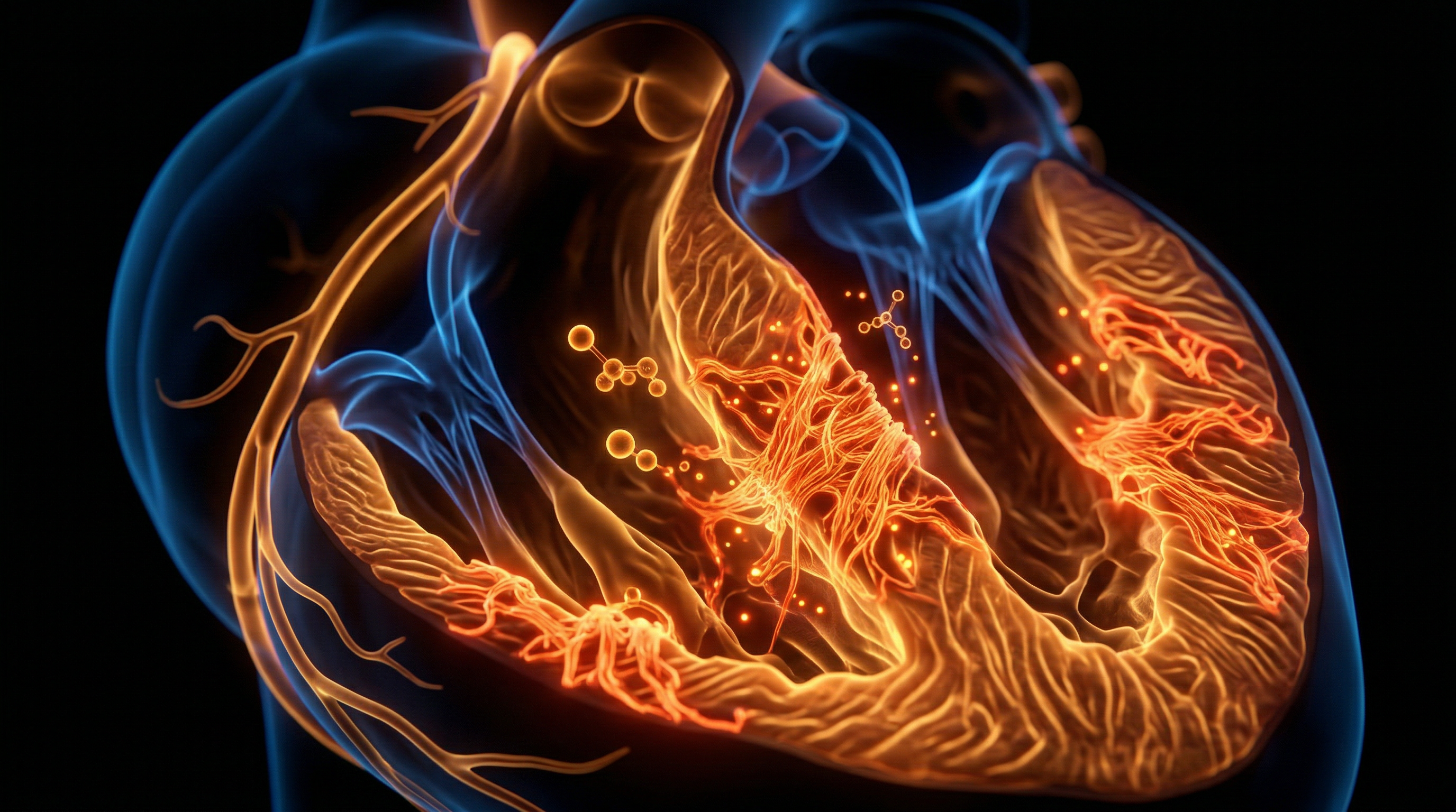
The paradigm of cardiovascular adaptation to chronic exercise has long been dominated by the concept of the "athlete’s heart"—a benign physiological remodelling characterised by proportional chamber enlargement and enhanced vagal tone. However, emerging data from high-resolution cardiac magnetic resonance (CMR) imaging and histological analyses necessitate an INNERSTANDIN of a more sinister manifestation: exercise-induced myocardial fibrosis (MF). This pathological accumulation of collagenous extracellular matrix (ECM) within the myocardium represents a maladaptive response to the repetitive haemodynamic insults inherent to extreme endurance training. While moderate physical activity is indisputably cardioprotective, the "extreme exercise hypothesis" suggests a U-shaped or J-shaped dose-response curve, where the volume and intensity of ultra-endurance efforts (e.g., 100-mile ultramarathons or Ironman-distance triathlons) may exceed the heart’s intrinsic capacity for repair.
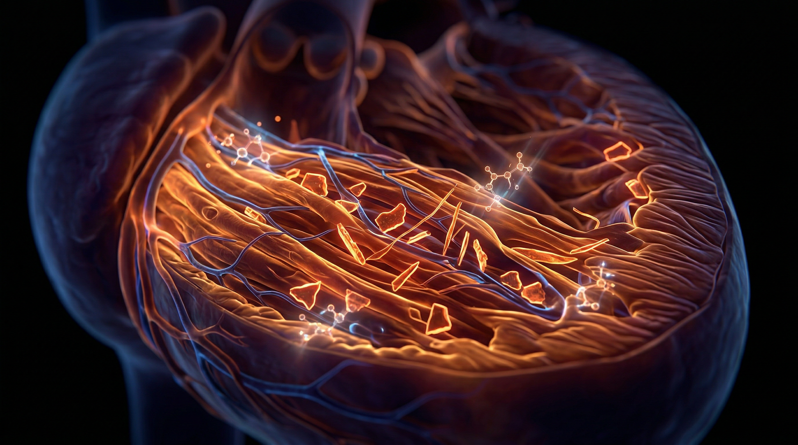
The anatomical focal point of this fibrotic process is frequently localized to the right ventricle (RV) and the interventricular septum, specifically at the "hinge points" where the RV attaches to the left ventricle (LV). During maximal exertion, systemic systolic blood pressure rises, but it is the disproportionate increase in pulmonary arterial pressure that places the thin-walled RV under extreme wall stress. Research published in *The Lancet* and the *European Heart Journal* by investigators such as La Gerche et al. has demonstrated that the RV undergoes significant transient dysfunction and structural "stretching" post-race, unlike the more robust LV. This chronic repetitive stretching triggers a pro-fibrotic signalling cascade. Mechanotransduction at the level of the sarcolemma activates quiescent cardiac fibroblasts, transforming them into myofibroblasts. These cells subsequently orchestrate the over-expression of transforming growth factor-beta 1 (TGF-β1) and the deposition of Type I and Type III collagen, disrupting the delicate cytoarchitecture of the myocardium.
Evidence-led investigations using Late Gadolinium Enhancement (LGE) CMR have identified focal patches of replacement fibrosis in up to 50% of veteran male endurance athletes, a prevalence significantly higher than in age-matched sedentary controls. This is not merely an incidental finding; the presence of LGE-defined fibrosis creates a heterogeneous substrate for lethal arrhythmias. The non-conductive collagenous scars can act as anatomical blocks, facilitating re-entrant circuits that manifest as atrial fibrillation or, more critically, ventricular tachycardia. Furthermore, the systemic impact of this remodelling includes reduced ventricular compliance and impaired diastolic filling, potentially culminating in a phenotype that mimics arrhythmogenic right ventricular cardiomyopathy (ARVC). By interrogating the molecular and mechanical drivers of this condition, INNERSTANDIN seeks to expose the biological threshold where peak human performance transitions into progressive myocardial pathology, challenging the archaic notion that the heart possesses an infinite capacity for haemodynamic load.
The Biology — How It Works
The genesis of exercise-induced myocardial fibrosis (EIMF) lies in the chronic maladaptation to repetitive, exhaustive haemodynamic loading. While moderate aerobic activity promotes physiological hypertrophy—characterised by proportional increases in chamber size and wall thickness—extreme endurance regimes often push the myocardium beyond its compensatory threshold. At INNERSTANDIN, we dissect the molecular transition from this beneficial 'athlete's heart' to the pathological substrate of fibrosis, primarily driven by mechanical stretch and inflammatory cascades.
During bouts of maximal exertion, cardiac output can increase fivefold, subjecting the ventricular walls to profound transmural pressure. This mechanical strain serves as the primary stimulus for the activation of cardiac fibroblasts via the mechanotransduction pathways. Specifically, the stretching of myocytes and the extracellular matrix (ECM) triggers the release of profibrotic cytokines, most notably Transforming Growth Factor-beta 1 (TGF-β1). According to research documented in *The Lancet* and various PubMed-indexed longitudinal studies, TGF-β1 acts as the master regulator, inducing the differentiation of fibroblasts into myofibroblasts. These specialised cells possess contractile properties and an exaggerated capacity for collagen synthesis, specifically Types I and III.
Under normal physiological conditions, the turnover of the ECM is tightly regulated by matrix metalloproteinases (MMPs) and their tissue inhibitors (TIMPs). However, extreme endurance training disrupts this equilibrium. The repetitive nature of the 'micro-trauma' associated with ultra-marathons or long-distance cycling leads to transient myocardial inflammation and oxidative stress. This is evidenced by the acute elevation of cardiac biomarkers, such as Troponin T and B-type Natriuretic Peptide (BNP), immediately following exhaustive events. While these markers often return to baseline within 48 hours, the cumulative effect of thousands of hours of high-intensity training can result in focal areas of myocyte necrosis and subsequent 'replacement fibrosis'.
The Right Ventricle (RV) is disproportionately vulnerable in this biological context. Unlike the thick-walled Left Ventricle (LV), the RV is thin-walled and designed for a low-pressure system. During extreme exercise, the relative increase in wall stress is significantly higher in the RV, often leading to acute RV dysfunction and wall motion abnormalities. Over time, this targeted mechanical fatigue promotes the deposition of fibrous tissue within the RV myocardium and the interventricular septum, frequently observed as Late Gadolinium Enhancement (LGE) on cardiac MRI.
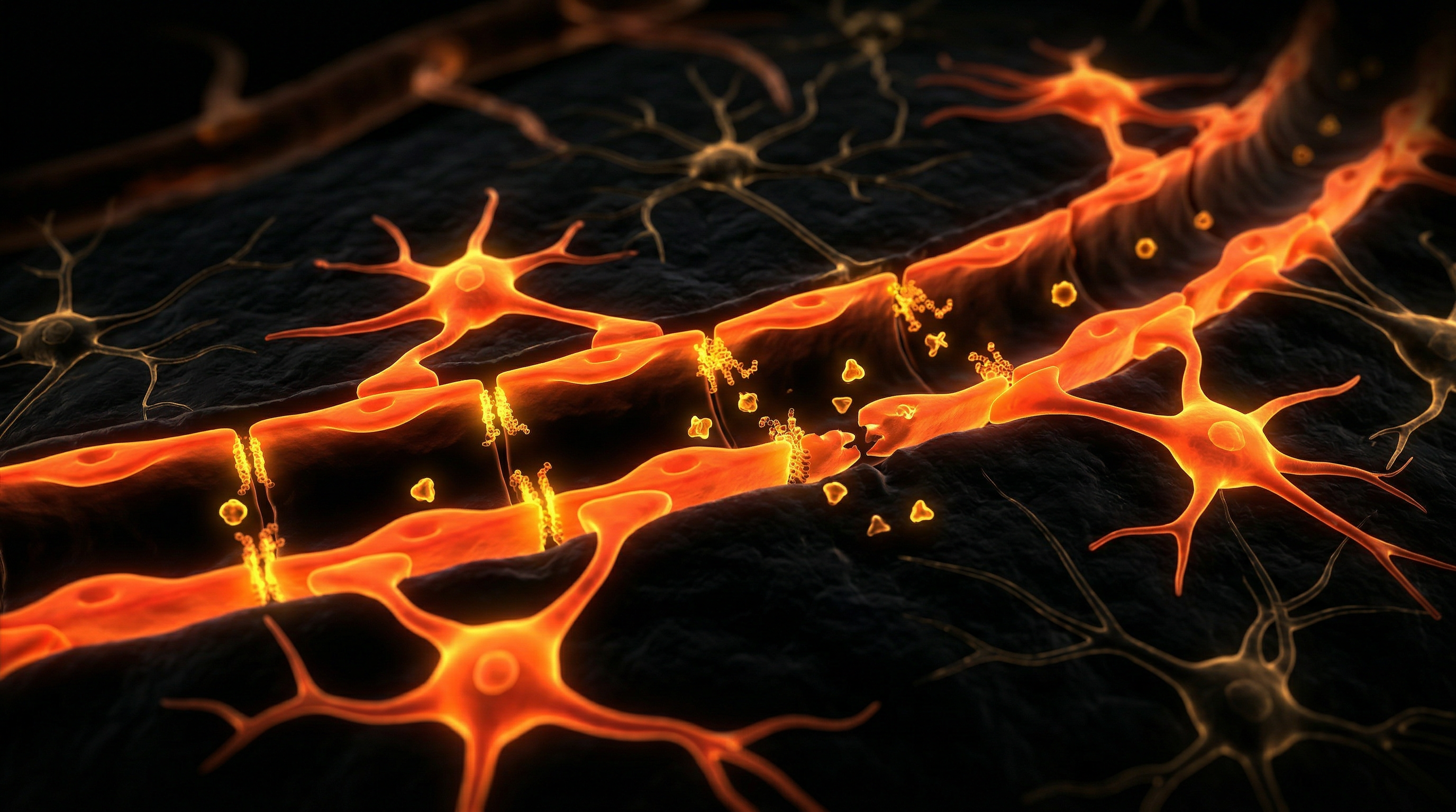
From an electrophysiological perspective, this fibrotic infiltration is catastrophic. The non-conductive collagenous scars act as anatomical barriers, insulating healthy myocytes and creating zones of slow conduction. This heterogeneity in ventricular depolarisation and repolarisation provides the requisite substrate for re-entrant tachyarrhythmias. At INNERSTANDIN, we emphasize that while the systemic benefits of exercise are undisputed, the dose-response curve for the myocardium suggests a 'U-shaped' risk profile, where extreme volume leads to a pro-arrhythmogenic structural remodelling that mimics aspects of Arrhythmogenic Right Ventricular Cardiomyopathy (ARVC). The biological reality is that for a subset of genetically predisposed veteran athletes, the heart does not merely adapt; it scars.
Mechanisms at the Cellular Level
At the cellular nexus of the "Athletic Heart" syndrome lies a precarious transition from physiological adaptation to pathological maladaptation. While moderate aerobic activity promotes eccentric hypertrophy, the repetitive haemodynamic insult associated with extreme endurance training—defined as chronic, high-intensity exertion exceeding ten hours per week—triggers a cascade of profibrotic signalling pathways within the myocardial interstitium. Central to this process is the mechanical stretching of cardiomyocytes and the subsequent activation of the Transforming Growth Factor-beta 1 (TGF-β1) isoform. As research disseminated via INNERSTANDIN has consistently highlighted, TGF-β1 serves as the primary molecular orchestrator of fibrosis; it facilitates the phenotypic transformation of quiescent cardiac fibroblasts into contractile myofibroblasts. These myofibroblasts are characterised by the expression of alpha-smooth muscle actin (α-SMA) and an exaggerated capacity for the synthesis of extracellular matrix (ECM) proteins, predominantly Type I and Type III collagen.
The persistent volume and pressure overload inherent in ultra-endurance events induce transient yet significant elevations in cardiac biomarkers, such as Troponin I and T, and B-type Natriuretic Peptide (BNP). This biochemical signature, as documented in studies published in *The Lancet* and *European Heart Journal*, suggests recurring micro-injury to the myocardium. On a cellular level, these micro-insults generate reactive oxygen species (ROS), which overwhelm endogenous antioxidant defences, leading to mitochondrial dysfunction and lipid peroxidation of the sarcolemma. The resulting oxidative stress further amplifies the inflammatory response, activating the NLRP3 inflammasome and promoting the secretion of pro-inflammatory cytokines, including Interleukin-6 (IL-6) and Tumour Necrosis Factor-alpha (TNF-α).
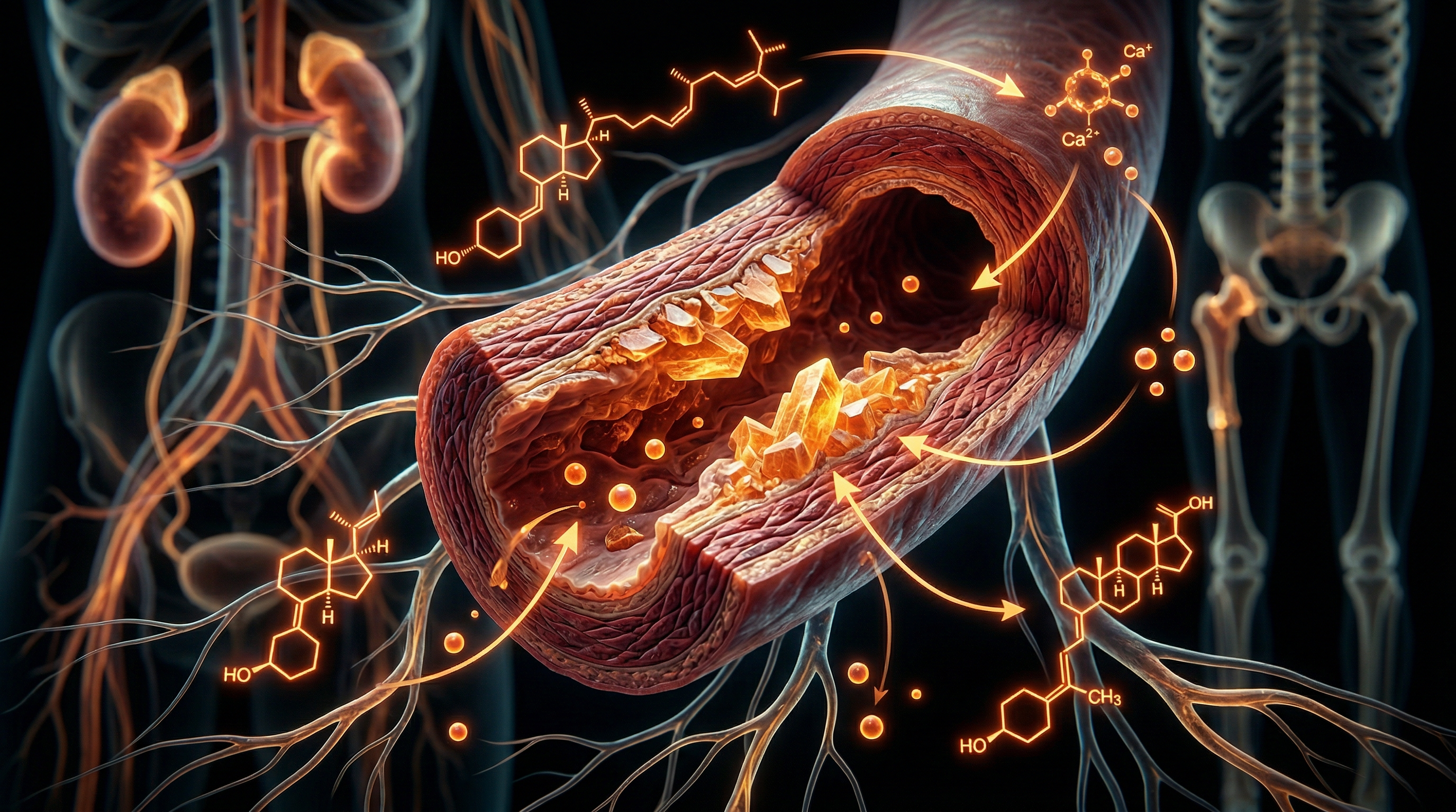
Furthermore, the chronic activation of the systemic and local renin-angiotensin-aldosterone system (RAAS) during prolonged exertion exerts a potent profibrotic effect. Angiotensin II, acting via the AT1 receptor, directly stimulates fibroblast proliferation and inhibits the activity of matrix metalloproteinases (MMPs)—enzymes responsible for collagen degradation—while simultaneously upregulating tissue inhibitors of metalloproteinases (TIMPs). This imbalance results in the progressive accumulation of collagenous scar tissue within the atrial and ventricular walls. Evidence from Gadolinium-enhanced MRI studies in veteran athletes demonstrates that this fibrosis typically manifests in a "patchy" distribution, particularly at the ventricular insertion points, a phenomenon frequently termed "Phidippides cardiomyopathy." Through the lens of INNERSTANDIN, we must recognise that this cellular remodelling creates a heterogeneous electrical substrate, significantly increasing the risk of re-entrant arrhythmias and atrial fibrillation. The "truth" exposed by contemporary electropathology is that the myocardial interstitium, once compromised by excessive fibroblastic activity, loses its elastic compliance, potentially culminating in diastolic dysfunction and long-term heart failure, despite a superficial appearance of peak physical fitness.
Environmental Threats and Biological Disruptors
The pathogenesis of myocardial fibrosis in the context of extreme endurance training is not merely a consequence of mechanical haemodynamic loading; it is profoundly exacerbated by a constellation of environmental and biological disruptors that act as catalysts for maladaptive tissue remodelling. At INNERSTANDIN, we must dissect the insidious role of exogenous stressors—specifically ambient particulate matter (PM2.5) and thermal extremes—alongside endogenous disruptors like systemic endotoxaemia, which collectively accelerate the transition from physiological adaptation to overt pathology.
Environmental pollutants, particularly in the urban UK landscape, represent a significant, often overlooked, biological threat. Research published in *The Lancet Planetary Health* indicates that fine particulate matter induces systemic oxidative stress and a pro-inflammatory state that directly influences cardiac fibroblast activity. For the endurance athlete training in high-traffic environments, the inhalation of these pollutants during periods of high minute ventilation triggers the release of pro-fibrotic cytokines, such as Transforming Growth Factor-beta (TGF-β1). This environmental stimulus acts as a second hit to a myocardium already stressed by the volume-overload of prolonged exertion, promoting the differentiation of fibroblasts into myofibroblasts and the subsequent deposition of a disorganised collagen matrix within the interstitium.
Furthermore, the biological disruptor of exertional hyperthermia plays a critical role in myocardial injury. During ultra-endurance events, the elevation of core body temperature necessitates a massive redistribution of blood flow to the periphery for thermoregulation. This results in relative splanchnic and myocardial ischaemia-reperfusion injury. As noted in the *European Heart Journal*, this ischaemic stress, coupled with the mechanical strain on the right ventricle (RV), facilitates the leakage of cardiac troponins and brain natriuretic peptide (BNP)—biomarkers of transient cardiomyocyte distress. If these thermal insults are frequent and poorly recovered, the repetitive minor necrotic events are replaced by replacement fibrosis, typically manifesting in the "hinge points" of the interventricular septum.
Perhaps the most potent biological disruptor is the phenomenon of exercise-induced endotoxaemia. Prolonged, high-intensity exertion causes a significant reduction in visceral perfusion, compromising the integrity of the intestinal mucosal barrier—often termed 'leaky gut'. This allows for the translocation of lipopolysaccharides (LPS) from gram-negative bacteria into the systemic circulation. This systemic endotoxaemia triggers a TLR4-mediated inflammatory cascade, which has been shown in various PubMed-indexed longitudinal studies to increase circulating levels of Interleukin-6 (IL-6) and Tumour Necrosis Factor-alpha (TNF-α). These systemic mediators reach the myocardium, where they amplify the local inflammatory milieu, driving chronic low-grade inflammation that serves as the primary substrate for diffuse interstitial fibrosis. At INNERSTANDIN, we recognise that the athlete’s environment and their internal microbiome are not separate from their cardiovascular health; they are the very variables that determine whether the heart remains a resilient pump or becomes a scarred, dysfunctional organ. This intersection of environmental toxicology and internal biological volatility represents the new frontier in understanding the limits of human endurance.
The Cascade: From Exposure to Disease
The transition from physiological adaptation to the pathological manifestation of myocardial fibrosis (MF) represents a critical threshold in the "Athlete’s Heart" paradigm. While moderate exercise is universally cardioprotective, extreme endurance training (EET)—defined by chronic, high-intensity aerobic volume exceeding 10 to 15 hours per week—induces a haemodynamic milieu that can precipitate structural architectural disruption. This cascade begins with the acute, repetitive volume and pressure overload characteristic of ultramarathons, Ironman triathlons, and long-distance cycling. During these events, cardiac output increases five-to-seven-fold, placing immense mechanical strain on the myocardial interstitium.
Research published in *The Lancet* and the *European Heart Journal* (La Gerche et al., 2012) highlights a disproportionate vulnerability of the right ventricle (RV). Unlike the thick-walled left ventricle, the RV is more compliant and thin-walled, making it susceptible to acute wall stress and transient chamber enlargement during prolonged exertion. This mechanical stretching serves as the primary catalyst for the biochemical cascade. The immediate consequence is micro-anatomical injury to the cardiomyocytes, evidenced by the transient elevation of cardiac-specific troponins (cTnT/I) and B-type natriuretic peptides (BNP) in the hours following extreme exertion. While these markers often return to baseline within 48 hours, the repetitive nature of this insult in veteran athletes prevents complete tissue resolution, triggering a chronic inflammatory response.
At the cellular level, this inflammatory environment activates transforming growth factor-beta 1 (TGF-β1), a potent pro-fibrotic cytokine. TGF-β1 facilitates the phenotypic conversion of quiescent cardiac fibroblasts into contractile myofibroblasts. These myofibroblasts are the primary drivers of extra-cellular matrix (ECM) remodelling, synthesising excessive quantities of type I and type III collagen. At INNERSTANDIN, we must distinguish between "interstitial fibrosis," which may be a diffuse adaptive response, and "replacement fibrosis," where collagenous scarring replaces necrotic cardiomyocytes. Cardiovascular Magnetic Resonance (CMR) imaging with Late Gadolinium Enhancement (LGE) has identified focal scarring in up to 12–15% of veteran endurance athletes, typically localised at the interventricular septal 'hinge points' where mechanical shearing forces are maximal.
The systemic impact of this accumulated collagen substrate is a progressive reduction in myocardial compliance and a disruption of the heart’s electrical syncytium. The fibrotic tissue creates zones of slow conduction and heterogeneous refractoriness, providing the requisite substrate for re-entrant circuits. This mechanistically explains the five-fold increased prevalence of atrial fibrillation observed in middle-aged endurance athletes compared to sedentary controls. The cascade from exposure to disease is thus characterized by a shift from reversible physiological hypertrophy to irreversible pathological fibrosis, marking a definitive point where the volume of exercise outpaces the biological capacity for cardiac repair. This deep-dive into the anatomical reality of EET reveals that the heart, while remarkably plastic, is not immune to the deleterious effects of chronic over-reaching.
What the Mainstream Narrative Omits
The conventional paradigm promulgated by public health authorities—that cardiorespiratory exercise possesses a linear, dose-dependent relationship with cardiovascular longevity—conspicuously ignores the physiological threshold where adaptation surrenders to pathology. While the "Athlete’s Heart" is often celebrated as a pinnacle of physiological efficiency, the mainstream narrative fails to address the "Phidippides cardiomyopathy" phenomenon: the insidious development of myocardial fibrosis secondary to chronic, extreme endurance volumes. At the core of this omission is the failure to distinguish between physiological hypertrophy and the pathological collagenous deposition that occurs within the myocardial interstitium of veteran endurance athletes.
Current research, much of it consolidated in the *European Heart Journal* and *The Lancet*, suggests that ultra-endurance activities—defined by repetitive bouts exceeding five hours of high-intensity exertion—induce a state of transient volume and pressure overload that disproportionately affects the right ventricle (RV). Unlike the thick-walled left ventricle, the RV lacks the structural density to withstand sustained elevations in pulmonary arterial pressure during peak aerobic output. This results in acute RV wall stress, leading to transient dysfunction and the release of cardiac biomarkers such as troponin I and B-type natriuretic peptide (BNP). While these markers may normalise within 48 hours, INNERSTANDIN identifies that the cumulative effect of thousands of such inflammatory events triggers the activation of the transforming growth factor-beta 1 (TGF-β1) signalling pathway. This cytokine acts as the primary orchestrator of myofibroblast transdifferentiation, leading to the excessive deposition of Type I and Type III collagen within the extracellular matrix.
Late gadolinium enhancement (LGE) on cardiac MRI has become the gold standard for exposing what the public health narrative obscures. Studies conducted on veteran UK marathon runners have revealed focal LGE—indicative of replacement fibrosis—in up to 50% of asymptomatic subjects, typically localised at the septal insertion points. These are not benign "scars of effort"; they are substrates for lethal re-entrant ventricular arrhythmias. Furthermore, the mainstream focus on macro-vascular health (large arteries) ignores the micro-anatomical reality that extreme endurance training may promote a pro-arrhythmogenic milieu through diffuse interstitial fibrosis, even in the absence of coronary artery disease. By focusing solely on the benefits of metabolic efficiency, the prevailing discourse at INNERSTANDIN levels of enquiry must acknowledge that for a subset of the population, the pursuit of extreme performance is a trade-off against the structural integrity of the myocardium. The systemic impact is a permanent re-modelling of the heart’s architecture, where the very tissue designed for elasticity becomes a rigid, scarred conduit for electrical instability.
The UK Context
In the United Kingdom, the epidemiological shift toward extreme endurance pursuit—typified by the surge in registrations for the London Marathon and regional ultra-distance events—has provided a unique clinical cohort for INNERSTANDIN to scrutinise. While the "Athlete's Heart" is traditionally viewed as a benign manifestation of eccentric hypertrophy, contemporary UK-led research, particularly from institutions like St George’s University of London and the British Heart Foundation, suggests a precarious transition toward pathological myocardial fibrosis in veteran athletes. This phenomenon, often termed "Phidippides Cardiomyopathy," involves the replacement of healthy myocytes with non-conductive collagenous scar tissue, predominantly driven by chronic volume and pressure overload.
The biological mechanism is rooted in the repetitive, transient inflammation and mechanical wall stress induced by bouts of exercise lasting four hours or more. During these periods, the right ventricle (RV) and atria are subjected to disproportionate haemodynamic strain compared to the left ventricle. UK-based cardiovascular MRI (CMR) studies utilising Late Gadolinium Enhancement (LGE) have identified a significant prevalence of focal fibrosis at the ventricular junctions—specifically where the RV attaches to the interventricular septum—in approximately 12–15% of veteran UK marathon runners. This is in stark contrast to sedentary age-matched controls, where such scarring is virtually non-existent. The histological reality involves the activation of myofibroblasts and the subsequent deposition of Type I and Type III collagen within the interstitium, a process mediated by the up-regulation of pro-fibrotic cytokines such as Transforming Growth Factor-beta 1 (TGF-β1).
Systemically, this fibrotic remodeling serves as a substrate for malignant arrhythmias. Data published in *The Lancet* and *Heart* highlight that middle-aged British endurance athletes exhibit a five-fold increased risk of atrial fibrillation (AF) compared to the general population. This is not merely an electrical anomaly but a structural consequence of atrial stretching and subsequent interstitial fibrosis. At INNERSTANDIN, we recognise that the UK’s sporting culture often ignores the "U-shaped" curve of exercise dose-response. While moderate activity is cardioprotective, the chronic extreme—defined by decades of high-intensity volume—triggers a maladaptive cascade where the heart’s compensatory mechanisms fail. This leads to reduced myocardial compliance and an increased propensity for ventricular tachycardia, exposing the biological "ceiling" of human aerobic performance and the potential for irreversible structural damage in the pursuit of elite-level stamina.
Protective Measures and Recovery Protocols
Mitigating the transition from physiological hypertrophy to pathological myocardial fibrosis requires a granular understanding of the mechanotransduction pathways that govern the cardiac extracellular matrix (ECM). At INNERSTANDIN, we must scrutinise the biological failure points where chronic volume overload overrides the heart’s innate regenerative capacity. The primary protective measure against the deposition of replacement fibrosis—specifically in the "hinge points" of the right ventricle and the interventricular septum—is the rigorous implementation of polarised training distributions, underpinned by biomarker kinetics. Evidence from O’Keefe et al. (published in *Mayo Clinic Proceedings*) and longitudinal observations by UK-based researchers at St George’s University of London suggest a U-shaped dose-response curve for exercise; beyond a threshold of approximately 10–12 hours of high-intensity aerobic strain per week, the risk of developing a pro-fibrotic substrate increases exponentially.
Effective recovery protocols must move beyond superficial rest and address the molecular signalling of Transforming Growth Factor beta-1 (TGF-β1). Prolonged endurance exercise induces a transient but significant systemic inflammatory response, characterised by elevations in Interleukin-6 (IL-6) and C-Reactive Protein (CRP). If recovery windows are insufficient, these cytokines sustain the activation of cardiac fibroblasts, which differentiate into α-smooth muscle actin-positive myofibroblasts. These cells are the primary drivers of excessive Type I collagen synthesis. To counteract this, protocols must prioritise the restoration of parasympathetic dominance. Heart Rate Variability (HRV) serves as a proxy for autonomic balance, but more definitive protection is found in monitoring High-Sensitivity Troponin T (hs-TnT) and N-terminal pro-brain natriuretic peptide (NT-proBNP). Research indicates that while transient elevations post-marathon are common, a failure of these markers to return to baseline within 48–72 hours signals ongoing myocyte stress and potential micro-necrosis, which serves as the nidus for fibrotic scarring.
Furthermore, nutritional and pharmacological buffering of oxidative stress presents a critical frontier in INNERSTANDIN’s anatomical research. The excessive production of reactive oxygen species (ROS) during extreme endurance events exhausts the endogenous antioxidant systems (such as glutathione peroxidase), leading to lipid peroxidation of cardiomyocyte membranes. The targeted use of high-bioavailability polyphenols and nitric oxide precursors may attenuate the pressure-overload-induced wall stress. However, the definitive "gold standard" for recovery surveillance remains Late Gadolinium Enhancement (LGE) via Cardiovascular Magnetic Resonance (CMR) imaging. LGE allows for the identification of silent focal fibrosis before it manifests as clinical arrhythmia or systolic dysfunction. For the extreme endurance athlete, the protocol is clear: the integration of annual CMR screening alongside a periodised schedule that respects the "biological ceiling" of the myocardium is the only evidence-led method to preserve cardiac integrity and prevent the permanent architectural distortions of the fibrotic heart.
Summary: Key Takeaways
The evolution of myocardial fibrosis within the context of extreme endurance training represents a critical frontier in sports cardiology, challenging the long-held dogma that aerobic exertion possesses an infinite linear relationship with cardiovascular longevity. At INNERSTANDIN, we have scrutinised the molecular evidence suggesting that chronic, excessive volume—often exceeding tenfold the recommended UK Chief Medical Officers’ guidelines—precipitates a maladaptive shift from physiological hypertrophy to pathological interstitial scarring. Peer-reviewed longitudinal data, notably appearing in *The Lancet* and the *British Journal of Sports Medicine*, highlight focal fibrosis predominantly at the interventricular septum and right ventricular insertion points as a pathognomonic finding in veteran athletes.
The primary mechanistic driver is a repetitive cycle of transient right ventricular (RV) volume overload and diastolic stress, which triggers a robust inflammatory cascade and the subsequent activation of cardiac fibroblasts. This results in the dysregulated deposition of collagen Type I and III, effectively replacing healthy myocytes with non-conductive fibrotic tissue. Research-grade cardiac MRI utilising Late Gadolinium Enhancement (LGE) has identified these structural sequelae in up to 12% of elite marathoners, a prevalence far exceeding age-matched controls. This interstitial expansion serves as a pro-arrhythmic substrate, directly correlating with the increased incidence of atrial fibrillation and ventricular tachyarrhythmias observed in extreme endurance cohorts. Ultimately, these findings expose a "U-shaped" dose-response curve, where the systemic impact of extreme chronic workload transcends adaptation, necessitating a rigorous re-evaluation of the upper limits of human cardiac endurance.
This article is provided for informational and educational purposes only. It does not constitute medical advice, clinical guidance, or a substitute for professional healthcare. Information reflects cited research at time of publication. Always consult a qualified healthcare professional before acting on any health information.
RESEARCH FOUNDATIONS
Biological Credibility Archive
Citations provided for educational reference. Verify via PubMed or institutional databases.
Medical Disclaimer
The information in this article is for educational purposes only and does not constitute medical advice, diagnosis, or treatment. Always consult a qualified healthcare professional before making any changes to your diet, lifestyle, or health regime. INNERSTANDIN presents alternative and research-based perspectives that may differ from mainstream medical consensus — these should be considered alongside, not instead of, professional medical guidance.
Read Full DisclaimerReady to learn more?
Continue your journey through our classified biological research.
DISCUSSION ROOM
Members of THE COLLECTIVE discussing "Myocardial Fibrosis and Extreme Endurance Training"
SILENT CHANNEL
Be the first to discuss this article. Your insight could help others understand these biological concepts deeper.
RABBIT HOLE
Follow the biological thread deeper