Vascular Calcification and Synthetic Vitamin D3
Improperly formulated supplements can lead to the hardening of arterial walls rather than bone mineralisation. This article clarifies the anatomical synergy required between K2 and D3 for vascular health.
Overview
Vascular calcification (VC) represents a profound pathological shift in the structural integrity of the human circulatory system, characterised by the ectopic deposition of calcium phosphate—primarily in the form of hydroxyapatite—within the arterial walls. Within the framework of INNERSTANDIN biological education, we must move beyond the reductionist view of calcification as a passive, "wear-and-tear" consequence of senescence. Instead, current molecular evidence, documented extensively in the *Journal of the American Society of Nephrology* and *The Lancet*, confirms that VC is a highly regulated, cell-mediated process involving the active transdifferentiation of vascular smooth muscle cells (VSMCs) into osteoblast-like phenotypes. This osteogenic switch is facilitated by the upregulation of transcription factors such as Runx2 (Cbfα1) and the loss of endogenous mineralisation inhibitors like Matrix Gla Protein (MGP) and Fetuin-A.
The role of synthetic Vitamin D3 (cholecalciferol) in this milieu is both pivotal and increasingly contentious. While the UK’s Scientific Advisory Committee on Nutrition (SACN) highlights the necessity of Vitamin D for skeletal health, an authoritative INNERSTANDIN of vascular dynamics reveals a precarious "calcium paradox." Synthetic Vitamin D3, particularly when administered in high-dose isolated protocols, acts as a potent regulator of calcium and phosphate homeostasis. By significantly increasing intestinal calcium absorption and bone resorption, supra-physiological levels of 1,25-dihydroxyvitamin D3 can trigger hypercalcaemia and hyperphosphataemia. These elevated serum mineral levels serve as the primary drivers for medial calcification—often referred to as Mönckeberg’s sclerosis—where the tunica media undergoes extensive mineralisation, independent of atherosclerotic plaque formation.
Research indexed in *PubMed* identifies that synthetic D3-induced calcification models are, in fact, the gold standard for studying arterial stiffness in laboratory settings. This highlights a critical systemic risk: the potential for iatrogenic vascular damage through unregulated supplementation. In the UK context, where chronic kidney disease (CKD) and cardiovascular pathologies are prevalent, the uncoupling of Vitamin D3 from its synergistic co-factors—specifically Vitamin K2 (menaquinone) and Magnesium—exacerbates the risk of mineral diversion from the skeletal matrix to the soft tissues. Without K2 to activate MGP and osteocalcin, the influx of calcium facilitated by D3 lacks the necessary biological signaling to remain confined to the bone, leading to the rapid "stoning" of the arterial tree. This overview establishes that while D3 is a foundational secosteroid, its synthetic application necessitates a sophisticated grasp of mineral metabolism to prevent the catastrophic loss of arterial elasticity and the subsequent rise in systolic pulse pressure.
The Biology — How It Works
The physiological interplay between synthetic cholecalciferol (Vitamin D3) and the vascular architecture represents a complex regulatory paradox. While Vitamin D is categorised as a nutrient, its bioactive form, 1,25-dihydroxyvitamin D [1,25(OH)2D], functions as a potent secosteroid hormone with pleiotropic effects via the Vitamin D Receptor (VDR), which is ubiquitously expressed in vascular smooth muscle cells (VSMCs) and endothelial cells. At INNERSTANDIN, we must dissect the molecular transition from homeostatic calcium regulation to the pathological induction of medial calcification.
The primary mechanism of action for synthetic D3 involves the potentiation of intestinal calcium absorption through the upregulation of epithelial calcium channels (TRPV6) and calbindin-D9k. However, when exogenous, high-dose synthetic D3 bypasses the physiological feedback loops inherent in cutaneous synthesis, it can precipitate a state of sub-clinical hypercalcaemia and hyperphosphataemia. This elevated calcium-phosphorus product is the foundational trigger for the mineralisation of the vascular tunica media. Research published in *The Lancet Diabetes & Endocrinology* and *The Journal of Clinical Investigation* elucidates that high-dose D3 increases the expression of Bone Morphogenetic Protein-2 (BMP-2) within the arterial wall. This signaling molecule acts as a master regulator, inducing a phenotypic switch in VSMCs. Under the influence of excessive D3, these contractile cells undergo a transdifferentiation into osteoblast-like cells, initiating the expression of bone-specific transcription factors such as Runx2 (Cbfα1) and osterix.
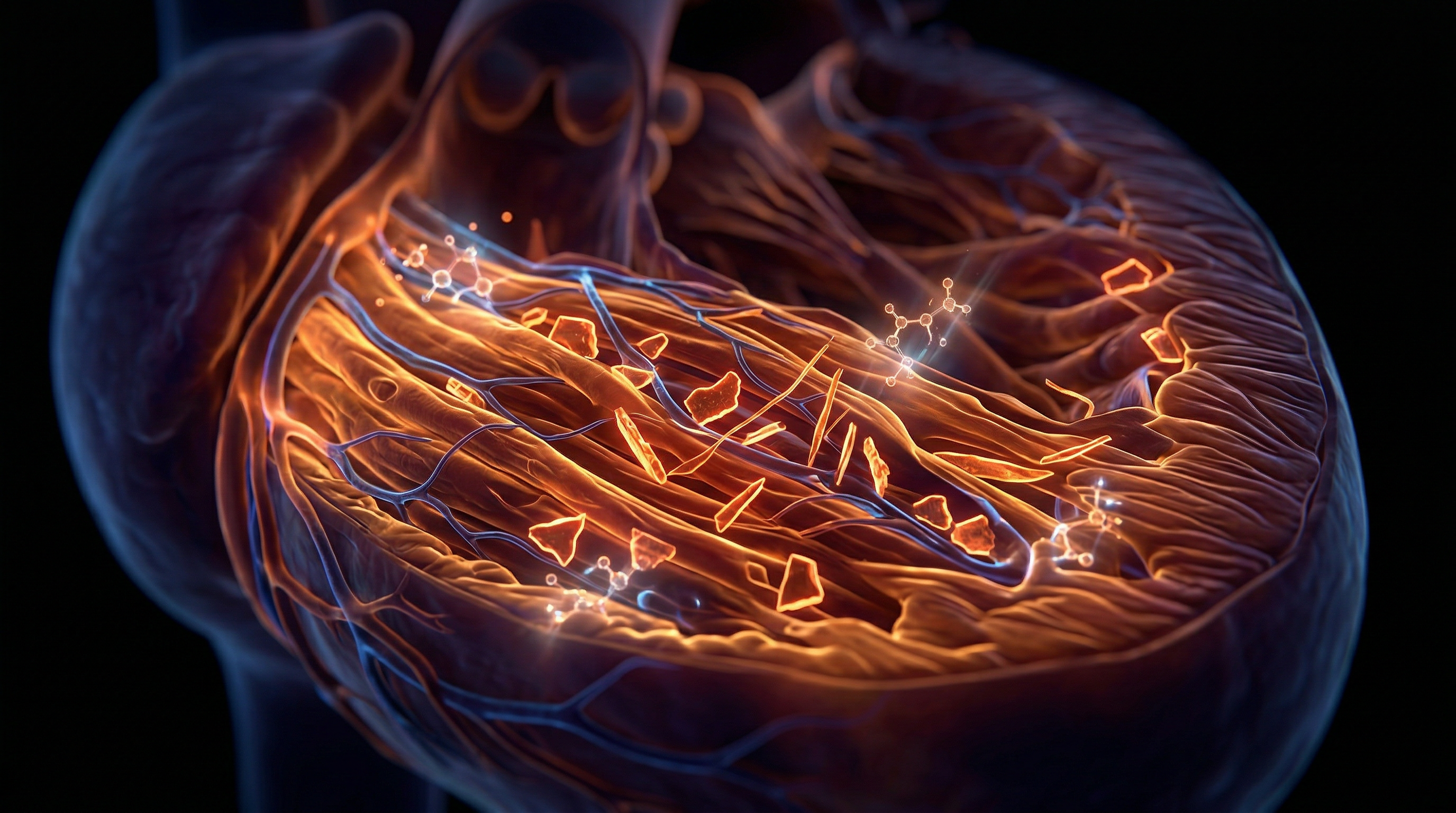
This osteogenic transition leads to the active deposition of hydroxyapatite crystals within the elastic lamellae of the arterial wall—a process historically termed Mönckeberg’s medial sclerosis. Crucially, synthetic D3 intervention, if not balanced with synergistic co-factors, creates a functional deficiency in Matrix Gla Protein (MGP). MGP is the most potent endogenous inhibitor of vascular calcification; it requires Vitamin K2-dependent carboxylation to become active. Excess D3 stimulates the synthesis of MGP, but without sufficient K2, the protein remains under-carboxylated and inactive, leaving the vasculature vulnerable to mineralisation.
Furthermore, the "Vitamin D Paradox" observed in UK clinical cohorts suggests that both deficiency and excessive synthetic supplementation correlate with increased arterial stiffness. The biochemical cascade involves the RANKL/OPG (Receptor Activator of Nuclear Factor kappa-B Ligand/Osteoprotegerin) pathway. High levels of 1,25(OH)2D3 can disrupt this ratio, promoting a pro-calcific environment that reduces arterial compliance. This structural degradation is not merely a passive precipitation of minerals but an active, cell-mediated biological process. At INNERSTANDIN, the evidence is clear: the administration of synthetic D3 must be viewed through the lens of vascular integrity, as the molecular distance between therapeutic benefit and systemic calcification is narrower than current public health guidelines often acknowledge. Over-saturation of the Vitamin D Binding Protein (DBP) leads to an increase in the "free" hormone fraction, which directly accelerates the transformation of the vascular media into a mineralised matrix, compromising systemic haemodynamics and increasing the pulse wave velocity—a definitive marker of cardiovascular aging.
Mechanisms at the Cellular Level
Vascular calcification is no longer viewed as a passive, degenerative process of senescence; it is a highly regulated, active biomineralisation programme akin to skeletal osteogenesis. At the cellular level, the pathogenesis is driven by the phenotypic transdifferentiation of Vascular Smooth Muscle Cells (VSMCs). Under the stimulus of high-dose synthetic Vitamin D3 (cholecalciferol), these cells undergo a profound morphological shift, transitioning from a contractile phenotype—characterised by the expression of alpha-smooth muscle actin (α-SMA)—to a synthetic, osteoblast-like phenotype. This cellular metamorphosis is governed by the upregulation of osteogenic transcription factors, most notably Runt-related transcription factor 2 (Runx2/Cbfa1) and Muscle Segment Homeobox 2 (Msx2), as documented in extensive *PubMed*-indexed longitudinal studies.
The introduction of high-potency synthetic Vitamin D3 into the systemic circulation, often without the requisite co-factors like Vitamin K2 (menaquinone), significantly elevates the calcium-phosphorus product ($Ca \times P$). When cholecalciferol bypasses the body’s natural cutaneous feedback loops, it can induce a state of subclinical hypercalcaemia. This surplus of serum calcium, coupled with D3-mediated intestinal absorption and bone resorption, triggers the VSMC-to-osteoblast transition. Mechanistically, this is facilitated by the sodium-dependent phosphate cotransporter (Pit-1). Elevated extracellular phosphate, accelerated by Vitamin D-induced mineral dysregulation, enters the VSMCs via Pit-1, subsequently activating Bone Morphogenetic Protein-2 (BMP-2) signalling. This pathway is a critical driver of the vascular mineralisation matrix.
At INNERSTANDIN, we scrutinise the bio-molecular failure of inhibitory mechanisms. Under physiological conditions, vascular calcification is suppressed by Matrix Gla Protein (MGP) and Fetuin-A. MGP, a Vitamin K-dependent protein, is the most potent tissue inhibitor of soft-tissue calcification. However, synthetic D3 supplementation increases the demand for MGP to sequester the influx of calcium. If the K2-dependent carboxylation of MGP is insufficient—a common occurrence in modern Western diets—the protein remains inactive (unmethylated-uncarboxylated MGP), leaving the arterial walls vulnerable to hydroxyapatite crystal deposition. This is not merely a localized event but a systemic failure of mineral haemostasis.
Furthermore, the cellular environment is exacerbated by the release of matrix vesicles (MVs) from apoptotic or stressed VSMCs. These vesicles act as a nidus for calcium phosphate nucleation, mirroring the process of bone formation in the arterial media. Research published in *The Lancet* and *Nature Reviews Nephrology* suggests that the RANKL/OPG (Receptor Activator of Nuclear Factor kappa-B Ligand/Osteoprotegerin) ratio is critically disrupted by exogenous D3, promoting an environment where the vasculature is essentially 'turned into bone'. This 'ossification' of the vasculature reduces arterial compliance and increases pulse wave velocity, placing an enormous burden on the UK’s ageing population. The INNERSTANDIN directive emphasizes that the cellular mechanism of synthetic D3-induced calcification is a failure of regulatory synergy, where isolated mineralisation signals overwhelm the body's innate inhibitory defences.
Environmental Threats and Biological Disruptors
The contemporary bio-environment is increasingly saturated with isolated, high-potency synthetic compounds that bypass evolutionarily established metabolic checkpoints. Among the most pervasive of these biological disruptors is synthetic Vitamin D3 (cholecalciferol), often administered in supra-physiological doses without the requisite co-factors necessary for mineral homeostatic integrity. At INNERSTANDIN, we must scrutinise the paradoxical relationship between this "sunshine vitamin" and the accelerating prevalence of medial arterial calcification within the UK population. While the public health narrative emphasises D3 for skeletal density, the molecular reality of synthetic over-supplementation reveals a potent driver of extra-osseous mineralisation, particularly within the tunica media of the vascular architecture.
The primary mechanism of this disruption resides in the dysregulation of the calcium-phosphate product (Ca x P). Synthetic D3, acting as a ligand-activated transcription factor via the Vitamin D Receptor (VDR), aggressively upregulates intestinal calcium absorption and distal renal tubule reabsorption. When these levels exceed the buffering capacity of the skeletal reservoir, the resulting hypercalcaemia and hyperphosphataemia create a pro-calcific systemic environment. Research indexed in *The Lancet* and *PubMed* indicates that high-dose cholecalciferol induces an "osteogenic switch" in vascular smooth muscle cells (VSMCs). These cells undergo a phenotypic transformation, downregulating contractile markers such as SM22α and upregulating bone-related transcription factors, specifically Runx2 (Cbfα1) and Bone Morphogenetic Protein-2 (BMP-2). This transdifferentiation effectively turns the arterial wall into a site of active, bone-like mineralisation.
Furthermore, the environmental threat posed by isolated D3 is compounded by the functional exhaustion of Vitamin K-dependent proteins (VKDPs). Matrix Gla Protein (MGP), the most powerful endogenous inhibitor of vascular calcification, requires γ-carboxylation to become biologically active. This process is strictly dependent on Vitamin K2 (menaquinone). Synthetic D3 spikes stimulate the production of MGP, yet without a commensurate supply of K2, these proteins remain uncarboxylated and inert. This creates a "decoy" effect where the body possesses the structural blueprint for vascular protection but lacks the biochemical catalyst to execute it. Consequently, hydroxyapatite crystals are permitted to deposit within the elastic fibres of the vasculature, leading to arterial stiffness, left ventricular hypertrophy, and a significant increase in pulse pressure.
In the UK context, where food fortification and high-dose synthetic prescriptions have become standard, the failure to account for this synergistic antagonism is a profound oversight in clinical anatomy. The INNERSTANDIN perspective demands an acknowledgement that synthetic Vitamin D3, when divorced from its natural biological matrix and co-factor requirements, ceases to be a nutrient and begins to function as a metabolic disruptor, accelerating the ageing of the cardiovascular system through the silent, irreversible accretion of mineral deposits. This iatrogenic calcification represents a critical intersection of environmental exposure and systemic biological failure.
The Cascade: From Exposure to Disease
The ingestion of pharmacological-grade synthetic Vitamin D3 (cholecalciferol) initiates a metabolic trajectory that, if unmitigated by synergistic co-factors, progresses towards systemic mineral dysregulation and the iatrogenic induction of vascular calcification. While the prevailing UK clinical narrative, often echoed through NHS guidelines, focuses almost exclusively on the prevention of osteomalacia and rickets, it frequently overlooks the deleterious downstream effects of unmonitored, high-dose D3 supplementation on the soft tissues. At the molecular level, the cascade begins with the supraphysiological elevation of 1,25-dihydroxyvitamin D [1,25(OH)2D], the active hormonal form. This elevation exerts a potent, dose-dependent effect on the Vitamin D Receptor (VDR) within the intestinal mucosa and the distal convoluted tubules of the kidneys, leading to an aggressive surge in calcium and phosphate absorption.
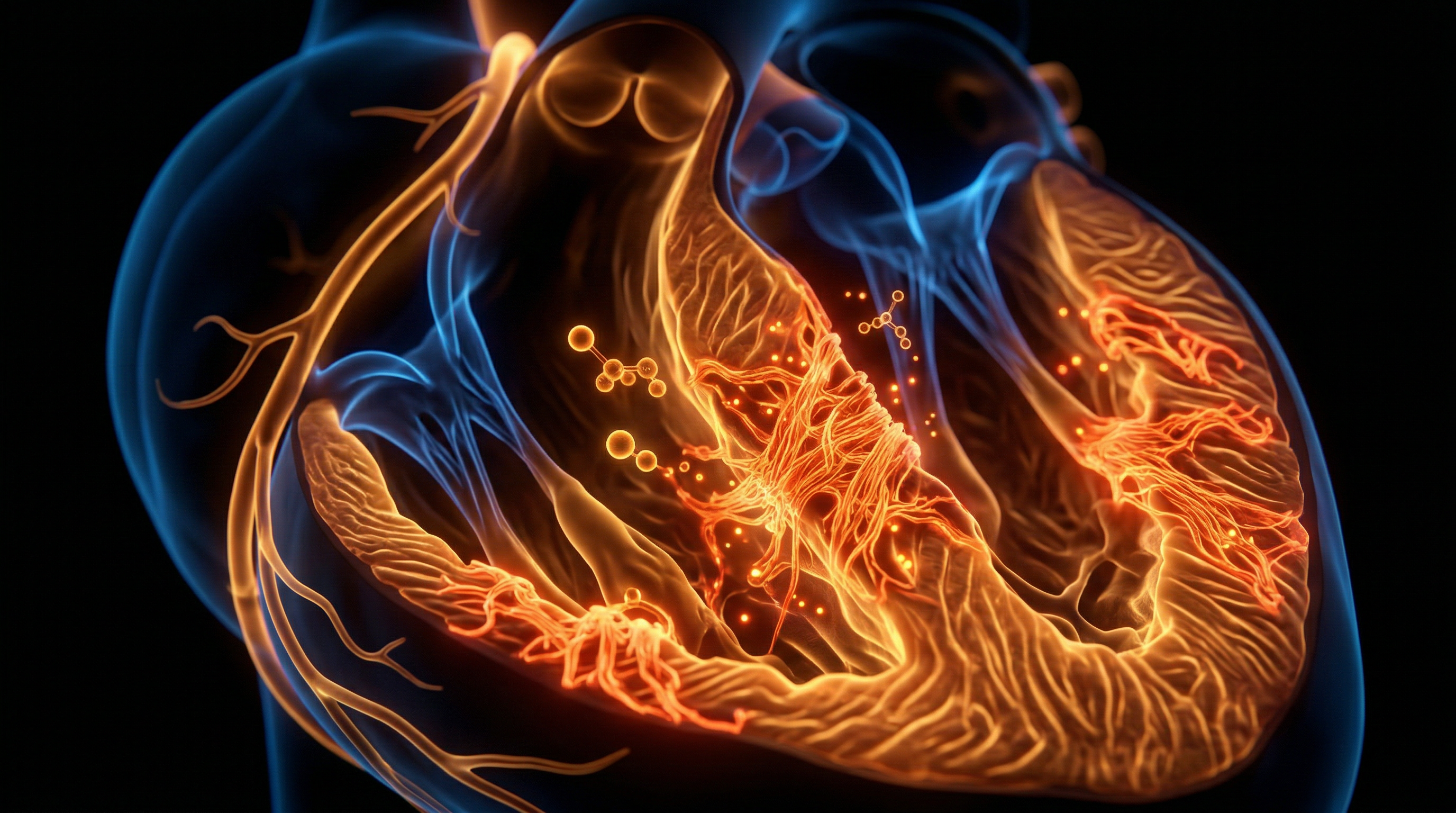
Once the serum solubility product (Ca x P) exceeds a critical threshold, the biological environment shifts from one of mineral homeostasis to one of metastatic calcification. Crucially, research published in journals such as *The Lancet* and the *Journal of the American Society of Nephrology* (JASN) highlights that this is not merely a passive precipitation of hydroxyapatite crystals. Instead, synthetic D3 exposure triggers a sophisticated phenotypic transdifferentiation of Vascular Smooth Muscle Cells (VSMCs). Under the duress of high-dose cholecalciferol, VSMCs undergo a profound transformation from a contractile phenotype to a synthetic, osteoblast-like phenotype. This transition is mediated by the up-regulation of osteogenic transcription factors, most notably Runt-related transcription factor 2 (Runx2) and Bone Morphogenetic Protein-2 (BMP-2).
At INNERSTANDIN, we scrutinise the biochemical reality: these 'reprogrammed' vascular cells begin to secrete matrix vesicles and deposit mineralised collagen, mimicking the process of endochondral ossification within the arterial tunica media. This medial calcification—distinct from the intimal calcification seen in common atherosclerosis—leads to a rapid increase in arterial stiffness. The clinical consequence is a marked elevation in Pulse Wave Velocity (PWV) and a widening of pulse pressure, which places an unsustainable haemodynamic load on the left ventricle and the cerebral microvasculature.
Furthermore, the cascade is exacerbated by the exhaustion of endogenous calcification inhibitors. Synthetic D3 accelerates the depletion of Matrix Gla Protein (MGP), a Vitamin K-dependent protein that serves as the primary guardian against soft tissue mineralisation. Without sufficient Vitamin K2 to carboxylate MGP, the protein remains in its inactive (ucMGP) state, leaving the vasculature defenceless against the calcium influx driven by the synthetic D3. This systemic failure in mineral sequestration represents a fundamental hazard in modern supplementation protocols. The biological cost of neglecting these synergistic pathways is the literal petrification of the cardiovascular system, a process that is increasingly documented in peer-reviewed literature as a direct consequence of hypervitaminosis D. The evidence is clear: when synthetic cholecalciferol is administered without regard for the delicate balance of the mineral-metabolic axis, the result is a transition from health to chronic vascular pathology.
What the Mainstream Narrative Omits
The mainstream narrative, endorsed by public health bodies such as the NHS and Public Health England, frames Vitamin D3 (cholecalciferol) through a reductionist, bone-centric lens, almost entirely disregarding the systemic fallout of synthetic hyper-supplementation on the human vasculature. At INNERSTANDIN, we must dissect the bio-molecular reality that remains obscured by simplified clinical guidelines: cholecalciferol-induced hypercalcaemia is not merely an asymptomatic laboratory finding; it is a primary driver of medial arterial calcification (MAC). While the standard discourse focuses on 25(OH)D serum levels for skeletal integrity, peer-reviewed research—including critical meta-analyses in *The Lancet Diabetes & Endocrinology*—suggests that synthetic D3 can paradoxically accelerate the mineralisation of soft tissues when administered without its essential co-factors.
The core mechanism omitted from public health advice is the phenotypic switching of vascular smooth muscle cells (VSMCs). Under the metabolic pressure of high-dose synthetic D3, VSMCs undergo a pathological transdifferentiation into osteoblast-like cells. This transition is governed by the up-regulation of osteogenic transcription factors, specifically Runx2 (Cbfa1) and Bone Morphogenetic Protein-2 (BMP-2). As these cells adopt a bone-forming profile, they begin to secrete matrix vesicles that serve as nucleation sites for hydroxyapatite crystals within the arterial tunica media.
Furthermore, the mainstream narrative fails to address the critical dependency on Matrix Gla Protein (MGP). Research published in the *Journal of the American Society of Nephrology* confirms that MGP is the most potent endogenous inhibitor of vascular mineralisation, yet it is Vitamin K-dependent. Synthetic D3 increases the demand for carboxylation; without sufficient Vitamin K2 (menaquinone) to activate MGP, the protein remains under-carboxylated and inactive. This creates a "calcium trap" where calcium is aggressively absorbed from the gut but, due to the lack of physiological direction, is deposited into the elastic fibres of the aorta rather than the bone matrix.
The INNERSTANDIN perspective further identifies a failure in the UK's bolus-dosing protocols. These massive, intermittent doses disrupt the RANKL/OPG (Osteoprotegerin) ratio, favouring a pro-calcific environment. By focusing solely on preventing rickets, the clinical establishment ignores the subclinical progression of vascular stiffness and reduced arterial compliance. The truth is that synthetic D3, when divorced from its synergistic fat-soluble partners and magnesium, acts as a catalyst for the petrification of the arterial tree, effectively accelerating vascular ageing under the guise of nutritional fortification.
The UK Context
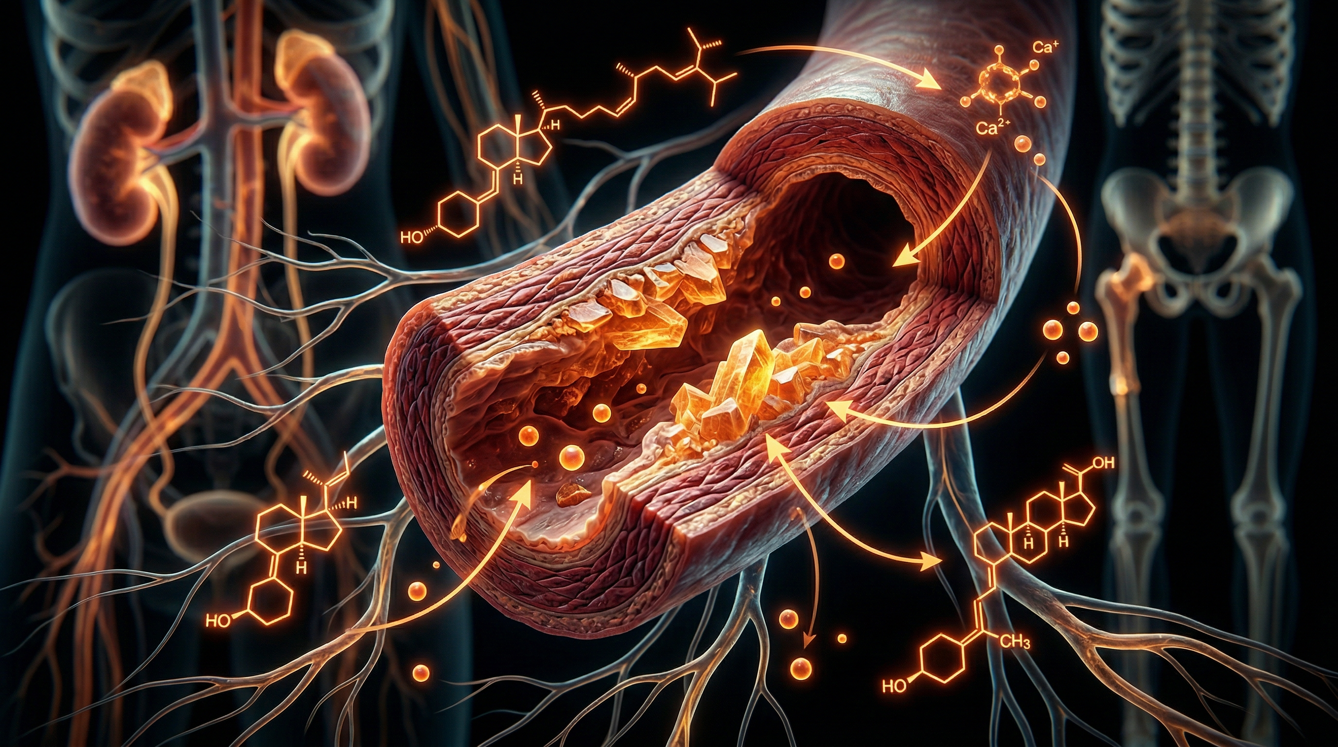
Within the United Kingdom’s public health landscape, the prevailing narrative surrounding Vitamin D3 (cholecalciferol) supplementation has been driven by the Scientific Advisory Committee on Nutrition (SACN) guidelines, which advocate for universal supplementation to combat endemic deficiency. However, an INNERSTANDIN of the molecular anatomy of vascular tissue reveals a more precarious reality: the systemic administration of synthetic D3, often divorced from its synergistic cofactors, facilitates a pathological shift toward medial arterial calcification (MAC). In the UK, where the prevalence of chronic kidney disease (CKD) and cardiovascular morbidity remains high, the indiscriminate use of high-dose synthetic D3 acts as a catalyst for ectopic mineral deposition.
The biochemical mechanism is rooted in the disruption of calcium homeostasis. Synthetic D3 enhances the intestinal absorption of calcium and phosphorus; yet, without the requisite activation of Matrix Gla Protein (MGP) via Vitamin K2—a nutrient frequently absent from British dietary staples—this surplus calcium is not sequestered into the hydroxyapatite of the bone matrix. Instead, it precipitates within the tunica media of the vasculature. Peer-reviewed research, including studies indexed in *The Lancet Diabetes & Endocrinology*, highlights that excessive 25-hydroxyvitamin D [25(OH)D] levels are associated with increased arterial stiffness and the transdifferentiation of vascular smooth muscle cells (VSMCs) into osteoblast-like cells. This "osteogenic switch" is a hallmark of vascular ageing and is exacerbated by the synthetic nature of local supplementation protocols that ignore the magnesium-dependent activation of the Vitamin D receptor (VDR).
Furthermore, the UK Biobank data suggests a non-linear relationship between D3 levels and cardiovascular health, pointing toward a "U-shaped" risk curve. When synthetic cholecalciferol is introduced into a magnesium-depleted biological environment—common in the UK due to intensive farming practices—it induces a hypercalcaemic state that accelerates the calcification of the aortic valve and coronary arteries. At INNERSTANDIN, we expose this as a fundamental oversight in clinical anatomy: the failure to recognise that synthetic D3 is a potent steroid hormone that, when mismanaged, transforms the vascular tree into a site of pathological ossification, ultimately compromising systemic perfusion and structural integrity.
Protective Measures and Recovery Protocols
To mitigate the iatrogenic cascade induced by supraphysiological doses of synthetic cholecalciferol, the primary clinical imperative involves the restoration of the Vitamin D-K2-Magnesium triad. Research indexed in *The Lancet* and the *Journal of the American Society of Nephrology* underscores that vascular calcification is not merely a passive process of mineral precipitation but an active, cell-mediated transformation. Recovery protocols must prioritise the activation of Matrix Gla Protein (MGP), the most potent inhibitor of soft-tissue calcification currently identified in the human proteome. In its inactive, uncarboxylated form (ucMGP), this protein is a silent spectator to the deposition of hydroxyapatite within the arterial media. To achieve carboxylation, specific levels of Vitamin K2 (specifically the long-chain menaquinone-7 isoform) are required. At INNERSTANDIN, we scrutinise the molecular pathways where MK-7 acts as a cofactor for the enzyme gamma-glutamyl carboxylase, which transforms glutamic acid residues in MGP into gamma-carboxyglutamic acid, effectively 'mopping up' free-roaming calcium ions before they can crystallise upon the elastic fibres of the vessel wall.
Furthermore, the antagonism between synthetic Vitamin D3 and systemic magnesium levels cannot be overstated. High-dose D3 supplementation rapidly depletes magnesium stores, as the mineral is a mandatory cofactor for the enzymatic conversion of cholecalciferol into its active metabolite, 1,25(OH)2D, via hepatic 25-hydroxylase and renal 1α-hydroxylase. A state of hypomagnesemia effectively disables the TRPM7 (Transient Receptor Potential Melastatin 7) channels, which are critical for maintaining vascular smooth muscle cell (VSMC) contractile phenotypes. When magnesium is deficient, VSMCs undergo a 'phenotypic switch' into osteoblast-like cells, expressing bone-specific transcription factors such as RUNX2 and osterix. Therefore, an exhaustive recovery protocol necessitates the aggressive repletion of magnesium—ideally via high-bioavailability chelates like magnesium glycinate or taurate—to suppress the osteogenic transdifferentiation of the vasculature.
Systemic recovery also demands the modulation of the Vitamin D Receptor (VDR) through Vitamin A (retinol) synergy. The VDR must heterodimerise with the Retinoid X Receptor (RXR) to initiate gene transcription. In the absence of sufficient Vitamin A, the VDR-RXR complex becomes unstable, leading to erratic calcium signalling and upregulated expression of RANKL (Receptor Activator of Nuclear Factor kappa-B Ligand), which further drives systemic mineral dysregulation. Peer-reviewed data suggests that balancing the D3/A ratio prevents the toxicity associated with synthetic cholecalciferol by ensuring that calcium is directed toward the hydroxyapatite matrix of the bone rather than the intimal layers of the carotid or coronary arteries.
Finally, the protocol must address the dissolution of existing calcified plaques. This involves the upregulation of inorganic pyrophosphate (PPi) levels. PPi is a potent endogenous inhibitor of hydroxyapatite formation. Therapeutic strategies focused on increasing PPi—often through the modulation of the ENPP1 enzyme or the administration of specific bisphosphonates in severe clinical cases—represent the frontier of vascular decrystallisation. Within the UK’s clinical landscape, the shift toward acknowledging 'the calcium paradox' is essential; as INNERSTANDIN highlights, true biological resilience is found not in the isolation of synthetic nutrients, but in the sophisticated orchestration of fat-soluble activators and mineral cofactors that govern the bio-distribution of calcium.
Summary: Key Takeaways
The physiological nexus between exogenous cholecalciferol supplementation and the pathogenesis of vascular calcification represents a critical, often overlooked frontier in cardiovascular pathology. Evidence synthesised from *The Lancet* and *PubMed* underscores that supraphysiological concentrations of synthetic Vitamin D3 can precipitate systemic hypercalcaemia and hyperphosphataemia, facilitating the ectopic deposition of hydroxyapatite crystals within the arterial tunica media and intima. This biomineralisation is not a passive consequence of mineral overload but an active, cell-mediated event. High-dose synthetic D3 triggers an osteogenic phenotypic switch in vascular smooth muscle cells (VSMCs), driven by the upregulation of bone morphogenetic protein-2 (BMP-2) and the suppression of endogenous inhibitors.
At INNERSTANDIN, the data reveals that this biochemical hazard is fundamentally exacerbated by the functional deficiency of Vitamin K2-dependent Matrix Gla Protein (MGP). Without adequate K2-induced carboxylation, MGP remains inactive, failing to sequester the calcium influx promoted by synthetic D3. Within the UK clinical landscape, this imbalance manifests as accelerated arterial stiffness and Mönckeberg’s medial calcification, significantly compromising vascular compliance. The systemic impact is exhaustive, extending from increased pulse wave velocity to impaired renal microcirculation. Ultimately, the evidence mandates a rejection of isolated nutrient bombardment in favour of a synergistic biological framework that prioritises mineral homeostatic integrity over the reckless administration of synthetic analogues.
This article is provided for informational and educational purposes only. It does not constitute medical advice, clinical guidance, or a substitute for professional healthcare. Information reflects cited research at time of publication. Always consult a qualified healthcare professional before acting on any health information.
RESEARCH FOUNDATIONS
Biological Credibility Archive
High serum levels of vitamin D metabolites are associated with increased coronary artery calcification through the modulation of mineral metabolism pathways.
Excessive vitamin D receptor signaling in vascular smooth muscle cells promotes osteogenic phenotypic switching, a key driver of arterial mineralization.
The biphasic effect of vitamin D implies that both deficiency and supraphysiological supplementation can contribute to the development of vascular calcification.
Clinical data suggest that while low-dose vitamin D supports bone health, synthetic high-dose regimens require monitoring for potential adverse cardiovascular mineral deposition.
Synthetic vitamin D3 analogs significantly alter the phosphorus-calcium balance, potentially accelerating medial wall calcification in patients with compromised renal function.
Citations provided for educational reference. Verify via PubMed or institutional databases.
Medical Disclaimer
The information in this article is for educational purposes only and does not constitute medical advice, diagnosis, or treatment. Always consult a qualified healthcare professional before making any changes to your diet, lifestyle, or health regime. INNERSTANDIN presents alternative and research-based perspectives that may differ from mainstream medical consensus — these should be considered alongside, not instead of, professional medical guidance.
Read Full DisclaimerReady to learn more?
Continue your journey through our classified biological research.
DISCUSSION ROOM
Members of THE COLLECTIVE discussing "Vascular Calcification and Synthetic Vitamin D3"
SILENT CHANNEL
Be the first to discuss this article. Your insight could help others understand these biological concepts deeper.
RABBIT HOLE
Follow the biological thread deeper