Necroptosis Pathways in Tissue Injury: Bridging Inflammatory Response and Programmed Necrosis
Discuss this resourceUpdated August 2026
An exploration of necroptosis, the regulated form of necrotic cell death, and its critical role in orchestrating inflammatory responses following tissue injury.
Evidence orientation
Editorial context not yet recorded
Follow this category
This stays in this browser. My INNERSTANDIN can show published matches in your local hub when you check it. It does not send email, push, or alert notifications.
Local learning review
A private browser aid for revisiting ideas. It is not an alert or a health recommendation.
Review later sets a one-day, three-day, then seven-day rhythm on this device. Choose it only when you want to revisit this article.
Introduction to Programmed Necrosis
In the landscape of cellular homeostasis, apoptosis is often framed as the 'quiet' death. However, when cells face extreme stress, viral invasion, or mechanical trauma, they may engage a more aggressive mechanism known as necroptosis. Unlike the clean breakdown of apoptosis, necroptosis is a form of programmed necrosis that triggers a robust inflammatory response. Understanding this pathway is central to grasping how the body manages chronic tissue injury and systemic inflammation.
The Molecular Architecture: The Necrosome
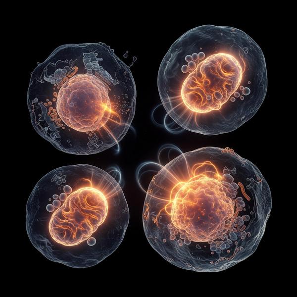
At the heart of the necroptotic process lies the necrosome—a multiprotein complex governed primarily by Receptor-Interacting Protein Kinases (RIPK1 and RIPK3) and the Mixed Lineage Kinase Domain-Like protein (MLKL). When caspase-8 activity is inhibited, the cell switches from an apoptotic program to a necroptotic one. RIPK1 phosphorylates RIPK3, which subsequently recruits and phosphorylates MLKL. This modified MLKL translocates to the plasma membrane, inducing pore formation and catastrophic osmotic lysis.
Bridging Inflammation and Injury
Necroptosis is not merely a death mechanism; it is a signal. As the cell membrane ruptures, it releases Damage-Associated Molecular Patterns (DAMPs) into the extracellular matrix. These DAMPs act as potent alarm signals to the immune system, recruiting macrophages and neutrophils to the site of injury. While this process is protective during viral infection, excessive necroptosis is a root cause of chronic tissue damage in conditions such as myocardial infarction, ischemic stroke, and inflammatory bowel disease.
Clinical Implications and Future Directions
Targeting the necroptotic pathway offers a novel therapeutic frontier for tissue preservation. By inhibiting RIPK1 or RIPK3 activity, researchers aim to dampen the inflammatory cascade that follows acute injuries. Our Innerstanding approach emphasizes that recognizing the transition from physiological death to pathogenic necroptosis is key to mitigating long-term organ damage and systemic dysfunction.
This article is provided for informational and educational purposes only. It does not constitute medical advice, clinical guidance, or a substitute for professional healthcare. Information reflects cited research at time of publication. Always consult a qualified healthcare professional before acting on any health information.
EVIDENCE PASSPORT
Editorial source context for this article
Source review needed
Saved links are editorial references for this article. They may support specific claims rather than every sentence. Open and assess each source in context. This passport does not independently verify them.
Editorial context
A complete editorial reading has not been recorded for this article, and no source links are saved on its record.
Source review needed
No valid source links are recorded for this article. Supplementary research, where available, is listed separately below.
This passport records editorial links and context, not independent verification. Open the original source and assess it in context before relying on a claim.
Medical Disclaimer
The information in this article is for educational purposes only and does not constitute medical advice, diagnosis, or treatment. Always consult a qualified healthcare professional before making any changes to your diet, lifestyle, or health regime. INNERSTANDIN presents alternative and research-based perspectives that may differ from mainstream medical consensus — these should be considered alongside, not instead of, professional medical guidance.
Read Full DisclaimerContinue the thread
Keep this question moving.
Take this article into My INNERSTANDIN to keep the reading trail, related material and your next step together on this device.
Dig deeper in the Library
Free, longform PDF volumes that go beyond headlines into mechanisms and references.
RABBIT HOLE
Follow the biological thread deeper