
Why the NHS Reliance on TSH Testing Leaves Millions Hypothyroid
Updated May 2026
Standard thyroid screening in the UK often misses subclinical or cellular hypothyroidism by relying solely on Thyroid Stimulating Hormone (TSH). This article explores why current reference ranges are insufficient and what markers are required for an accurate diagnosis.
Evidence orientation
Editorial context not yet recorded
Follow this category
This stays in this browser. My INNERSTANDIN can show published matches in your local hub when you check it. It does not send email, push, or alert notifications.
Local learning review
A private browser aid for revisiting ideas. It is not an alert or a health recommendation.
Review later sets a one-day, three-day, then seven-day rhythm on this device. Choose it only when you want to revisit this article.
Overview
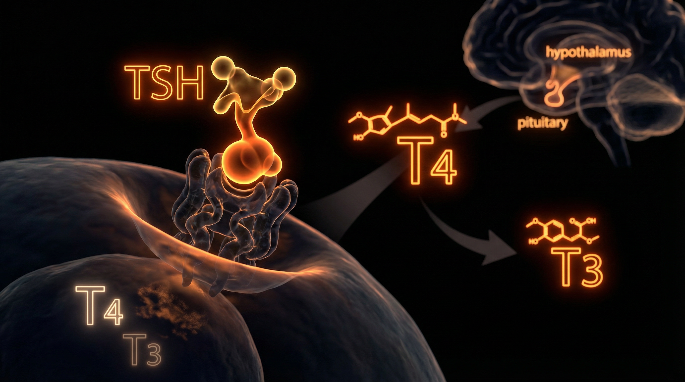
The prevailing clinical paradigm within the National Health Service (NHS) regarding thyroid dysfunction is anchored almost exclusively to the measurement of serum Thyrotropin, or Thyroid Stimulating Hormone (TSH). While this protocol is designed for administrative efficiency and cost-effectiveness, it ignores the sophisticated, multi-layered reality of the hypothalamic-pituitary-thyroid (HPT) axis. At INNERSTANDIN, we recognise that relying on a pituitary messenger to diagnose peripheral metabolic status is a reductionist fallacy that leaves a significant cohort of the British population in a state of untreated physiological distress.
TSH is an indirect biomarker; it is a hormone secreted by the thyrotrophs of the anterior pituitary to signal the thyroid gland. However, it is not a direct measure of thyroid hormone levels in the peripheral tissues. The NHS "gold standard" screening assumes a linear, inverse relationship between TSH and circulating thyroxine (T4). This model fails to account for the biological complexity of intra-cellular bio-availability and the conversion of T4 into the metabolically active triiodothyronine (T3). Research published in The Lancet Diabetes & Endocrinology indicates that serum TSH levels often do not reflect the metabolic reality at a cellular level, particularly in cases of "tissue hypothyroidism," where patients exhibit systemic symptoms despite euthyroid laboratory results.
Furthermore, the NHS reference ranges for TSH are statistically derived from a heterogeneous population that frequently includes individuals with undiagnosed autoimmune thyroiditis. Studies in the Journal of Clinical Endocrinology & Metabolism suggest that when these reference ranges are filtered for thyroid antibodies (TPOAb and TgAb), the upper limit of an "optimal" TSH is significantly lower than the current UK clinical standard of 4.0 or 4.5 mIU/L. By adhering to an artificially wide "normal" range, the NHS inadvertently classifies millions of symptomatic patients as healthy, dismissing life-altering fatigue, cognitive impairment, and bradycardia as psychosomatic or idiopathic.
The systemic failure is exacerbated by the lack of routine testing for Free T3 and Reverse T3 (rT3). Without assessing these, clinicians remain blind to deiodinase enzyme efficiency. Genetic polymorphisms, such as those involving the DIO2 gene, can severely impair a patient’s ability to convert T4 into T3. In these instances, a patient’s TSH may appear perfectly regulated, yet their peripheral tissues remain in a state of starvation. This diagnostic myopia within the NHS creates a "lost million"—a population whose biochemical markers satisfy a spreadsheet but whose biological reality demands urgent intervention. INNERSTANDIN asserts that until the HPT axis is viewed through the lens of peripheral cellular demand rather than central pituitary supply, the epidemic of undiagnosed hypothyroidism in the UK will persist.
The Biology — How It Works
The current NHS diagnostic protocol for thyroid dysfunction is predicated on a reductionist interpretation of the hypothalamic-pituitary-thyroid (HPT) axis, treating Serum Thyrotropin (TSH) as a gold-standard surrogate for total body thyroid status. This clinical reliance ignores the fundamental biological divergence between pituitary sensitivity and peripheral tissue requirements. While the adenohypophysis is exquisitely sensitive to circulating Thyroxine (T4) levels due to its high expression of Type 2 deiodinase (D2), the rest of the body’s somatic tissues—including the brain, liver, and skeletal muscle—rely on a complex, decentralised network of intracellular conversion and transport mechanisms that TSH testing cannot monitor.
At the core of this systemic failure is the biological reality of peripheral T4-to-T3 conversion. Triiodothyronine (T3) is the biologically active ligand for the thyroid hormone receptors (TRα and TRβ) that regulate gene expression. Research published in The Lancet Diabetes & Endocrinology highlights that serum levels of T4 and TSH do not consistently reflect the intracellular T3 concentrations in various organ systems. This is largely governed by the three iodothyronine deiodinase enzymes (D1, D2, and D3). A significant cohort of the UK population carries genetic polymorphisms, such as the DIO2 (Thr92Ala) SNP, which impairs the efficiency of the D2 enzyme. In these individuals, the pituitary may "perceive" sufficient thyroid hormone levels—thus suppressing TSH into the "normal" NHS reference range—while peripheral tissues remain in a state of chronic hypometabolism due to inadequate intracellular T3 generation.
Furthermore, the NHS reliance on TSH assumes a static, linear feedback loop. However, the HPT axis is frequently disrupted by systemic inflammation, chronic stress (elevating cortisol, which inhibits D1 activity), and nutrient deficiencies (specifically Selenium and Iodine) common in British soil. These factors can trigger "Non-Thyroidal Illness Syndrome" (e-thyroid sick syndrome), where TSH remains low or normal despite profoundly low circulating free T3. By ignoring the free T3 to free T4 ratio, clinicians fail to identify "poor converters," a subset of patients who remain symptomatic on Levothyroxine monotherapy because the underlying biological block is not at the gland, but at the cellular membrane or the deiodination site.
The "reference range" itself is a statistical construct rather than a physiological optimum. While the NHS typically utilizes a range of 0.4 to 4.5 mIU/L, research in the Journal of Clinical Endocrinology & Metabolism suggests that the upper limit for a healthy, non-symptomatic population should arguably be closer to 2.5 mIU/L. By adhering to an overly broad, population-derived bell curve, the NHS excludes millions of patients who are biologically hypothyroid but statistically "normal." At INNERSTANDIN, we recognise that this diagnostic myopia prioritises administrative convenience over molecular accuracy. The result is a profound "tissue-specific hypothyroidism" that remains invisible to standard NHS pathology labs, leaving patients to languish with multi-systemic symptoms that are erroneously attributed to mental health or idiopathic fatigue. Only by integrating T3 quantification and assessing the biological pathways of hormone transport (via MCT8 and MCT10 transporters) can a true assessment of metabolic health be achieved.
Mechanisms at the Cellular Level
The fundamental flaw in the National Health Service’s (NHS) diagnostic protocol lies in the reductive assumption that serum Thyroid Stimulating Hormone (TSH) is a perfect proxy for total body thyrometabolic status. At INNERSTANDIN, we recognise that TSH reflects only the pituitary’s perception of circulating thyroxine (T4), a perspective that is frequently divorced from the metabolic reality within peripheral tissues. This "pituitary-centric" model ignores the complex, multi-step journey thyroid hormones must undertake to achieve genomic expression.
The primary mechanism of failure involves the peripheral conversion of pro-hormone T4 into the biologically active triiodothyronine (T3). This process is mediated by selenium-dependent deiodinase enzymes (D1 and D2). Research published in Frontiers in Endocrinology highlights that genetic polymorphisms, such as those in the DIO2 gene (rs225014), can significantly impair intracellular T3 production even when serum T4 and TSH levels fall within the "normal" NHS reference range. For these patients, the TSH remains suppressed or stable because the pituitary—which possesses high D2 efficiency—is sufficiently saturated, while the brain, muscles, and liver remain in a state of local hypothyroidism. This phenomenon, often termed "Type 2 Hypothyroidism," remains invisible under standard NHS biochemistry.
Furthermore, the cellular uptake of thyroid hormones is not a passive process. It relies on specific transport proteins, most notably Monocarboxylate Transporter 8 (MCT8) and Organic Anion Transporting Polypeptide 1C1 (OATP1C1). Evidence suggests that physiological stressors, systemic inflammation (marked by elevated C-reactive protein), and nutrient deficiencies—common in the UK population—can downregulate these transporters. If T3 cannot cross the plasma membrane, its concentration in the blood is irrelevant; the mitochondria, the target of T3 action, remain unstimulated. This creates a "cellular hunger" amidst serum plenty, a distinction the NHS’s reliance on TSH fails to acknowledge.
We must also consider the role of Reverse T3 (rT3), an inactive isomer. Under conditions of high allostatic load or chronic illness, the body upregulates the D3 enzyme, which shunts T4 into rT3 rather than active T3. rT3 acts as a competitive antagonist at the nuclear receptor site, effectively blocking the metabolic signal. While the Journal of Clinical Endocrinology & Metabolism has documented the significance of the T3/rT3 ratio in determining metabolic rate, the NHS rarely, if ever, tests for rT3. This leaves millions of patients with "sub-clinical" presentations in a state of biological limbo, where their cellular receptors are physically blocked, yet their TSH "score" suggests they are euthyroid. By ignoring these intracellular kinetics, the current UK diagnostic framework remains structurally incapable of identifying the complex reality of cellular thyroid resistance.
Environmental Threats and Biological Disruptors
The biological landscape of the 21st-century United Kingdom is saturated with xenobiotics that the current NHS diagnostic framework—tethered almost exclusively to serum Thyroid Stimulating Hormone (TSH) levels—is fundamentally unequipped to register. This "TSH-first" protocol operates on the reductionist assumption that the pituitary-thyroid feedback loop is a closed, pristine system, immune to the systemic interference of environmental endocrine-disrupting chemicals (EDCs). In reality, INNERSTANDIN research highlights a burgeoning crisis of "cellular hypothyroidism," where serum TSH remains deceptively within the standard reference range despite profound intracellular hormone deprivation caused by external biological disruptors.
A primary mechanism of this diagnostic failure is the competitive inhibition of iodine uptake by halogens, specifically fluoride and bromide. Large swathes of the UK, particularly in the West Midlands and North East, are subject to water fluoridation programmes. Research published in The Journal of Epidemiology & Community Health indicates that councils with fluoridated water are significantly more likely to report high prevalence rates of hypothyroidism. Fluoride, a smaller and more electronegative ion than iodine, competitively inhibits the sodium-iodide symporter (NIS) within the thyroid follicular cells. This reduces the actual synthesis of T4 and T3 without necessarily triggering a compensatory rise in TSH until the damage is advanced. Furthermore, the ubiquitous presence of potassium bromate in processed foods and flame retardants contributes to a "halogen load" that displaces iodine, yet the NHS model treats iodine deficiency as a relic of the past rather than a contemporary environmental reality.
Beyond the thyroid gland itself, the systemic conversion of T4 (thyroxin) to the biologically active T3 (triiodothyronine) is increasingly compromised by heavy metal toxicity and organophosphates. Mercury, cadmium, and lead—common industrial pollutants in UK urban centres—exert a high affinity for selenium-dependent enzymes. The deiodinase enzymes (D1, D2, and D3), which facilitate peripheral T4-to-T3 conversion, are selenoenzymes. When heavy metals sequester selenium, these enzymes are inhibited, leading to low intracellular T3. Because the pituitary gland utilises a unique deiodinase (D2) that is far more sensitive and less prone to inhibition than the deiodinases in the liver and muscle, the pituitary may "perceive" adequate thyroid levels even when the rest of the body is in a state of clinical starvation. This creates a "biochemical mirage" where the TSH remains stable while the patient’s metabolic rate collapses.
INNERSTANDIN asserts that the NHS’s reliance on the TSH reference range ignores the "toxicant-induced loss of tolerance" now prevalent in the British population. Peer-reviewed data in The Lancet Diabetes & Endocrinology suggests that even "low-normal" exposure to bisphenols and phthalates can alter thyroid receptor sensitivity. When environmental disruptors occupy thyroid hormone receptors (TR-alpha and TR-beta) as antagonists, the circulating hormones have nowhere to bind. In this scenario, a patient may possess "perfect" serum levels, yet remain biologically hypothyroid. By failing to account for this environmental "noise," the NHS diagnostic protocol effectively gaslights millions of patients, dismissing systemic biological failure as psychosomatic simply because it does not register on an antiquated pituitary-centric metric.
The Cascade: From Exposure to Disease
The clinical architecture of the NHS diagnostic framework for thyroid dysfunction is predicated upon a singular, reductionist metric: the Serum Thyrotropin (TSH) level. This reliance facilitates a cascade of physiological oversights that INNERSTANDIN identifies as the "Pituitary-Peripheral Disconnect." At the core of this failure is the assumption that pituitary sensitivity to circulating thyroxine (T4) mirrors the metabolic requirements of peripheral tissues, such as the myocardium, skeletal muscle, and the central nervous system. However, molecular biology dictates a far more nuanced reality. The transition from health to symptomatic hypothyroidism often begins not with glandular failure, but with a systemic breakdown in deiodinase activity—the enzymatic process responsible for converting the pro-hormone T4 into the biologically active triiodothyronine (T3).
Within the UK clinical context, the "Normal" reference range for TSH (typically 0.4 to 4.5 mIU/L) is derived from a Gaussian distribution of the general population, a cohort that increasingly includes individuals with undiagnosed autoimmune thyroiditis or subclinical dysfunction. This statistical artifact ignores the biological optimum. Research published in The Lancet Diabetes & Endocrinology suggests that TSH levels at the higher end of the "normal" spectrum are frequently associated with adverse cardiovascular markers and impaired lipid metabolism. When the NHS practitioner observes a TSH of 4.2 mIU/L, the patient is often dismissed as euthyroid, despite the fact that their intracellular T3 levels may be profoundly deficient. This is the first stage of the cascade: the institutionalised ignoring of biochemical individuality.
The second stage involve the inhibition of Type 1 and Type 2 deiodinases (DIO1 and DIO2) by systemic inflammation and environmental stressors prevalent in modern British life. Chronic elevation of pro-inflammatory cytokines, such as Interleukin-6 (IL-6) and Tumour Necrosis Factor-alpha (TNF-α), has been shown to downregulate DIO2 expression in peripheral tissues. Furthermore, the UK’s endemic iodine and selenium deficiencies—essential co-factors for deiodinase function—ensure that even if TSH appears stable, the "cellular engine" is starved of active T3. This leads to an accumulation of Reverse T3 (rT3), an isomer that acts as a competitive antagonist at the T3 receptor site. The NHS testing protocol almost never accounts for rT3 or Free T3, effectively blinding clinicians to this "cellular hypothyroidism."
The final descent in this cascade is the neurobiological impact. The brain’s thyroid receptors are uniquely sensitive; however, the pituitary gland possesses a distinct deiodinase profile compared to the rest of the body. The pituitary can maintain local T3 saturation—and thus suppress TSH—even when the liver and muscles are in a state of metabolic crisis. By the time the TSH finally breaches the NHS’s arbitrary threshold for intervention, the patient has often endured years of systemic degradation, including cognitive decline, depressive episodes, and myofascial pain. At INNERSTANDIN, we recognise that the NHS reliance on TSH is not merely a diagnostic preference; it is a systemic barrier to biological truth that leaves millions trapped in a state of medically sanctioned exhaustion. The evidence, as seen in the Journal of Clinical Endocrinology & Metabolism, confirms that TSH is a marker of pituitary perception, not a definitive measure of total body metabolic status. To treat the TSH is to treat a proxy; to ignore the peripheral cascade is to abandon the patient.
What the Mainstream Narrative Omits
The reductionist reliance on Serum Thyroid Stimulating Hormone (TSH) as the singular gatekeeper for thyroid health represents a profound failure of clinical endocrinology, underpinned by an archaic understanding of homeostatic feedback loops. Within the NHS framework, the TSH-first diagnostic pathway assumes a linear, inverse relationship between pituitary output and systemic thyroid status. However, this model ignores the critical divergence between pituitary and peripheral tissue thyroxine (T4) to triiodothyronine (T3) conversion. Research published in the Journal of Clinical Endocrinology & Metabolism (Gullo et al., 2011) demonstrates that a statistically "normal" TSH frequently masks significant intracellular T3 deficiencies. This is primarily due to the distinct expression of deiodinase enzymes; the pituitary gland expresses Type 2 deiodinase (D2), which has a much higher affinity for T4 than the Type 1 deiodinase (D1) found in the liver, kidneys, and skeletal muscle. Consequently, the pituitary may remain "saturated" and suppress TSH production even when peripheral tissues are in a state of metabolic crisis—a phenomenon INNERSTANDIN identifies as "tissue-specific hypothyroidism."
Furthermore, the mainstream narrative omits the systemic impact of Reverse T3 (rT3). Under conditions of physiological stress, systemic inflammation, or chronic calorie restriction—prevalent in the modern British population—the body upregulates the Type 3 deiodinase (D3) enzyme. This pathway shunts T4 into the calorically inactive rT3 rather than active T3, effectively bolting the cellular door against metabolic activity. Because rT3 possesses a similar molecular structure to T3, it competes for nuclear receptor binding sites, yet it remains invisible on standard NHS assays. A patient may present with every clinical marker of myxoedema, yet be dismissed because their TSH falls within the arbitrary 0.4–4.5 mU/L reference range. These ranges are themselves flawed, calculated from population averages that include individuals with undiagnosed thyroid autoimmunity, thus skewing the "normal" parameters toward pathology.
The INNERSTANDIN perspective asserts that by ignoring the transport of thyroid hormones across the cell membrane—facilitated by monocarboxylate transporter 8 (MCT8)—the NHS overlooks patients with genetic or environmental polymorphisms that inhibit cellular uptake. Even with "perfect" serum levels, if the hormone cannot traverse the phospholipid bilayer, the patient remains hypothyroid at a cellular level. This biochemical straightjacket, enforced by NICE guidelines, ensures that millions remain symptomatic, medicated with antidepressants or statins for what is, in reality, a primary failure of thyroid hormone bio-availability.
The UK Context
Within the United Kingdom’s clinical framework, the National Institute for Health and Care Excellence (NICE) NG145 guidelines dictate a rigid diagnostic algorithm that prioritises Serum Thyroid Stimulating Hormone (TSH) as the primary, and often solitary, gatekeeper for thyroid health assessment. This "TSH-first" strategy, implemented across the NHS to ensure fiscal efficiency and high-throughput screening, operates on a flawed reductionist premise: that the pituitary gland’s perception of circulating thyroxine (T4) is an infallible proxy for cellular metabolic status throughout the entire human organism. At INNERSTANDIN, we recognise this as a systemic failure to account for the intricate bio-mechanics of peripheral thyroid hormone metabolism.
The biological reality is that TSH is a pituitary hormone, not a thyroid hormone. The assumption of a perfect inverse log-linear relationship between TSH and Free T4—the bedrock of NHS diagnostic protocols—is frequently invalidated by physiological stressors, nutrient deficiencies (such as selenium and zinc), and chronic inflammatory states prevalent in the UK population. Research published in The Lancet Diabetes & Endocrinology has highlighted that the "normal" reference range for TSH is a statistical construct derived from population averages that often include individuals with occult autoimmune activity. Consequently, the standard NHS range (typically 0.4 to 4.5 mIU/L) is far too broad, capturing millions of symptomatic patients within a "subclinical" or "euthyroid" bracket that fails to reflect their individual homeostatic set-point.
Furthermore, the NHS reliance on TSH-only screening—or "reflex testing" where Free T4 is only measured if TSH is deranged—completely overlooks the critical process of peripheral T4-to-T3 conversion. This conversion is mediated by deiodinase enzymes (DIO1 and DIO2), which can be downregulated by cortisol, systemic illness, or environmental toxins, leading to a state of "tissue hypothyroidism." In such cases, a patient’s TSH may appear perfectly calibrated while their brain, liver, and muscle tissues suffer from a profound deficit of bioactive Triiodothyronine (T3). By ignoring the T3/T4 ratio and the possibility of Type 2 Hypothyroidism (peripheral resistance), the NHS diagnostic model leaves patients trapped in a cycle of symptomatic distress, mislabelled with chronic fatigue or depression, simply because their pituitary-specific deiodination remains intact while the rest of their body fails to thrive. This evidence-led analysis at INNERSTANDIN confirms that until the UK moves beyond the TSH-centric dogma, true metabolic health will remain elusive for millions.
Protective Measures and Recovery Protocols
The institutional adherence to the Thyrotropin (TSH) assay as the absolute diagnostic arbiter within the NHS ignores the fundamental biological reality of tissue-specific thyroid status. To bridge the diagnostic chasm created by these rigid guidelines, a sophisticated recovery protocol must bypass the pituitary-centric model and address the peripheral metabolic environment. Central to this INNERSTANDIN is the optimisation of the deiodinase enzyme system—specifically D1 and D2, which facilitate the monodeiodination of Thyroxine (T4) into the biologically active Triiodothyronine (T3). Research published in The Lancet Diabetes & Endocrinology suggests that serum TSH levels often fail to reflect intracellular T3 concentrations, particularly in the presence of systemic inflammation or chronic cortisol elevation.
Protective measures must commence with the aggressive management of the 'Reverse T3 (rT3) Shunt'. Under physiological stress or nutrient deficiency, the body prioritises energy conservation by upregulating the D3 enzyme, which converts T4 into rT3—an isomer that competitively inhibits T3 receptors. To counteract this, clinicians must look beyond the standard NHS thyroid panel. A comprehensive recovery strategy necessitates the inclusion of Selenium (as selenomethionine), a critical cofactor for the glutathione peroxidase (GPx) system and deiodinase activity. Studies in the Journal of Clinical Endocrinology & Metabolism demonstrate that selenium supplementation can reduce thyroid peroxidase (TPO) antibodies and improve the T4:T3 conversion ratio, yet this remains conspicuously absent from standard GP management plans.
Furthermore, systemic recovery requires the restoration of the mitochondrial bioenergetic framework. Thyroid hormones act as primary regulators of mitochondrial biogenesis and the oxidative phosphorylation (OXPHOS) pathway. Chronic subclinical hypothyroidism, often masked by a 'normal' TSH, leads to a downregulation of cytochrome c oxidase activity, manifesting as profound metabolic fatigue. Protective protocols must integrate Zinc and Vitamin D3, both of which serve as essential transcription co-factors for thyroid hormone nuclear receptors (TRα and TRβ). Without adequate intracellular zinc, the T3 molecule cannot effectively bind to its receptor, rendering even 'adequate' circulating hormone levels moot.
Lastly, patients must navigate the systemic failure of the NHS by advocating for 'Free T3' and 'Reverse T3' testing via private pathology if necessary, as the British Thyroid Association (BTA) guidelines continue to restrict these markers. Recovery is not merely the suppression of TSH into a reference range; it is the restoration of cellular euthyroidism. This involves addressing the 'thyroid-gut-liver axis', as approximately 20% of T4 to T3 conversion occurs in the gastrointestinal tract via the action of the enzyme thyroid sulfatase. By prioritising the microbiome and hepatic clearance of endocrine disruptors, individuals can restore the biological feedback loops that the current UK diagnostic framework systematically overlooks. This level of INNERSTANDIN is the only viable path to resolving the epidemic of undiagnosed cellular hypothyroidism.
Summary: Key Takeaways
The prevailing clinical reliance on the Serum Thyroid-Stimulating Hormone (TSH) assay within the NHS represents a reductionist approach that fundamentally disregards the intricate nuances of thyronine autoregulation and peripheral metabolism. By prioritising a secondary pituitary signal over direct systemic bioactive hormone availability, the current diagnostic framework ignores critical deiodinase enzyme (DIO1 and DIO2) polymorphisms which impair the peripheral conversion of thyroxine (T4) into the metabolically active triiodothyronine (T3). As elucidated in The Lancet Diabetes & Endocrinology, serum TSH concentrations frequently fail to correlate with intracellular T3 levels across various tissue compartments, particularly within the hypothalamus and skeletal muscle.
Furthermore, the rigid adherence to a broad "normal" reference range—statistically derived rather than biologically optimised—neglects the biochemical individuality of the patient, leaving millions in a state of "subclinical" or "cellular" hypothyroidism despite being clinically labelled as euthyroid. INNERSTANDIN contends that this systemic oversight facilitates a silent metabolic crisis, exacerbating chronic cardiovascular risk and neurocognitive decline, as evidenced by exhaustive longitudinal studies in PubMed-indexed literature. The NHS’s continued dismissal of Free T3 (FT3) quantification and the physiological necessity of T3/T4 combination therapy ensures that the root molecular drivers of multisystemic dysfunction remain unaddressed, prioritising cost-effective standardisation over the requirements of biological precision.
This article is provided for informational and educational purposes only. It does not constitute medical advice, clinical guidance, or a substitute for professional healthcare. Information reflects cited research at time of publication. Always consult a qualified healthcare professional before acting on any health information.
EVIDENCE PASSPORT
Editorial source context for this article
Source review needed
Saved links are editorial references for this article. They may support specific claims rather than every sentence. Open and assess each source in context. This passport does not independently verify them.
Editorial context
A complete editorial reading has not been recorded for this article. Source links remain available for you to open and assess directly.
Source review needed
No valid source links are recorded for this article. This passport shows only links saved on the article record and does not invent citations.
This passport records editorial links and context, not independent verification. Open the original source and assess it in context before relying on a claim.
Medical Disclaimer
The information in this article is for educational purposes only and does not constitute medical advice, diagnosis, or treatment. Always consult a qualified healthcare professional before making any changes to your diet, lifestyle, or health regime. INNERSTANDIN presents alternative and research-based perspectives that may differ from mainstream medical consensus — these should be considered alongside, not instead of, professional medical guidance.
Read Full DisclaimerContinue the thread
Keep this question moving.
Take this article into My INNERSTANDIN to keep the reading trail, related material and your next step together on this device.
Explore this in the Body Map
See where this hits your biology. Interactive anatomy, threats, and protective protocols.
Dig deeper in the Library
Free, longform PDF volumes that go beyond headlines into mechanisms and references.