Oxidative Stress and the Breakdown of the Pericardial Mesothelial Barrier
Discuss this resourceUpdated August 2026
An in-depth exploration of how oxidative stress compromises the pericardial mesothelium, leading to inflammation and systemic cardiac vulnerability.
Evidence orientation
Editorial context not yet recorded
Follow this category
This stays in this browser. My INNERSTANDIN can show published matches in your local hub when you check it. It does not send email, push, or alert notifications.
Local learning review
A private browser aid for revisiting ideas. It is not an alert or a health recommendation.
Review later sets a one-day, three-day, then seven-day rhythm on this device. Choose it only when you want to revisit this article.

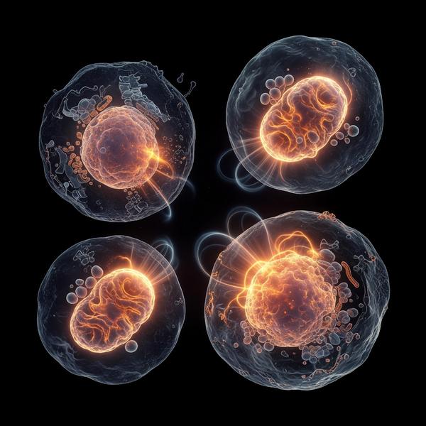
The pericardium is far more than a simple protective sac for the heart; it is a complex, biologically active organ essential to cardiovascular homeostasis. Central to its function is the mesothelial layer—a single sheet of specialized cells that produce lubricative fluid, reducing friction during the cardiac cycle. When oxidative stress disrupts this delicate barrier, the consequences reach far beyond localized irritation, potentially triggering chronic systemic inflammation.
The Role of Oxidative Stress
Oxidative stress occurs when there is an imbalance between the production of reactive oxygen species (ROS) and the body's ability to neutralize them via antioxidant defenses. Within the pericardial space, ROS are often produced by infiltrating immune cells or metabolic dysfunction. When these molecules accumulate, they initiate lipid peroxidation, damaging the cell membranes of the mesothelial lining.
Mechanisms of Barrier Breakdown
The mesothelial cells act as a semi-permeable gatekeeper. Under oxidative insult, tight junction proteins such as occludin and claudin are downregulated or degraded. This 'leaky' barrier allows inflammatory cytokines, fibrous proteins, and inflammatory cells to infiltrate the pericardial space. This process often marks the transition from acute irritation to chronic adhesive pericarditis, where the heart becomes tethered by fibrotic adhesions, significantly impairing its filling and pumping mechanics.
The Root Cause Perspective
At INNERSTANDING, we look beyond the symptoms. Oxidative stress in the pericardium is rarely an isolated event. It is often fueled by systemic metabolic dysregulation, chronic low-grade inflammation, or environmental toxic burdens that the body cannot adequately process. Mitochondrial dysfunction within the mesothelial cells themselves reduces their regenerative capacity, creating a self-perpetuating cycle of cellular stress.
Restoring Mesothelial Integrity
Healing requires a multifaceted approach focused on reducing the oxidative burden and supporting cellular repair pathways. This includes stabilizing metabolic health, optimizing antioxidant status, and managing systemic inflammatory triggers. By reducing the ROS load at the root, the body can begin the essential work of repairing the mesothelial barrier, restoring the frictionless environment the heart requires for optimal performance.
This article is provided for informational and educational purposes only. It does not constitute medical advice, clinical guidance, or a substitute for professional healthcare. Information reflects cited research at time of publication. Always consult a qualified healthcare professional before acting on any health information.
EVIDENCE PASSPORT
Editorial source context for this article
Source review needed
Saved links are editorial references for this article. They may support specific claims rather than every sentence. Open and assess each source in context. This passport does not independently verify them.
Editorial context
A complete editorial reading has not been recorded for this article, and no source links are saved on its record.
Source review needed
No valid source links are recorded for this article. Supplementary research, where available, is listed separately below.
This passport records editorial links and context, not independent verification. Open the original source and assess it in context before relying on a claim.
Medical Disclaimer
The information in this article is for educational purposes only and does not constitute medical advice, diagnosis, or treatment. Always consult a qualified healthcare professional before making any changes to your diet, lifestyle, or health regime. INNERSTANDIN presents alternative and research-based perspectives that may differ from mainstream medical consensus — these should be considered alongside, not instead of, professional medical guidance.
Read Full DisclaimerContinue the thread
Keep this question moving.
Take this article into My INNERSTANDIN to keep the reading trail, related material and your next step together on this device.
Explore this in the Body Map
Explore related body structures and their educational pages in the interactive Body Map.
Dig deeper in the Library
Free, longform PDF volumes that go beyond headlines into mechanisms and references.
RABBIT HOLE
Follow the biological thread deeper