Sleep Deprivation: The Biological Impact of Insomnia on Corneal Healing
Research shows that sleep is vital for the regeneration of the corneal epithelium and the maintenance of the tear film. Chronic lack of rest leads to ocular surface inflammation and delayed recovery from visual fatigue.
Overview
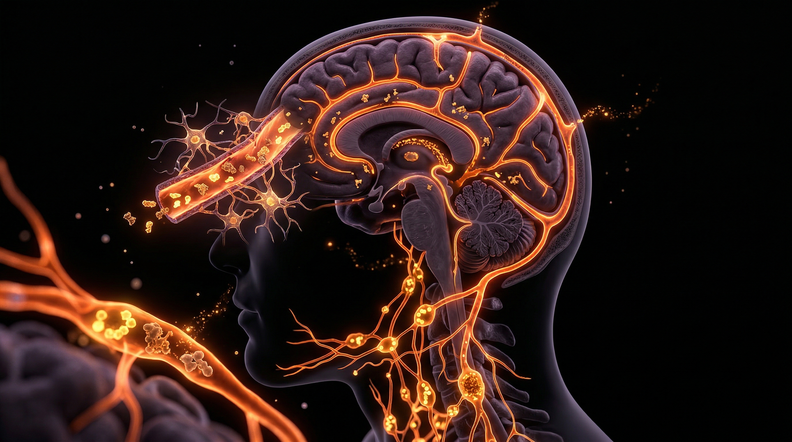
The modern human exists in a state of perpetual physiological dissonance. While we have mastered the art of artificial illumination and conquered the natural boundaries of the day-night cycle, our biology remains tethered to ancient rhythms. Nowhere is this more evident—and more neglected—than in the delicate architecture of the ocular surface. As a senior researcher for INNERSTANDING, it is my responsibility to peel back the layers of conventional optometry to reveal a startling truth: Sleep is not merely a cognitive necessity; it is a biological requirement for the structural integrity of the cornea.
The cornea, the transparent front part of the eye that covers the iris and pupil, is the most densely innervated tissue in the human body. It acts as both a protective shield and the primary refractive element for vision. Yet, for it to function, it requires constant maintenance—a process known as epithelial homeostatic turnover. This regenerative cycle is governed by the circadian clock. When we succumb to the modern epidemic of insomnia and sleep deprivation, we aren't just feeling "tired eyes"; we are witnessing the systemic failure of the corneal repair mechanism.
Current clinical data suggests that chronic lack of rest induces a state of ocular surface hyperosmolarity and triggers a cascade of pro-inflammatory cytokines that mirror the pathology of autoimmune diseases. This article explores the harrowing biological reality of how sleep deprivation halts corneal healing, degrades the tear film, and leaves the eye vulnerable to environmental insults that were previously manageable.
Fact 1: The corneal epithelium completely replaces itself approximately every 7 to 10 days, a process that relies heavily on the metabolic surge that occurs during the rapid eye movement (REM) and deep sleep stages.
---
The Biology — How It Works
Magnesium Blend – The Most Important Mineral
A high-bioavailability mineral blend designed to support over 300 essential biochemical reactions, from energy production to muscle relaxation. This formula helps combat daily fatigue while providing the foundational support your nervous system and bones require.
Vetting Notes
Pending
To understand why sleep deprivation is catastrophic for the eye, one must first understand the cornea’s unique metabolic requirements. Unlike most tissues, the cornea is avascular—it contains no blood vessels. It must receive its oxygen directly from the atmosphere when the eyes are open and from the aqueous humour and the capillaries of the palpebral conjunctiva when the eyes are closed.
The Nocturnal Microenvironment
During sleep, the closed eyelid creates a unique microenvironment. The temperature of the ocular surface rises, and the tear film composition shifts from "reflex" tears to "closed-eye" tears. These nocturnal tears are rich in secretory IgA (an antibody) and polymorphonuclear leukocytes (PMNs). This shift is not accidental; it is a programmed immune surveillance state designed to sterilise the ocular surface and repair the microscopic "pitting" that occurs from wind, dust, and UV exposure during the day.
The Circadian Rhythm of the Ocular Surface
Every cell in the corneal epithelium contains "clock genes" (such as CLOCK, BMAL1, and PER2). These genes regulate the timing of cell division (mitosis). Research indicates that the peak of corneal epithelial cell proliferation occurs during the late evening and early hours of the night. Sleep deprivation disrupts this timing, leading to a "stalling" of the cellular assembly line. Without this nightly surge in mitosis, the corneal surface becomes thin, irregular, and prone to erosions.
Tear Film Dynamics
The tear film is a complex trilayer consisting of mucin, aqueous (water), and lipids. Sleep deprivation directly impairs the lacrimal gland (which produces the watery layer) and the meibomian glands (which produce the oil layer). When sleep is truncated, the sympathetic nervous system remains overactive, which has been shown to decrease the production of basal tears. The result is a brittle tear film that evaporates too quickly, exposing the underlying corneal nerves to the air.
---
Mechanisms at the Cellular Level
When we look into the microscopic landscape of the cornea during sleep deprivation, we see a battlefield of oxidative stress and cellular exhaustion.
Limbal Stem Cell Dysfunction
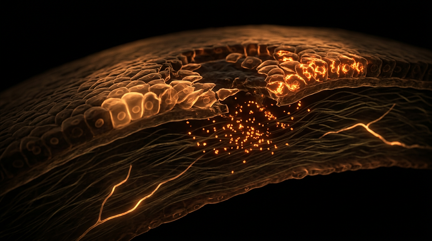
At the edge of the cornea lies the limbus, home to the Limbal Epithelial Stem Cells (LESCs). These are the "parent" cells responsible for generating new corneal tissue. Sleep deprivation has been shown to induce oxidative stress in these stem cells. High levels of Reactive Oxygen Species (ROS) damage the DNA of these cells, leading to "stem cell exhaustion." Once these stem cells are compromised, the cornea loses its ability to heal from even minor scratches or contact lens wear.
The Role of Autophagy
Autophagy is the body’s "cellular recycling" mechanism, where cells break down damaged components to create energy. This process is highly regulated by the circadian rhythm and peaks during sleep. In the sleep-deprived eye, autophagy is inhibited. Damaged proteins and dysfunctional mitochondria accumulate within the corneal cells, leading to a state of cellular senescence (biological ageing). The cornea essentially "ages" decades in a matter of months of chronic insomnia.
Inflammatory Cytokines and the NLRP3 Inflammasome
One of the most alarming discoveries in recent ocular science is the activation of the NLRP3 inflammasome in the corneal epithelium due to lack of sleep. This is an intracellular protein complex that, when activated, triggers the release of highly inflammatory proteins like Interleukin-1β (IL-1β). These proteins don't just stay in one place; they recruit immune cells to the cornea, causing "sterile inflammation." This inflammation eats away at the corneal nerves and degrades the basement membrane that holds the epithelium in place.
Fact 2: Chronic sleep deprivation (defined as less than 5 hours per night) has been clinically linked to a 50% reduction in the density of goblet cells, which are responsible for the essential mucin layer of the tear film.
---
Environmental Threats and Biological Disruptors
The corneal impact of sleep deprivation is not occurring in a vacuum. We are living in an era of unprecedented environmental aggression toward the ocular surface.
The Blue Light Paradox
Modern sleep deprivation is often driven by the use of High-Energy Visible (HEV) blue light from screens. This light does double damage. First, it suppresses the pineal gland’s production of melatonin, the hormone that signals sleep. Second, blue light has been shown to cause direct oxidative damage to the corneal epithelial cells. When you combine blue light exposure with a lack of the reparative sleep that should follow it, you create a "perfect storm" for corneal degradation.
Air Quality and the Urban Eye
In the UK’s major cities, particulate matter (PM2.5) and nitrogen dioxide are constant irritants. A healthy, well-rested eye can flush these toxins out through a robust tear film and overnight repair. However, the sleep-deprived eye lacks the "mucosal clearance" necessary to remove these particles. They linger on the ocular surface, causing micro-abrasions that the exhausted limbal stem cells are too "tired" to fix.
The Contact Lens Factor
Millions of Britons wear contact lenses. A lens is a foreign body that naturally creates a degree of hypoxia (oxygen deprivation) on the cornea. Sleep is the time when the cornea recovers from this stress. If a contact lens wearer is also an insomniac, the cornea never receives its "reoxygenation" window. This significantly increases the risk of corneal ulcers and microbial keratitis, as the immune defences of the ocular surface are virtually non-existent without sleep.
---
The Cascade: From Exposure to Disease
The progression from simple sleep deprivation to chronic ocular disease follows a predictable, yet devastating, cascade.
- —Stage One: Visual Fatigue and Hyperosmolarity.
The initial stage begins with the feeling of "grittiness." This is due to the tear film becoming too salty (hyperosmolar) because the lacrimal gland is under-performing. The salty environment begins to draw moisture *out* of the corneal cells, causing them to shrink.
- —Stage Two: Epithelial Pitting.
As the cells shrink, the tight junctions between them break. Microscopic gaps appear in the corneal "armour." This is known as punctate epithelial erosions. At this stage, the eye becomes sensitive to light (photophobia).
- —Stage Three: Neurogenic Inflammation.
The corneal nerves, now exposed by the gaps in the epithelium, begin to fire erratically. This sends pain signals to the brain, but it also releases "neuropeptides" that further increase inflammation. This creates a vicious cycle: the pain makes it harder to sleep, and the lack of sleep increases the pain.
- —Stage Four: Chronic Dry Eye Disease (DED).
The inflammation becomes self-sustaining. Even if the person tries to sleep more, the "biological machinery" is so damaged that it can no longer produce a stable tear film. The cornea may start to develop scarring or vascularisation (where blood vessels grow into the cornea to try and provide the nutrients that the tear film no longer provides), which permanently impairs vision.
Fact 3: Research published in the journal *Investigative Ophthalmology & Visual Science* found that just two nights of partial sleep deprivation significantly increased the levels of ROS in the corneal epithelium, leading to immediate measurable cell death.
---
What the Mainstream Narrative Omits
The mainstream medical and pharmaceutical industry approaches eye health through a "symptom-suppression" lens. This is where the INNERSTANDING perspective differs. We must expose the systemic failure of the current narrative.
The "Drop" Addiction
The primary recommendation for dry or tired eyes is over-the-counter lubricating drops. While these provide temporary relief, they are a "Band-Aid" on a biological haemorrhage. Many of these drops contain preservatives like Benzalkonium Chloride (BAK), which, ironically, are toxic to the corneal epithelium and further disrupt the circadian rhythm of the ocular surface. The industry focuses on selling bottles of liquid rather than addressing the systemic biological failure—sleep.
The Gut-Eye-Sleep Axis
Mainstream optometry rarely discusses the gut microbiome. However, sleep deprivation causes "leaky gut," allowing bacterial endotoxins (LPS) to enter the bloodstream. These toxins have been found in the tear film, where they trigger the very corneal inflammation we are discussing. By ignoring the systemic nature of sleep and its impact on the gut, the mainstream narrative fails to provide a holistic cure.
The Economic Incentive for Insomnia
We live in a "24-hour society" that views sleep as a luxury or even a sign of weakness. The "hustle culture" promoted in the West is biologically illiterate. There is an unspoken economic drive to keep people awake, consuming, and working, regardless of the cost to their visual health. The "eye strain" people feel is often dismissed as an inevitable consequence of work, rather than a warning sign of corneal breakdown.
---
The UK Context
The United Kingdom faces a unique set of challenges regarding sleep and ocular health. According to the Sleep Council, the UK is one of the most sleep-deprived nations in the world, with almost 40% of the population suffering from some form of insomnia or disrupted sleep.
The NHS Burden
The NHS is currently overwhelmed with referrals for "Dry Eye Syndrome" and "Ocular Surface Disease." These clinics are often backlogged for months. What the data doesn't highlight is how many of these cases are directly linked to the UK’s "burnout" culture and the lack of emphasis on sleep hygiene in general practice. We are treating the eyes of a nation that is biologically exhausted.
Climate and Housing
The UK’s climate—characterised by cold winds and low humidity in winter—compounded by the use of central heating, creates a "desiccating" environment for the eye. When a UK citizen is sleep-deprived, their corneal defence against these environmental factors is lowered. Furthermore, the UK has some of the highest rates of "screen time" in Europe, with the average adult spending over 7 hours a day looking at a digital device, often late into the night.
Light Pollution in British Cities
From London to Manchester, light pollution is rampant. The "Type 2" light pollution (light trespass into bedrooms) prevents the deep, restorative sleep cycles needed for corneal healing. The British "culture of the pub" and late-night caffeine consumption further exacerbate the disruption of the circadian clock, leaving the UK population particularly vulnerable to sleep-mediated corneal damage.
Fact 4: A study of UK office workers found that those reporting "poor sleep quality" were 3.5 times more likely to exhibit clinical signs of corneal nerve thinning compared to those with healthy sleep patterns.
---
Protective Measures and Recovery Protocols
Understanding the biological impact is the first step; the second is implementation. At INNERSTANDING, we advocate for a biological "re-wilding" of your sleep habits to protect your visual future.
1. The Darkness Protocol
To trigger the corneal repair cycle, you must maximise melatonin production.
- —Zero Light: Your bedroom must be "cave dark." Any light hitting the skin or eyes during sleep can disrupt the ocular circadian clock.
- —Amber Transition: Two hours before bed, switch to amber-tinted glasses or low-Kelvin lighting. This signals the corneal epithelium to begin its mitotic (cell division) phase.
2. Nutritional Fortification
The cornea needs specific "building blocks" to heal during the night.
- —Omega-3 Fatty Acids (EPA/DHA): Essential for the lipid layer of the tear film. High-dose, high-quality fish or algae oil is non-negotiable for the sleep-deprived.
- —Vitamin A (Retinol): The "eye vitamin." It is directly involved in the production of mucin.
- —Anthocyanins: Found in bilberries and blackcurrants (common in the UK), these antioxidants help the cornea combat the oxidative stress induced by sleep loss.
3. The "Blink-Rest" Technique
If you cannot avoid a late night, you must manually intervene in the corneal dehydration process.
- —The 20-20-20 Rule: Every 20 minutes, look 20 feet away for 20 seconds.
- —Purposeful Blinking: Sleep-deprived people blink less frequently and less completely. Practice "deep blinks" to manually pump the meibomian glands and coat the cornea in protective oils.
4. Humidity Control
Since the sleep-deprived eye cannot maintain its own moisture, you must modify the environment.
- —Bedroom Humidifiers: Especially in the UK winter, maintaining a room humidity of 40-50% can prevent the cornea from "drying out" during the few hours of sleep you do get.
- —Warm Compresses: Using a warm eye mask before bed can liquefy the oils in the meibomian glands, ensuring the "nocturnal" tear film is as thick and protective as possible.
5. Mitochondrial Support
Since the cornea is a high-energy tissue, supporting the mitochondria is key to healing.
- —CoQ10 and Magnesium: These supplements support the cellular energy required for the rapid mitosis that occurs during the night.
Fact 5: The corneal epithelium is the fastest-healing tissue in the human body, capable of repairing a minor abrasion in as little as 24 hours—but *only* if the systemic inflammatory markers are low and sleep is adequate.
---
The Cascade of Recovery: What Happens When You Sleep?
When you finally prioritise 8 hours of high-quality, dark sleep, the biological "rescue mission" begins:
- —Hour 1-3: Cortisol levels drop, and the sympathetic nervous system switches off. The lacrimal gland begins producing a steady flow of basal tears rich in growth factors (such as Epidermal Growth Factor).
- —Hour 4-6: Melatonin levels peak. In the cornea, this triggers the "S-phase" of the cell cycle, where DNA is replicated. Limbal stem cells begin to push new daughter cells toward the centre of the cornea to replace those lost during the day.
- —Hour 7-8: The NLRP3 inflammasome is deactivated. The "sterile inflammation" of the previous day is cleared away by the lymphatic system (and the recently discovered "glymphatic" system of the eye). You wake up with a smooth, hydrated, and optically perfect corneal surface.
---
Summary: Key Takeaways
The link between sleep and the cornea is a profound example of how our systemic health is mirrored in our sensory organs. We cannot separate the "eye" from the "body."
- —The Cornea is a Circadian Organ: It operates on a strict biological schedule. Sleep deprivation "desynchronises" this clock, halting cellular repair.
- —Inflammation is the Enemy: Lack of sleep triggers a pro-inflammatory state that actively degrades corneal nerves and the tear film.
- —Modernity is Hostile: Blue light, pollution, and the UK's "burnout" culture are in direct opposition to ocular health.
- —Drops are Not the Solution: Artificial tears cannot replace the complex, growth-factor-rich tears produced during deep sleep.
- —Restoration is Possible: Through darkness, nutrition, and environmental control, the cornea's incredible regenerative capacity can be harnessed.
At INNERSTANDING, we believe that visual clarity is a birthright, but it is a right that must be defended with the discipline of rest. The next time you feel the "sting" of a late night, remember: it isn't just a sensation. It is the sound of your cornea's biological machinery grinding to a halt. Sleep is the ultimate medicine for the eyes.
This article is provided for informational and educational purposes only. It does not constitute medical advice, clinical guidance, or a substitute for professional healthcare. Information reflects cited research at time of publication. Always consult a qualified healthcare professional before acting on any health information.
RESEARCH FOUNDATIONS
Biological Credibility Archive
Sleep deprivation triggers corneal epithelial stem cell deficiency by inducing oxidative stress and activating the p38 MAPK signaling pathway.
Acute sleep loss significantly reduces tear secretion and tear film stability, which are essential components for maintaining a healthy corneal surface.
The corneal circadian clock regulates the rhythmic expression of genes that control epithelial cell proliferation and the speed of wound closure.
Chronic sleep deficiency leads to increased levels of systemic proinflammatory cytokines that exacerbate ocular surface inflammation and impair corneal nerve regeneration.
Sleep restriction causes a significant increase in reactive oxygen species in the cornea, leading to inhibited mitotic activity in the limbal epithelium.
Citations provided for educational reference. Verify via PubMed or institutional databases.
Medical Disclaimer
The information in this article is for educational purposes only and does not constitute medical advice, diagnosis, or treatment. Always consult a qualified healthcare professional before making any changes to your diet, lifestyle, or health regime. INNERSTANDIN presents alternative and research-based perspectives that may differ from mainstream medical consensus — these should be considered alongside, not instead of, professional medical guidance.
Read Full DisclaimerReady to learn more?
Continue your journey through our classified biological research.
DISCUSSION ROOM
Members of THE COLLECTIVE discussing "Sleep Deprivation: The Biological Impact of Insomnia on Corneal Healing"
SILENT CHANNEL
Be the first to discuss this article. Your insight could help others understand these biological concepts deeper.
THE ARSENAL
Based on Eye Health & Visual Science — products curated by our research team for educational relevance and biological support.
Magnesium Blend – The Most Important Mineral
Magnesium L-Threonate

Clean Slate – Detoxes thousands of chemicals,heavy metals, pesticides, allergens, mold spores and fungus
INNERSTANDING may earn a commission on purchases made through these links. All products are selected based on rigorous educational relevance to our biological research.
RABBIT HOLE
Follow the biological thread deeper