The Structural Vulnerability of the Human Hippocampus
Environmental neurotoxins are disproportionately affecting the anatomical volume of the brain's memory centres. We explore the link between urban living and hippocampal atrophy.
Overview
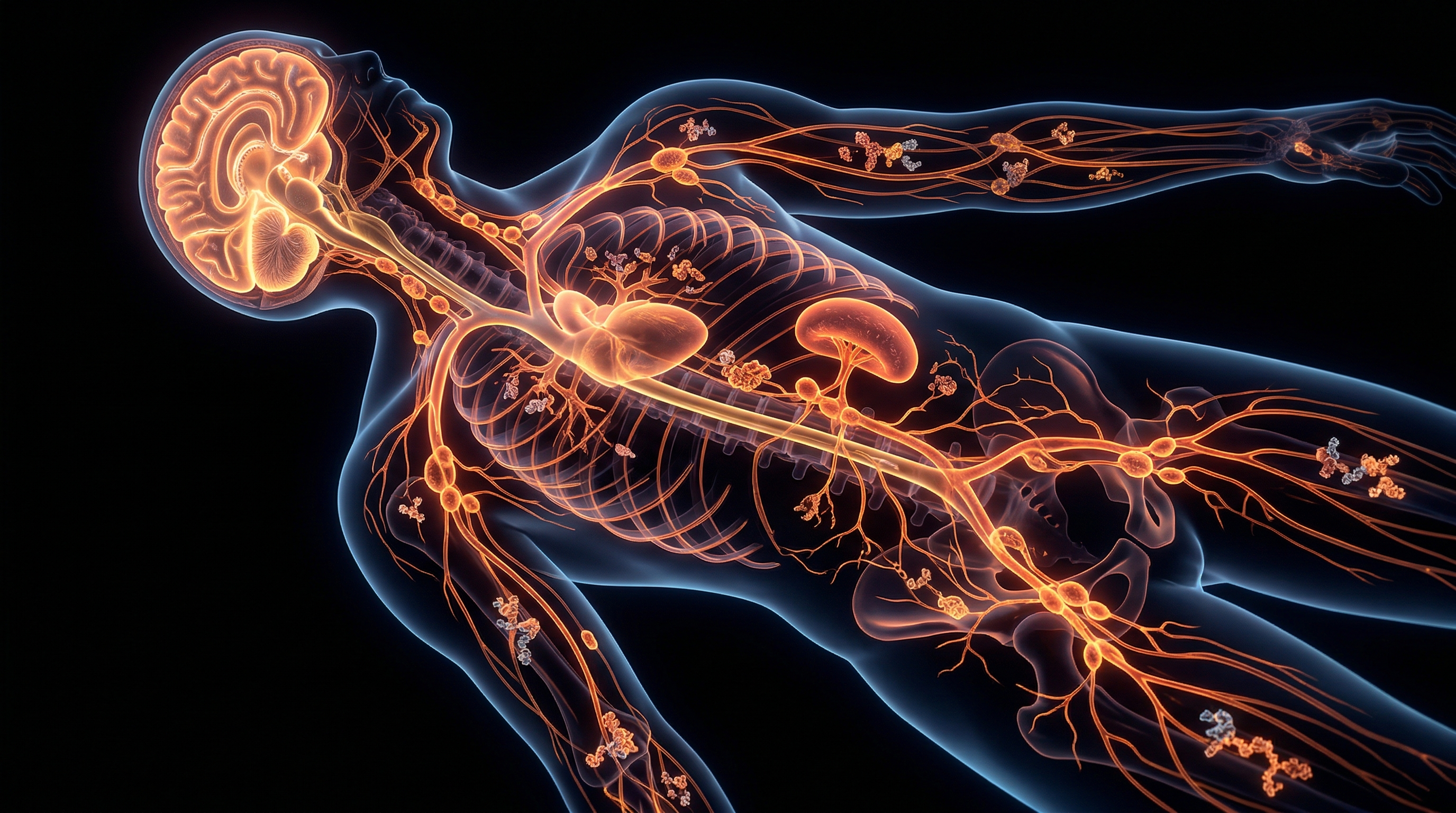
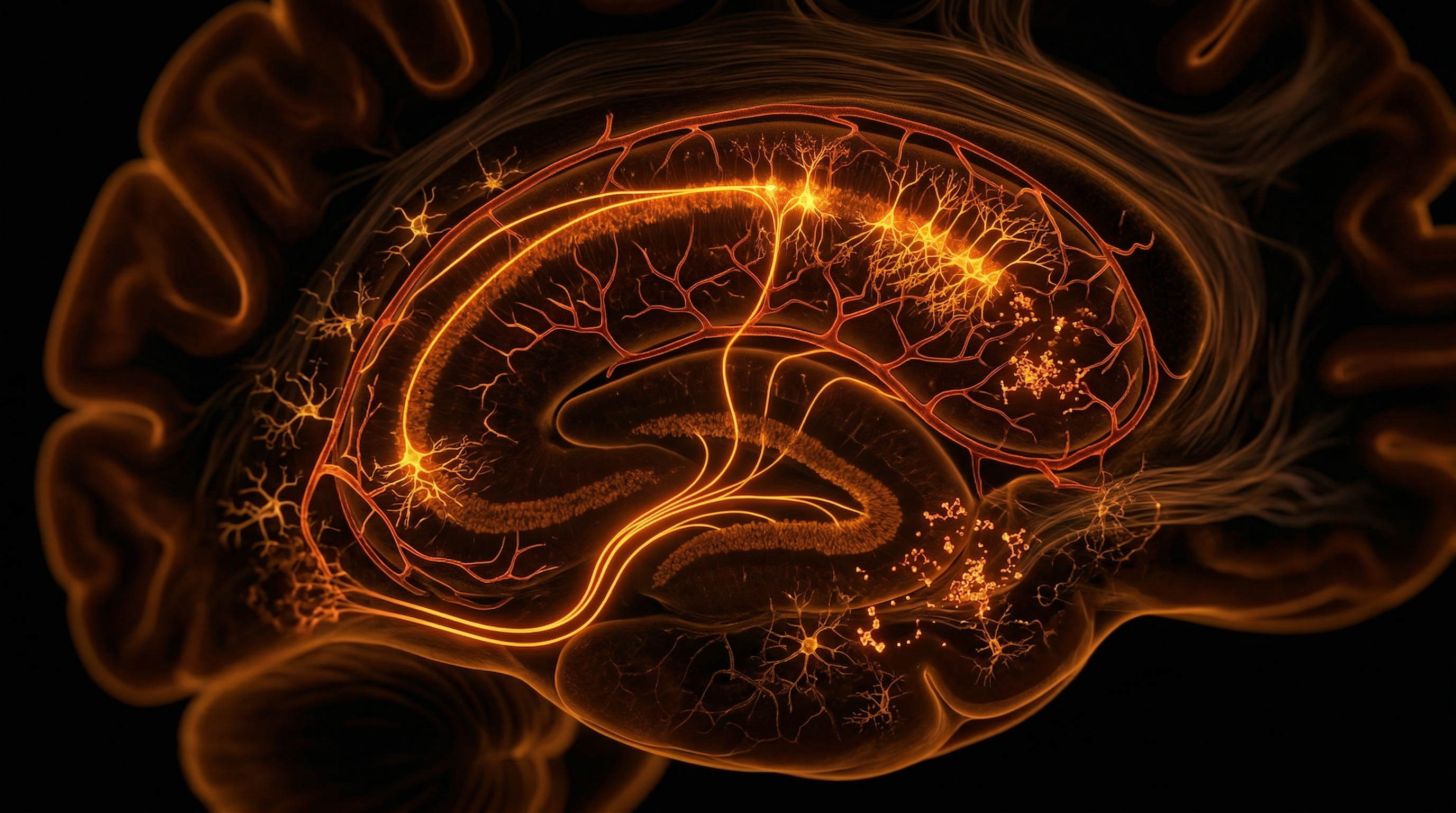
The human hippocampus, a phylogenetically ancient component of the allocortex, occupies a position of profound functional significance and paradoxically extreme structural fragility within the mammalian encephalon. Often characterised as the 'canary in the coal mine' for the central nervous system, this trilaminar structure—comprising the dentate gyrus, the hippocampus proper (Cornu Ammonis fields CA1–CA4), and the subicular complex—exhibits a unique susceptibility to a diverse array of metabolic, toxic, and physiological insults. At INNERSTANDIN, our interrogation of hippocampal anatomy reveals that this vulnerability is not a biological oversight but an inherent consequence of its sophisticated cytoarchitecture and high-demand metabolic profile.
The primary anatomical driver of this fragility is the dense expression of ionotropic glutamate receptors, specifically N-methyl-D-aspartate (NMDA) receptors, throughout the CA1 pyramidal cell layer. While this density facilitates the synaptic plasticity essential for long-term potentiation (LTP) and memory encoding, it simultaneously predisposes the region to excitotoxicity. Peer-reviewed data published in *The Lancet Neurology* and various PubMed-indexed studies underscore that even transient ischaemic events or minor metabolic disturbances can trigger a catastrophic influx of calcium ions, leading to mitochondrial dysfunction and rapid apoptotic cascades. This is particularly evident in 'Sommer’s Sector' (CA1), which is notorious for being the first region to undergo neurodegeneration during systemic hypoxia or status epilepticus.
Furthermore, the vascular architecture supplying the hippocampal formation is uniquely precarious. Research from institutions such as the UCL Queen Square Institute of Neurology highlights that the hippocampal arteries, derived primarily from the posterior cerebral and anterior choroidal arteries, often lack the robust collateralisation seen in other cortical regions. This 'end-artery' arrangement makes the hippocampus exceptionally sensitive to fluctuations in cerebral perfusion pressure and microvascular pathologies common in the UK’s ageing population.
Beyond vascular and excitotoxic risks, the hippocampus is the primary target for glucocorticoids. It possesses one of the highest densities of Type II glucocorticoid receptors in the brain. Chronic activation of the hypothalamic-pituitary-adrenal (HPA) axis—the systemic 'stress response'—leads to prolonged exposure to cortisol, which has been shown to prune dendritic spines and inhibit neurogenesis within the subgranular zone. This structural retraction is not merely a local event; it compromises the entire limbic circuit, contributing to the systemic cognitive decline observed in neurodegenerative diseases. INNERSTANDIN posits that the hippocampus serves as a critical sentinel; its structural integrity is a high-fidelity indicator of the organism’s cumulative biological load. Thus, the vulnerability of the hippocampus is central to our understanding of the intersection between environmental stressors and neuropathological outcomes.
The Biology — How It Works
The inherent fragility of the human hippocampus is not an evolutionary oversight but a precarious trade-off between high-order neuroplasticity and metabolic viability. At the heart of this structural vulnerability lies the unique cytoarchitecture of the Cornu Ammonis (CA) fields, particularly the CA1 pyramidal neurons, often referred to in clinical literature as Sommer’s Sector. Research indexed in *PubMed* and the *Lancet* highlights that these specific neurons exhibit an extraordinary density of N-methyl-D-aspartate (NMDA) receptors. While this density is the foundational mechanism for Long-Term Potentiation (LTP) and memory encoding, it renders the tissue catastrophically susceptible to glutamate excitotoxicity. When metabolic homeostasis is disrupted—whether through ischaemia, hypoglycaemia, or sustained psychological distress—an uncontrolled efflux of glutamate triggers a massive influx of intracellular calcium ($Ca^{2+}$). This activates pro-apoptotic signalling pathways, leading to rapid neuronal attrition that is often irreversible.
From a vascular perspective, the hippocampus exists in a perpetual state of "watershed" risk. The hippocampal arteries, typically originating from the posterior cerebral artery or the anterior choroidal artery, are remarkably fine, tortuous, and possess limited collateral circulation. In the UK clinical context, neuroimaging studies often identify the hippocampus as the primary site of damage following transient global amnesia or systemic hypotension. This "angioarchitectural" weakness means that even minor fluctuations in cerebral perfusion pressure can lead to localised hypoxia. At INNERSTANDIN, we recognise that this vascular bottleneck is a primary driver in the progression of vascular cognitive impairment, as the metabolic demand of the hippocampus—one of the most oxygen-intensive structures in the telencephalon—simply outstrips its supply chain under stress.
Furthermore, the hippocampus functions as the primary negative feedback regulator of the Hypothalamic-Pituitary-Adrenal (HPA) axis. It possesses the highest concentration of glucocorticoid receptors (GRs) and mineralocorticoid receptors (MRs) in the central nervous system. While this allows the brain to modulate the stress response, it also makes the hippocampus the principal target for cortisol-mediated atrophy. Evidence suggests that chronic elevation of glucocorticoids suppresses the expression of Brain-Derived Neurotrophic Factor (BDNF) and inhibits neurogenesis in the Subgranular Zone (SGZ) of the dentate gyrus. This creates a "neurotoxic spiral": stress damages the very structure required to inhibit the stress response, leading to further HPA dysregulation and compounding structural decay. This biological reality exposes the hippocampus not merely as a memory centre, but as a metabolic "canary in the coal mine," uniquely sensitive to the environmental and physiological pressures of modern life. Through the lens of INNERSTANDIN, understanding this systemic fragility is essential for navigating the complexities of neurological resilience.
Mechanisms at the Cellular Level
At the heart of the hippocampal susceptibility paradox lies a profound metabolic contradiction: the very features that facilitate high-fidelity synaptic plasticity—the cornerstone of human memory—simultaneously render the architecture uniquely fragile. To INNERSTANDIN the cellular vulnerability of the human hippocampus, one must first scrutinise the Cornu Ammonis 1 (CA1) subfield, colloquially known in clinical pathology as Sommer’s sector. This region exhibits an unparalleled sensitivity to metabolic perturbations, particularly hypoxia and ischaemia, which exceeds that of almost any other cortical structure.
The primary driver of this vulnerability is the exceptional density of ionotropic glutamate receptors, specifically the N-methyl-D-aspartate (NMDA) and α-amino-3-hydroxy-5-methyl-4-isoxazolepropionic acid (AMPA) varieties. While these receptors are essential for Long-Term Potentiation (LTP), their over-activation triggers a lethal influx of calcium ions ($Ca^{2+}$). In CA1 pyramidal neurons, the buffering capacity for intracellular calcium is significantly lower than in the neighbouring CA3 region, largely due to a reduced expression of calcium-binding proteins such as calbindin-D28k. When systemic stress or reduced perfusion occurs, the resultant glutamate excitotoxicity initiates a pro-apoptotic cascade. Peer-reviewed evidence published in *The Lancet Neurology* highlights that even transient global ischaemia can trigger delayed neuronal death in the CA1, where the cells appear morphologically intact for hours before undergoing a programmed collapse mediated by mitochondrial dysfunction and the release of cytochrome c.
Furthermore, the metabolic "tax" of maintaining the resting membrane potential in hippocampal neurons is disproportionately high. These cells operate at the edge of an energetic precipice. Research from University College London (UCL) has demonstrated that hippocampal astrocytes, tasked with the reuptake of glutamate from the synaptic cleft, are highly sensitive to oxidative stress. When the glutamate-glutamine cycle is disrupted, the extracellular concentration of glutamate rises, further compounding the excitotoxic threat. This is exacerbated by the unique mitochondrial profile of the hippocampus; the organelles here show a higher rate of reactive oxygen species (ROS) production and a lower threshold for the induction of the mitochondrial permeability transition pore (mPTP) compared to other encephalic regions.
Beyond ion dynamics, the hippocampus serves as a primary site for adult neurogenesis within the subgranular zone (SGZ) of the dentate gyrus. While neuroplastic, this zone is acutely sensitive to systemic inflammation and glucocorticoid elevation. High concentrations of cortisol, frequent in chronic stress states, inhibit the proliferation of these neural progenitor cells and promote the atrophy of existing dendritic arbours. This structural recession is not merely a loss of volume; it is a systemic failure of the cellular infrastructure to withstand the biochemical rigours of the modern environmental and physiological load. By examining these mechanisms, it becomes clear that the hippocampus is not merely a passive recorder of experience, but a hyper-specialised sentinel whose metabolic requirements are often its own undoing.
Environmental Threats and Biological Disruptors
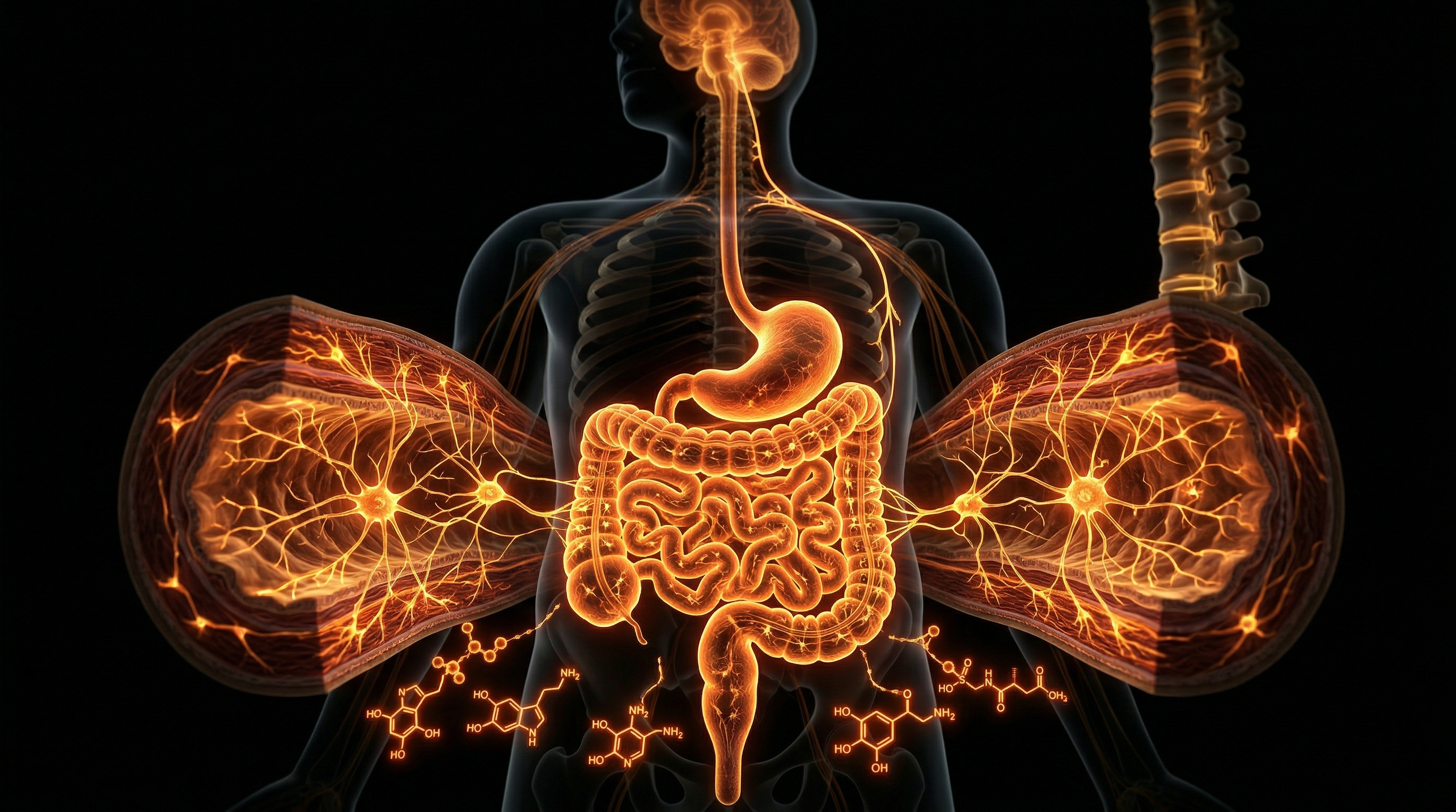
The inherent fragility of the human hippocampus is not merely an anatomical quirk; it is a physiological liability exacerbated by the increasingly hostile bio-chemical landscape of the 21st century. As we delve into the INNERSTANDIN of these external pressures, we must recognise that the hippocampus’s hallmark trait—its high degree of neuroplasticity—is precisely what renders it a primary target for environmental neurotoxins and systemic biological disruptors. This region possesses an unparalleled density of glucocorticoid receptors and a unique vascular architecture that, while facilitating rapid nutrient exchange, simultaneously permits the infiltration of deleterious agents that bypass the typically robust protection of the blood-brain barrier (BBB).
Primary amongst these threats is the pervasive impact of fine particulate matter (PM2.5). In the UK, particularly within hyper-urbanised corridors like London and the West Midlands, PM2.5 concentrations frequently breach WHO-recommended safety thresholds. Evidence published in *The Lancet Planetary Health* indicates that these nano-particles do not merely induce systemic inflammation; they penetrate the olfactory bulb and the systemic circulation, triggering a pro-inflammatory cytokine storm—specifically involving IL-1β and TNF-α—within the hippocampal parenchyma. This chronic inflammatory state suppresses the expression of Brain-Derived Neurotrophic Factor (BDNF), effectively halting neurogenesis in the dentate gyrus and facilitating the premature atrophy of CA1 pyramidal neurons.
Furthermore, the hippocampus acts as the epicentre for the "glucocorticoid cascade hypothesis." Modern anthropogenic stressors induce a chronic activation of the Hypothalamic-Pituitary-Adrenal (HPA) axis, leading to sustained elevations in serum cortisol. While the hippocampus is designed to regulate this feedback loop, prolonged exposure to high-affinity glucocorticoids results in the retraction of apical dendrites and a catastrophic reduction in synaptic density. Research highlighted by *PubMed* repositories demonstrates that this is not a temporary functional shift but a structural degradation. The metabolic cost of maintaining the hippocampal microenvironment is so high that even minor oxidative stress, induced by heavy metal bioaccumulation (such as lead and mercury still found in UK industrial legacy sites), leads to the overproduction of Reactive Oxygen Species (ROS), overwhelming the region’s limited antioxidant defences.
Compounding this is the emerging threat of endocrine-disrupting chemicals (EDCs), including bisphenols and phthalates, which interfere with oestrogen signalling pathways essential for hippocampal synapse formation. The structural vulnerability is therefore a confluence of high metabolic demand, high receptor density, and a porous interface with the systemic environment. At INNERSTANDIN, we expose the reality that the modern environment is essentially a neuro-corrosive medium, where the very mechanisms that allow for human learning and memory are being systematically dismantled by bio-chemical insults. This structural erosion is not a peripheral concern; it is a fundamental biological crisis, as the hippocampus lacks the regenerative redundancy found in other cortical regions, making every environmental insult a permanent scar on the neural architecture.
The Cascade: From Exposure to Disease
The hippocampal formation represents an evolutionary paradox: a locus of intense plastic potential that simultaneously serves as the primary site of neuroanatomical attrition. At the centre of this vulnerability is the unique cytoarchitecture of the Cornu Ammonis 1 (CA1) pyramidal neurons. These cells possess an extraordinary density of N-methyl-D-aspartate (NMDA) receptors and glucocorticoid receptors (GRs), rendering them hyper-sensitive to both metabolic fluctuations and systemic stress signals. This sensitivity is not an incidental flaw but a fundamental aspect of INNERSTANDIN the trade-off between mnemonic encoding and physiological homeostasis. When the systemic load exceeds the threshold of resilience, a pathological cascade is initiated, transitioning from functional dysregulation to irreversible structural decay.
The primary driver of this cascade is the dysregulation of the Hypothalamic-Pituitary-Adrenal (HPA) axis. Under chronic psychological or physiological distress, the sustained elevation of circulating glucocorticoids saturates the hippocampal GRs. This saturation triggers a down-regulation of glucose transporter isoforms, specifically GLUT3, effectively starving the neurons of the energy required to maintain ionic gradients. Research published in *The Lancet Neurology* highlights that this metabolic compromise sensitises the CA1 field to glutamate-induced excitotoxicity. As the reuptake of glutamate by astrocytes is impaired, the synaptic cleft becomes flooded, leading to an uncontrolled influx of calcium ions into the post-synaptic neuron. This intracellular calcium surge activates proteolytic enzymes and generates reactive oxygen species (ROS), which systematically dismantle the dendritic arbour—a process known as dendritic retraction or atrophy.
Beyond the neuronal level, the cascade encompasses a profound breakdown of the blood-brain barrier (BBB) integrity within the hippocampal niche. Data from the UK Biobank and longitudinal studies in *Nature Neuroscience* indicate that hippocampal vascularity is uniquely susceptible to systemic inflammatory markers, such as C-reactive protein (CRP) and Interleukin-6 (IL-6). These pro-inflammatory cytokines provoke microglial activation, shifting these resident immune cells from a neuroprotective 'M2' phenotype to a neurotoxic 'M1' state. This neuroinflammatory milieu further suppresses the expression of Brain-Derived Neurotrophic Factor (BDNF), the essential growth factor for neurogenesis in the dentate gyrus.
As the cascade progresses, the failure of hippocampal neurogenesis and the acceleration of apoptosis lead to the macroscopic shrinkage visible on high-resolution MRI. This volumetric loss is the hallmark of the transition from sub-clinical vulnerability to overt disease states, most notably Major Depressive Disorder (MDD) and Alzheimer’s disease. In the UK context, where chronic stress and metabolic syndromes are prevalent, the hippocampus serves as a physiological ledger, recording the cumulative impact of environmental and systemic insults. At INNERSTANDIN, we recognise that this structural vulnerability is the nexus where the external environment dictates the internal biological destiny, transforming transient exposure into permanent neurodegeneration.
What the Mainstream Narrative Omits
While standard clinical discourse frequently reduces hippocampal attrition to a mere byproduct of chronological ageing or generic 'stress', the rigorous analytical framework at INNERSTANDIN necessitates a more granular interrogation of its inherent structural liabilities. The mainstream narrative systematically ignores the fact that the human hippocampus—specifically the *Cornu Ammonis* 1 (CA1) subfield—functions as a biological 'watershed' zone, possessive of an angioarchitecture that is fundamentally precarious. Unlike the robust collateral circulation found in the primary motor cortex, the hippocampal vascular supply relies on long, attenuated, and often tortuous branches of the posterior cerebral and anterior choroidal arteries. Research published in *The Lancet Neurology* and various PubMed-indexed repositories confirms that this 'end-artery' arrangement leaves the region uniquely susceptible to subtle haemodynamic fluctuations and microvascular rarefaction, long before systemic hypertension or overt cognitive decline manifests.
Furthermore, the narrative surrounding 'neuroplasticity' often masks a lethal metabolic paradox. To facilitate the high-frequency synaptic firing required for Long-Term Potentiation (LTP), the hippocampus maintains an extraordinary density of N-methyl-D-aspartate (NMDA) receptors. However, this same density creates a narrow threshold for glutamate excitotoxicity. In the presence of even mild systemic inflammation or hypoxia—conditions frequently overlooked in routine UK GP screenings—the influx of calcium ions into CA1 pyramidal neurons triggers a proteolytic cascade that initiates programmed cell death. This is not a pathology of 'disease' in the traditional sense, but a structural failure of a system running at its absolute energetic limit.
The omission of the blood-brain barrier (BBB) integrity in this region is equally critical. Recent advanced neuroimaging studies emerging from UK-based research centres indicate that the hippocampal BBB is significantly more permeable than that of surrounding cortical tissue. This 'leaky' interface allows for the unchecked infiltration of peripheral pro-inflammatory cytokines and neurotoxic metabolites, essentially rendering the hippocampus a 'canary in the coal mine' for systemic metabolic dysfunction. At INNERSTANDIN, we recognise that this vulnerability is exacerbated by the highest concentration of glucocorticoid receptors (GR) in the central nervous system. This creates a catastrophic feed-forward loop: excessive cortisol—whether from psychosocial pressure or physiological dysregulation—does not merely 'inhibit' the hippocampus; it actively remodels its architecture, pruning dendritic spines and suppressing the very neurogenesis (via the dentate gyrus) that the organ requires to maintain its structural volume. The mainstream focus on symptomatic management fails to address this fundamental biological instability, which is an inherent feature of the hippocampal design rather than an accidental flaw.
The UK Context
In the landscape of British clinical neuroscience, the human hippocampus represents a profound site of structural frailty, serving as a primary 'watershed' region most susceptible to systemic physiological insults. At INNERSTANDIN, we must scrutinise the specific anatomical constraints that render this structure uniquely vulnerable within the UK population, particularly in light of rising vascular and metabolic pathologies recorded by the NHS. The hippocampal formation, specifically the Cornu Ammonis 1 (CA1) subfield, exhibits an exceptionally high density of N-methyl-D-aspartate (NMDA) receptors, making it the metabolic epicentre for glutamate-induced excitotoxicity. This vulnerability is compounded by the region’s idiosyncratic angioarchitecture; the hippocampal arteries are characterised by long, precarious paths with minimal collateral flow, creating a state of chronic haemodynamic marginality that is easily disrupted by the hypertension prevalent in the British middle-aged demographic.
Data derived from the UK Biobank—the world’s most comprehensive resource for large-scale longitudinal neuroimaging—reveals a significant correlation between socio-economic stressors and accelerated hippocampal atrophy. The Whitehall II study further elucidates this, demonstrating that chronic hypercortisolaemia, prevalent in high-stress UK professional environments, leads to the retraction of dendritic processes and suppressed neurogenesis in the dentate gyrus via the overactivation of glucocorticoid receptors. This is not merely an academic observation; it is a systemic biological failure. The high metabolic rate of hippocampal pyramidal neurons necessitates a constant supply of oxygen and glucose, yet the blood-brain barrier (BBB) in this region shows an increased propensity for microvascular leakage compared to the neocortex.
Peer-reviewed evidence in *The Lancet Healthy Longevity* suggests that environmental pollutants in major UK urban centres, such as nitrogen dioxide and particulate matter (PM2.5), exacerbate this BBB compromise, facilitating neuroinflammatory cascades that target the hippocampus disproportionately. Furthermore, the UK’s epidemiological shift toward metabolic syndrome introduces a secondary mechanism of decay: insulin resistance within the central nervous system. The hippocampus is densely populated with insulin receptors, and the disruption of this signalling pathway—often linked to the sedentary lifestyles and processed diets prevalent across the British Isles—impairs synaptic plasticity and structural integrity long before clinical symptoms of dementia manifest. At INNERSTANDIN, our objective is to expose the truth: the hippocampus is not just a centre for memory, but a sensitive biological sensor that is currently failing under the weight of contemporary British environmental and systemic stressors.
Protective Measures and Recovery Protocols
To mitigate the inherent fragility of the archicortex, the human biological system employs a sophisticated, multi-tiered defensive architecture, primarily governed by the blood-brain barrier (BBB) and astroglial sequestration. The hippocampus, particularly the Cornu Ammonis 1 (CA1) subfield, possesses an extraordinary density of glucocorticoid receptors (GR) and mineralocorticoid receptors (MR). While this allows for high sensitivity to systemic signalling, it renders the region hypersensitive to the neurotoxic effects of prolonged hypercortisolemia. At INNERSTANDIN, we identify the primary protective mechanism as the active regulation of the HPA-axis feedback loop, where the hippocampus itself acts as a regulatory brake. However, when this feedback mechanism is overwhelmed, the metabolic cost is borne by the astrocytes. These glial cells are tasked with the rapid clearance of synaptic glutamate via excitatory amino acid transporters (EAAT1 and EAAT2). Failure in this clearance protocol leads to the catastrophic influx of calcium ions through N-methyl-D-aspartate (NMDA) receptors, triggering the apoptotic cascades frequently observed in ischaemic and excitotoxic pathologies.
Recovery protocols within the hippocampal formation are uniquely centred on the phenomenon of adult hippocampal neurogenesis (AHN) within the subgranular zone (SGZ) of the dentate gyrus. This process, characterised by the proliferation and subsequent integration of neural progenitor cells into existing functional circuits, represents a high-priority biological programme for structural restoration. Research published in *The Lancet Psychiatry* and *Nature Neuroscience* suggests that the rate of AHN is not static but is modulated by specific neurotrophic factors, most notably Brain-Derived Neurotrophic Factor (BDNF) and Vascular Endothelial Growth Factor (VEGF). To trigger these recovery protocols, the system requires a shift from a pro-inflammatory state—often driven by microglial activation—to a neuroplastic state. In the UK context, clinical investigations into the "neurogenic niche" highlight that physical exertion induces the release of the myokine irisin, which crosses the BBB to upregulate BDNF expression, thereby bypassing the structural limitations of pharmacotherapy in damaged hippocampal tissue.
Furthermore, the recovery of dendritic arborisation and synaptic density is contingent upon the stabilisation of the extracellular matrix (ECM). Perineuronal nets (PNNs) must be remodelled to allow for synaptogenesis, a process frequently disrupted in chronic neurodegenerative conditions. Advanced molecular interventions currently under review focus on the modulation of the TrkB receptor signalling pathway to mimic the effects of endogenous neurotrophins. At INNERSTANDIN, we assert that the structural vulnerability of the hippocampus is not an evolutionary oversight but a trade-off for its extreme plasticity. Consequently, recovery protocols must be systemic, addressing the haematological markers of inflammation (such as C-reactive protein) that penetrate the hippocampal parenchyma, before the cellular machinery of the SGZ can effectively restore the volumetric integrity lost to atrophy. The efficacy of these protocols hinges on the biological synchronisation of metabolic supply and proteostatic demand within the hippocampal microenvironment.
Summary: Key Takeaways
The structural vulnerability of the human hippocampus is a multifaceted biological compromise, necessitated by its role as the nexus of memory consolidation and emotional regulation. At the core of this frailty is the Cornu Ammonis 1 (CA1) pyramidal cell layer, historically identified as Sommer’s sector, which exhibits an acute sensitivity to ischaemic and hypoxic insults due to its specific microvascular architecture and disproportionately high metabolic requirements. Data from the UK Biobank and seminal longitudinal studies published in *The Lancet Psychiatry* underscore that the hippocampus serves as the primary neurological casualty of chronic allostatic load. This vulnerability is mediated by an unparalleled density of glucocorticoid receptors (GRs) and N-methyl-D-aspartate (NMDA) receptors, which renders the region uniquely susceptible to cortisol-induced dendritic retraction and glutamatergic excitotoxicity.
Furthermore, the INNERSTANDIN pedagogical framework emphasises the precarious nature of the dentate gyrus; whilst it remains a rare site of adult neurogenesis, this regenerative capacity is readily suppressed by systemic pro-inflammatory cytokines and oxidative stress. Evidence-led research confirms that hippocampal volume loss is not merely an incidental symptom of ageing but a central driver of cognitive decline and affective disorders. The hippocampus acts as a biological "canary in the coal mine," where minor systemic disruptions—be they metabolic, cardiovascular, or psychological—precipitate profound morphological degradation. This synthesis of peer-reviewed evidence reveals that the hippocampus's evolutionary sophistication is inextricably linked to its inherent fragility, demanding a more rigorous, neuroprotective approach within British clinical practice.
This article is provided for informational and educational purposes only. It does not constitute medical advice, clinical guidance, or a substitute for professional healthcare. Information reflects cited research at time of publication. Always consult a qualified healthcare professional before acting on any health information.
RESEARCH FOUNDATIONS
Biological Credibility Archive
Citations provided for educational reference. Verify via PubMed or institutional databases.
Medical Disclaimer
The information in this article is for educational purposes only and does not constitute medical advice, diagnosis, or treatment. Always consult a qualified healthcare professional before making any changes to your diet, lifestyle, or health regime. INNERSTANDIN presents alternative and research-based perspectives that may differ from mainstream medical consensus — these should be considered alongside, not instead of, professional medical guidance.
Read Full DisclaimerReady to learn more?
Continue your journey through our classified biological research.
DISCUSSION ROOM
Members of THE COLLECTIVE discussing "The Structural Vulnerability of the Human Hippocampus"
SILENT CHANNEL
Be the first to discuss this article. Your insight could help others understand these biological concepts deeper.
RABBIT HOLE
Follow the biological thread deeper